小麦分蘖形态学特征X射线-CT无损检测
2017-11-24 06:07杨万能牛智有黄成龙
农业工程学报 2017年14期
吴 迪,杨万能,2,牛智有,黄成龙※
(1. 华中农业大学工学院,武汉 430070;2. 华中农业大学作物遗传改良国家重点实验室,武汉 430070)
小麦分蘖形态学特征X射线-CT无损检测
吴 迪1,杨万能1,2,牛智有1,黄成龙※1
(1. 华中农业大学工学院,武汉 430070;2. 华中农业大学作物遗传改良国家重点实验室,武汉 430070)
针对传统小麦分蘖形态学特征需采用人工接触或有损方法获取,不仅过程繁琐、主观性强而且会影响小麦后续生长。为实现小麦分蘖性状快速、准确、无损测量,该文提出一种基于X射线断层成像的小麦分蘖形态学特征提取方法。首先构建了小麦分蘖X-ray CT断层扫描成像系统,采用滤波反投影(filtered back projection,FBP)断层重建算法和图形处理单元(graphics processing unit,GPU)加速算法快速获取小麦分蘖茎秆断层图像,设计了专门的分蘖图像分析算法实现对小麦分蘖形态学特征参数(分蘖数、分蘖角度、分蘖茎粗和分蘖壁厚)的无损检测。该研究对107株小麦植株测量结果表明:分蘖数测量准确率可达100%,分蘖角度、茎粗和壁厚的平均测量误差分别为 3.65%,4.84%和7.86%。该技术相较于人工测量方法和石蜡切片方法,能够对小麦分蘖形态学特征进行较为精准无损检测,实现单株测量效率约200 s,对于小麦功能基因组和抗倒伏能力品种的筛选具有重要的研究意义。
X射线;断层重建;图像处理;小麦分蘖;无损检测
0 引 言
小麦是中国乃至世界最主要的粮食作物之一[1-3]。小麦是人类淀粉的主要摄取来源,同时富含蛋白质、脂肪、矿物质等丰富的营养[4-5]。为进一步提高小麦产量,小麦形态改良[6]逐渐从矮化育种转向增加株高方向进行育种[7-8]。小麦分蘖起到了养分输送与抗倒伏的作用,分蘖性状与产量密切相关[9],小麦分蘖数与产量直接相关,分蘖角度影响小麦的株型结构,分蘖茎粗、壁厚决定小麦的抗倒伏能力[10-11],因此小麦分蘖性状研究对小麦育种具研究具有十分重要的意义[12]。
随着小麦功能基因组及分子育种的飞速发展,需要对大量品种进行表型鉴定。传统的小麦分蘖检测手段主要依靠人工完成,通过计量工具测量并计算水稻分蘖的分蘖数量、分蘖角度、分蘖茎粗、分蘖壁厚[13-14]。人工检测为有损离体测量,具有测量精度低、可重复性差等缺点,已经远远不能满足当前小麦育种的需求。因此研发一种无损且精度高的小麦分蘖性状提取技术,是小麦功能基因组发展急需解决的一个问题[15-16]。
近年来,机器视觉技术因其无损性和高效性正广泛应用于农产品检测[17-18]。杜光源等[19]利用核磁共振可以测量小麦叶片衰老态势,邓继忠等[20]采用图像识别技术可以对小麦腥黑穗病进行特征提取与分类,张云鹤等[21]利用可见光视觉技术研发了作物分蘖直径变化测量仪。Bauriegel等[22]采用高光谱成像技术可以对小麦镰刀霉病进行早期预测。综上所述,基于可见光、高光谱、核磁共振等反射成像技术,只能获取对小麦表观性状的信息,无法用于获取分蘖内结构信息。而X射线作为一种透射成像技术,目前已经应用于土壤孔隙检测[23]、植物根系研究[24],木材内部裂纹、孔洞缺陷检测[25],水稻分蘖数测量[26],为小麦分蘖性状获取提供了一种可行的途径。
本文通过采用自动控制系统、微型CT(computerized tomography)技术及图像处理技术相结合的方式,研制了一套自动化的表型提取设备,以完成对小麦分蘖数、分蘖角度、分蘖茎粗、分蘖壁厚等形态学特征性状的在体无损检测。
1 小麦CT系统组成和成像结构
1.1 系统组成
本文设计的CT系统主要由计算机1(双核CPU 3.2 GHz,内存2.98 GB,显卡ATI Radeon HD 5450)、PLC控制器 2(programmable logic controller)(CP1H,OMRON,日本)、微焦斑射线源3(Nova 600,OXFORD,英国)、旋转平台4、平板探测器5(2520DX,VARIAN,美国)等5个功能模块组成[27-28],系统结构示意图如图1所示。先将待测样本小麦放置在载物旋转台上,微焦斑射线源发射X射线穿透小麦样本,通过计算机系统发送指令给PLC,PLC控制载物旋转台实现样本的等间距旋转,利用平板探测器获取每个角度下的X射线吸收面阵列图像,取同一高度的全部角度图像组成新的图像,即投影正弦图,再结合滤波反投影 FBP(filtered backprojection)算法[29]和图形处理GPU(graphics processing unit)加速算法[30]得到断层重建图像,最后基于图像处理技术对断层重建图像进行图像分析与处理,测算并提取出小麦分蘖的性状参数[31]。CT重建的原理源于X射线通过物体时衰减的物理规律[32-33],即郎伯定律如式(1),然后结合Radon变换得到投影重建图像。

式中I为平板探测器探测到的X射线的强度,I0为入射X射线的强度(Ci),µ为成像物质的吸收系数,L为物体的厚度(mm)。
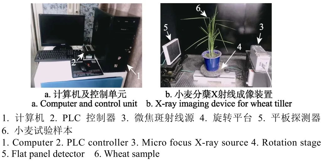
图1 CT系统组成示意图Fig.1 Schematic diagram of CT system
本试验的研发主要是基于美国国家仪器公司(National Instruments)的LabVIEW软件、VARIAN公司的VIVA软件、美国微软公司的Microsoft Visual Studio软件。首先通过 VIVA软件完成平板探测器的偏移校准和增益校准。然后通过LabVIEW软件完成X射线图像采集和载物台旋转控制功能。再利用Microsoft Visual Studio软件来实现断层重建和分蘖图像处理算法,并生成动态链接库(dynamic link library,DLL)。最后利用LabVIEW将上述功能集成,实现小麦分蘖图像校准、采集、处理一体化。
1.2 成像结构
由于微焦斑射线源的出射角为33°,结合平板探测器的尺寸(195 mm×244 mm),可计算出当射线源与平板探测器之间的距离为634 mm时射线源发出的X射线刚好全部覆盖平板探测器,考虑到小麦样本的实际尺寸并获取尽可能高的空间分辨率,本试验中选取的微焦斑射线源至试验样本的距离为305 mm,基于该成像结构可获取的X-ray投影及重建分辨率可达61μm × 61μm。试验选用的具体性能参数如表1所示。
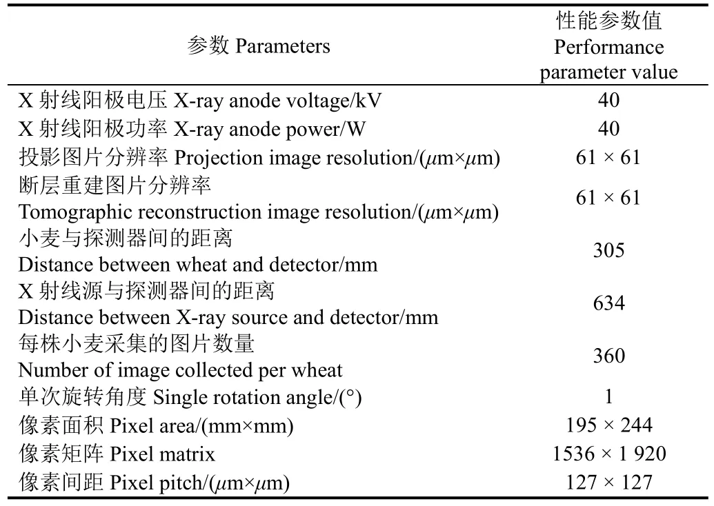
表1 小麦CT系统性能参数Table 1 CT system performance parameters for wheat
2 试验材料及方法
2.1 试验材料
本试验一共选取5个不同的小麦品种(华麦2668,华麦 2533,华麦 166,华麦 2566,华麦 2153),每个品种有25株,一共125株小麦样本,试验样本盆栽种植。用于种植试验样本小麦的土壤先在太阳下晒干,然后将晒干的土壤初步碾碎,通过8 mm的网筛去除土壤中的石头和杂草,保证土壤的均一性,最后将氮、磷、钾肥料按照 2∶1∶1的配比搅拌均匀。在小麦生长过程中提供正常的水肥管理,待小麦播种90 d后开始进行小麦分蘖形态参数的无损检测及人工验证。
2.2 试验具体步骤
CT系统具体的控制操作步骤如下:1)打开X射线源冷却装置,使得射线源温度控制在 20 ℃以内;2)打开射线源和探测器,对系统进行校准;3)探测器采集图片,并判断采集的图片信噪比是否符合要求;4)采集完一株样本后,放置下一株样本继续采集,直至全部样本采集完成;5)关闭射线源、探测器和计算机。
由于天气等外在原因,最终用于试验的小麦样本为107株小麦。人工测量的具体测量步骤为:1)2名试验人员分别对每株小麦数取分蘖数量,用量角器分别获取分蘖角度,取 2人所得值的平均值做为分蘖数和分蘖角度的人工测量值;2)对每株小麦进行CT系统检测,采集投影图片;3)将每株小麦剪剩至1根分蘖为止(方便后期与人工值进行匹配对应),对其再次进行 CT系统检测,采集投影图片;4)2名试验人员利用游标卡尺,分别对剩下的1根分蘖测量分蘖茎粗和分蘖壁厚,取2人所得值的平均值做为分蘖茎粗和分蘖壁厚的人工测量值。
2.3 试验方法和性状提取
本文提出的基于X射线微型CT的小麦分蘖性状无损提取方法,技术路线如图 2所示,具体效果如图 3所示。
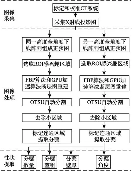
图2 小麦分蘖性状提取技术路线Fig.2 Flow chart of technical route for wheat tiller traits extraction
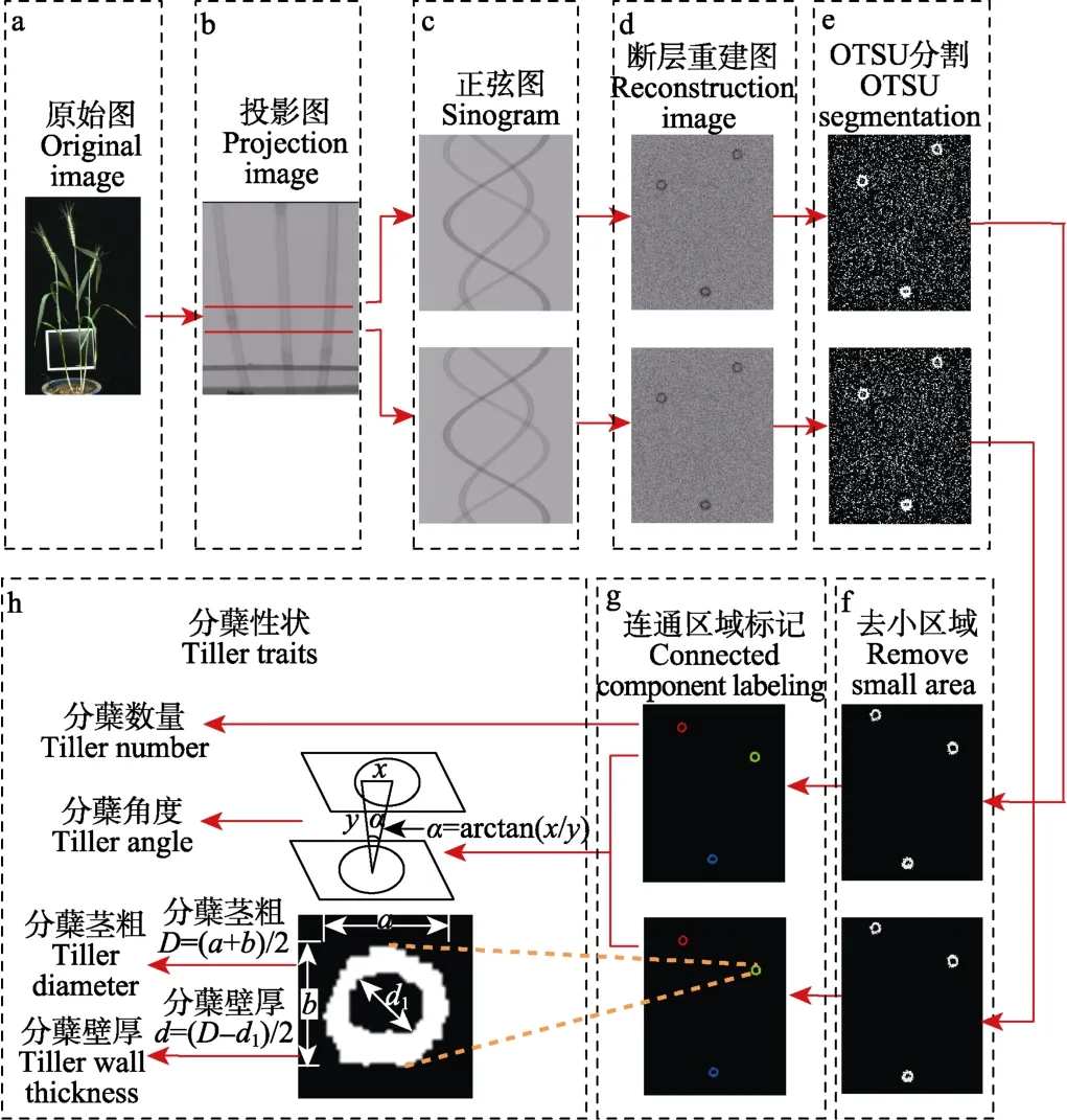
图3 图像处理及分蘖性状提取流程Fig.3 Flow chart of image processing and tiller traits extraction
2.3.1 小麦分蘖断层结构快速重建
对于小麦植株如图 3a,首先利用 CT系统获取小麦分蘖X-ray投影图像如图3b,然后选取两个不同高度及360个角度下的投影图像组成正弦图如图3c,再基于滤波反投影FBP算法,并结合平板探测器的偏移校准、增益校准降噪方法来获取清晰的小麦分蘖断层结构,其重建效果如图3d所示。
本研究还利用感兴趣区域(region of interest,ROI)算法和图像处理单元加速算法,进一步减少CT断层重建所消耗的时间,实现小麦分蘖内部断层结构的快速重建。
2.3.2 小麦分蘖内部性状的提取
小麦分蘖断层图像处理及性状提取步骤如下所述。
针对小麦分蘖断层重建图像,首先采用大津算法(OTSU)自动计算分割阈值,利用该阈值将图像进行二值化[32](如图3e),然后利用去除小区域算法消除背景噪声(如图3f),再采用连通区域标记方法识别出单个小麦分蘖区域(如图3g),最后计算出小麦分蘖性状的分蘖数、分蘖茎粗、分蘖壁厚和分蘖角度(如图3h),具体计算方法如下。
分蘖数量的计算:数取连通区域的个数;
分蘖角度α的计算:对同一对象,分别在两个不同高度的断层面取质心,投影到同一平面后,计算平面内 2点的欧氏距离x,并且系统已知两个断层面的垂直距离y,因此可以计算出分蘖角度α(°)。

分蘖茎粗D的计算:将单个分蘖先进行旋转,再测量长轴a与短轴b,从而计算出茎粗

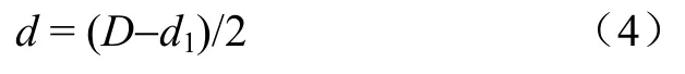
分蘖壁厚d的计算:按茎粗的计算方法先获取内部髓腔的直径d1,从而计算出壁厚
2.3.3 系统评估
基于上述图像分析过程,将系统测量的 107份小麦植株分蘖数、分蘖角度、分蘖茎粗、分蘖壁厚与人工测量值对比来评价该方法的精度。测量结果所得的平均绝对百分比误差(mean absolute percentage error,MAPE)和标准差(root mean square error,RMSE)的计算方法为
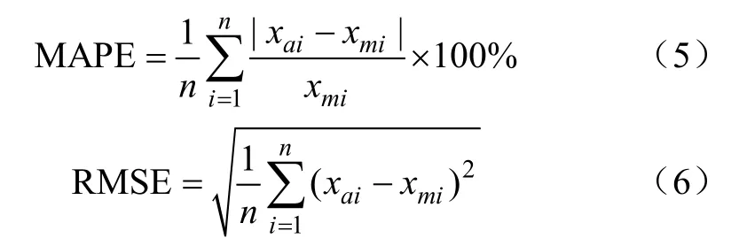
式中n为样本数,xai为系统测量值,xmi为人工测量值。
3 试验结果与分析
3.1 系统测量精度
基于上述小麦分蘖重建和图像分析过程,本试验对107份小麦植株提取的所有分蘖内部性状进行分析。将本系统测量的分蘖数、分蘖角度、茎粗和壁厚与人工测量值对比来评价本系统的精度。其中,人工测量小麦样本的植株位置与系统测量高度基本一致(均为距离土面5 cm处),以减少不同高度下所产生的分蘖角度、茎粗和壁厚误差。
图4a是分蘖数的系统测量值与人工测量值的对比散点图,可看出R2= 1,MAPE = 0%,RMSE = 0,本系统测量分蘖数的准确性为100%。图4b是分蘖角度的系统测量值与人工测量值的对比散点图,可看出R2= 0.77,MAPE = 3.65%,RMSE = 2.96°。图4c是茎粗的系统测量值与人工测量值的对比散点图,可看出R2= 0.91,MAPE =4.84%,RMSE = 0.17 mm,散点大部分落1∶1线右侧,说明大部分系统测量值较人工测量大,这可能是由于试验人员用游标卡尺对分蘖茎粗进行测量时,卡尺用力将茎秆夹紧时,茎秆变细导致的人工测量误差。图4d是壁厚的系统测量值与人工测量值的对比散点图,可看出R2= 0.87,MAPE = 7.86%,RMSE=0.12 mm。结果表明本系统具有较高的测量精度,系统测量值与人工测量值具有较好的一致性,本研究结果说明基于X-ray CT技术可以准确的测量小麦分蘖形态学特征。
3.2 系统测量效率
本套系统的图像采集时间大约200 s/株,图像重建、图像分析和性状提取时间大约120 s/株,由于图像重建、图像分析和性状提取可以与图像采集并行执行,故只需要考虑二者最长的时间,即本系统的测量效率为200 s/株。如该系统一天连续工作24 h,理论上1 d可以测量432株小麦。
人工测量效率会受到分蘖复杂程度的影响,分蘖越复杂,人工测量效率越慢,可重复性差,容易产生测量误差,但系统不会受到分蘖复杂程度的影响,能准确的测量小麦分蘖形态学特征。
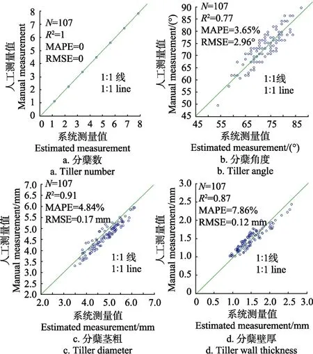
图4 系统测量与人工测量结果Fig.4 Results of system measurement and manual measurement
3.3 系统对比分析
本研究系统与其它测量方法的对比结果如表2所示。本方法与人工测量方法相比,首先可以实现无损表型性状的获取,其次在测量效率上人工处理该107株小麦植株花费时间大约为 30 h,平均每株测量效率约为1 000 s,而本系统大约只需要6 h,测量效率提高约5倍,在测量精度上本系统可以与人工测量相比具有较高的一致性,并能克服人工测量上存在主观、易疲劳的缺点。与石蜡切片法相比,同样可以克服有损的缺点并与其报道的测量效率相比提高约 9倍[34]。与基于多光谱的可见光预测方法相比,本方法不但可以获取小麦分蘖的内部信息如分蘖壁厚,还能克服小麦分蘖之间的交叠提供更精准的小麦分蘖性状,以分蘖数测量为例可见光预测方法能达到的最高决定系数为0.85[35],而本系统可达到1。
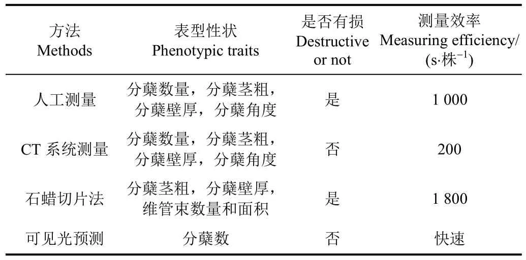
表2 该系统与其它测量方法的性能对比Table 2 Performance comparison between CT system and other methods
4 结论与讨论
小麦分蘖为小麦生长提供营养输送作用,其茎粗、壁厚与抗倒伏能力密切相关,分蘖数、分蘖角度直接影响小麦的株型,小麦分蘖形态学特征性状研究对小麦遗传育种及功能基因研究具有要用意义,本研究提出了一种基于X-CT测量小麦分蘖形态的方法。
1)本文针对小麦分蘖形态学特征性状获取,构建了一套X射线断层成像系统,通过该系统可以无损、快速获取小麦茎秆的断层图像,最后采用图像分析算法实现对小麦分蘖形态学特征的在体、无损、自动检测。针对107株小麦植株进行分蘖断层重建,自动获取分蘖性状参数包括分蘖数、分蘖角度、茎粗和壁厚,试验结果显示测量精度(mean absolute percentage error,MAPE)分别为100%、96.35%、95.16%、92.14%;标准差(root mean square error,RMSE)分别为 0、2.96°、0.17 mm、0.12 mm。以上结论表明本套系统具有较高的测量精度。
2)本套系统方便扩展应用于水稻、玉米、油菜等其他作物,该技术可与现有植物表型提取技术(可见光、近红外、红外成像等)相集成,为植物表型性状获取提供一种新的技术手段。
3)传统的测量小麦分蘖形态学特征主要有可见光检测、人工有损检测和石蜡切片电镜检测。其中可见光只能获取作物的表面信息,分蘖之间的遮挡对测量精度影响较大,且无法测量壁厚,后两种方法虽然可以实现本研究目标,但均为有损检测,不仅效率低下且无法重复。因此本研究为小麦分蘖形态测量提供了一种新的方法。
[1] Qin X, Zhang F, Liu C, et al. Wheat yield improvements in China: Past trends and future directions[J]. Field Crops Research, 2015, 177(6): 117-124.
[2] Slafer G A, Savin R, Sadras V O. Coarse and fine regulation of wheat yield components in response to genotype and environment[J]. Field Crops Research, 2014, 157(2): 71-83.
[3] 钱永兰,毛留喜,周广胜. 全球主要粮食作物产量变化及其气象灾害风险评估[J]. 农业工程学报,2016,32(1):226-235.Qian Yonglan, Mao Liuxi, Zhou Guangsheng. Changes in global main crop yields and its meteorological risk assessment[J]. Transactions of the Chinese Society of Agricultural Engineering (Transactions of the CSAE), 2016,32 (1): 226-235. (in Chinese with English abstract)
[4] Shangguan Z, Shao M, Dyckmans J. Effects of nitrogen nutrition and water deficit on net photosynthetic rate and chlorophyll fluorescence in winter wheat[J]. Journal of Plant Physiology, 2000, 156(1): 46-51.
[5] Annison G. The role of wheat non-starch polysaccharides in broiler nutrition[J]. Crop and Pasture Science, 1993, 44(3):405-422.
[6] Gupta P K, Varshney R K, Sharma P C, et al. Molecular markers and their applications in wheat breeding[J]. Plant breeding, 1999, 118(5): 369-390.
[7] Lupton F G H. History of wheat breeding[M]. Springer Netherlands, 1987: 51-70.
[8] Zuber U, Winzeler H, Messmer M M, et al. Morphological traits associated with lodging resistance of spring wheat(Triticum aestivum L.)[J]. Journal of Agronomy and Crop Science, 1999, 182(1): 17-24.
[9] Kipp S, Mistele B, Baresel P, et al. High-throughput phenotyping early plant vigour of winter wheat[J]. European Journal of Agronomy, 2014, 52(2): 271-278.
[10] 袁志华,冯宝萍,赵安庆,等. 作物茎秆抗倒伏的力学分析及综合评价探讨[J]. 农业工程学报,2002,18(6):30-31.Yuan Zhihua, Feng Baoping, Zhao Anqing, et al. Dynamic analysis and comprehensive evaluation of crop-stem lodging resistance[J]. Transactions of the Chinese Society of Agricultural Engineering (Transactions of the CSAE), 2002,18(6): 30-31. (in Chinese with English abstract)
[11] 胡婷,付志一,焦群英. 小麦茎秆抗弯性能研究[J]. 农业工程学报,2006,22(10):31-35.Hu Ting, Fu Zhiyi, Jiao Qunying. Experimental study on the bending mechanical properties of wheat stalk[J]. Transactions of the Chinese Society of Agricultural Engineering(Transactions of the CSAE), 2006, 22(10): 31-35. (in Chinese with English abstract)
[12] Deiss L, Moraes A, Pelissari A, et al. Oat tillering and tiller traits under different nitrogen levels in an eucalyptus agroforestry system in Subtropical Brazil[J]. Ciência Rural,2014, 44(1): 71-78.
[13] Yang W, Duan L, Chen G, et al. Plant phenomics and high-throughput phenotyping: accelerating rice functional genomics using multidisciplinary technologies[J]. Current Opinion in Plant Biology, 2013, 16(2): 180-187.
[14] Yang W, Guo Z, Huang C, et al. Genome-wide association study of rice (Oryza sativaL.) leaf traits with a highthroughput leaf scorer[J]. Journal of Experimental Botany,2015, 66(18): 5605-5615.
[15] Cabrera‐Bosquet L, Crossa J, von Zitzewitz J, et al.High-throughput phenotyping and genomic selection: The frontiers of crop breeding converge[J]. Journal of Integrative Plant Biology, 2012, 54(5): 312-320.
[16] Andrade-Sanchez P, Gore M A, Heun J T, et al. Development and evaluation of a field-based high-throughput phenotyping platform[J]. Functional Plant Biology, 2014, 41(1): 68-79.
[17] 丁为民,赵思琪,赵三琴,等. 基于机器视觉的果树树冠体积测量方法研究[J]. 农业机械学报,2016,47(6):1-10.Ding Weimin, Zhao Siqi, Zhao Sanqin, et al. Measurement methods of fruit tree canopy volume based on machine vision[J]. Transactions of the Chinese Society for Agricultural Machinery, 2016, 47(6): 1-10. (in Chinese with English abstract)
[18] 伍艳莲,曹卫星,汤亮,等. 基于OpenGL的小麦形态可视化技术[J]. 农业工程学报,2009,25(1):121-126.Wu Yanlian, Cao Weixing, Tang Liang, et al. Open GL-based visual technology for wheat morphology[J]. Transactions of the Chinese Society of Agricultural Engineering(Transactions of the CSAE), 2009, 25(1): 121-126. (in Chinese with English abstract)
[19] 杜光源,唐燕,张嵩午,等. 小麦叶片衰老态势核磁共振分析[J]. 农业机械学报,2014,45(4):264-270.Du Guangyuan, Tang Yan, Zhang Songwu, et al.Investigating senescence status of wheat leaves by nuclear magnetic resonance[J]. Transactions of the Chinese Society for Agricultural Machinery, 2014, 45(4): 264-270. (in Chinese with English abstract)
[20] 邓继忠,李敏,袁之报,等. 基于图像识别的小麦腥黑穗病害特征提取与分类[J]. 农业工程学报,2012,28(3):172-176.Deng Jizhong, Li Min, Yuan Zhibao, et al. Feature extraction and classification of Tilletia diseases based on image recognition[J]. Transactions of the Chinese Society of Agricultural Engineering (Transactions of the CSAE), 2012,28(3): 172-176. (in Chinese with English abstract)
[21] 张云鹤,乔晓军,王成,等. 基于机器视觉的作物茎秆直径变化测量仪研发[J]. 农机化研究,2011,33(12):158-160.Zhang Yunhe, Qiao Xiaojun, Wang Cheng, et al.Development of diameter micro-variation measurement instrument for plant stem based on machine vision technology[J]. Journal of Agricultural Mechanization Research,2011, 33(12): 158-160. (in Chinese with English abstract)
[22] Bauriegel E, Giebel A, Geyer M, et al. Early detection of Fusarium infection in wheat using hyper-spectral imaging[J].Computers and Electronics in Agriculture, 2011, 75(2):304-312.
[23] 王金满,郭凌俐,白中科,等. 基于CT分析露天煤矿复垦年限对土壤有效孔隙数量和孔隙度的影响[J]. 农业工程学报,2016,32(12):229-236.Wang Jinman, Guo Lingli, Bai Zhongke, et al. Effects of land reclamation time on soil pore number and porosity based on computed tomography (CT) images in opencast coal mine dump[J]. Transactions of the Chinese Society of Agricultural Engineering (Transactions of the CSAE), 2016, 32(12):229-236. (in Chinese with English abstract)
[24] 罗锡文,周学成,严小龙,等. 基于XCT技术的植物根系原位形态可视化研究[J]. 农业机械学报,2004,35(2):104-106.Luo Xiwen, Zhou Xuecheng, Yan Xiaolong,et al.Visualization of plant root morphology in situ based on X-ray CT imaging technology[J]. Transactions of the Chinese Society for Agricultural Machinery, 2004, 35(2): 104-105.(in Chinese with English abstract)
[25] 葛浙东,侯晓鹏,鲁守银,等. 基于反投影坐标快速算法的木材CT检测系统研究[J]. 农业机械学报,2016,47(3):335-341.Ge Zhedong, Hou Xiaopeng, Lu Shouyin, et al. Wood CT Detection system based on fast algorithm of inverse projection coordinate[J]. Transactions of the Chinese Society for Agricultural Machinery, 2016, 47(3): 335-341. (in Chinese with English abstract)
[26] Yang W, Xu X, Duan L, et al. High-throughput measurement of rice tillers using a conveyor equipped with x-ray computed tomography[J]. Review of Scientific Instruments, 2011, 82(2):761-793.
[27] 方正.适于小动物研究的μCT系统开发研制. [D]. 武汉:华中科技大学,2008.Fang Zheng. Development of µCT System for Small Animal Study[D]. Wuhan: Huazhong University of Science and Technology, 2008. (in Chinese with English abstract)
[28] 汤漾平,张振峰, 冯清秀.基于PLC的微型CT自动扫描系统[J].机电工程,2010,27(9):46-48.Tang Yangping, Zhang Zhenfeng, Feng Qingxiu. PLC-based automatic scanning system of micro-CT[J]. Journal of Mechanical & Electrical Engineering, 2010,27(9):46-48.(in Chinese with English abstract)
[29] Mayo S C, Chen F, Evans R. Micron-scale 3D imaging of wood and plant microstructure using high-resolution X-ray phase-contrast microtomography[J]. Journal of structural biology, 2010, 171(2): 182-188.
[30] Jia X, Lou Y, Lewis J, et al. GPU-based fast low-dose cone beam CT reconstruction via total variation[J]. Journal of X-ray Science and Technology, 2011, 19(2): 139-154.
[31] Miller N D, Haase N J, Lee J, et al. A robust, highthroughput method for computing maize ear, cob, and kernel attributes automatically from images[J]. Plant Journal, 2017,89(1): 169-178.
[32] Paulus M J, Gleason S S, Kennel S J, et al. High resolution X-ray computed tomography: An emerging tool for small animal cancer research[J]. Neoplasia, 2000, 2(1/2): 62-70.
[33] Otsu N. A threshold selection method from gray-level histograms[J]. Automatica, 1975, 11 (285/296): 23-27.
[34] 王秀文. 石蜡切片法中染色技术的改良[J]. 植物研究,2015,35(1):158-160.Wang Xiuwen. The improved stained methods of paraffin sections production[J]. Bulletin of Botanical Research, 2015,35(1): 158-160. (in Chinese with English abstract)
[35] 张猛,孙红,李民赞,等. 基于4波段作物光谱测量仪的小麦分蘖数预测[J]. 农业机械学报,2016,47(9):341-347.Zhang Meng, Sun Hong, Li Minzan, et al. Prediction of winter wheat tiller number based on 4 waveband crop monitor with spectral reflectance[J]. Transactions of the Chinese Society for Agricultural Machinery, 2016, 47(9):341-347. (in Chinese with English abstract)
Non-destructive detection of wheat tiller morphological traits based on X-ray CT technology
Wu Di1, Yang Wanneng1,2, Niu Zhiyou1, Huang Chenglong1※
(1.College of Engineering, Huazhong Agricultural University, Wuhan430070,China;2.National Key Laboratory of Crop Genetic Improvement, Huazhong Agricultural University, Wuhan430070,China)
Wheat tillers play an important role for nutrition transport to support the wheat growth. The wheat stem diameter and thickness are closely related to lodging resistance. Meanwhile tiller number and tiller angle directly determine the plant type of wheat. Therefore, the morphological trait extraction of wheat tillers is very important to the study of wheat genetics research, breeding improvement and functional genes location. With the development of the wheat cultivation and genetic breeding, the fast and accurate measurement of morphological traits for wheat tillers is imperative. However, the traditional method for tiller trait measurement is still manual, which is destructive and time consuming. Although a lot of efforts had been made to extract the tillers traits generally based on visible light, it is not able to acquire the inner information of wheat tiller and is affected seriously by tillers overlap. To solve the problem, a nondestructive technology for wheat tillers measurement was proposed and equipped with X-ray CT imaging device. In this study, the X-ray CT imaging system was constructed with the Micro-focus X-ray source and flat detector, which was used to obtain the sinogram images of wheat tiller with the spatial resolution 61 μm by 61 μm, and totally 360 images were collected for every one degree rotation for each plant. Then the FBP and GPU algorithms were adopted to reconstruct the tomography image of wheat tillers based on the sinogram images,and the inner information of wheat tiller was visible in the image. Moreover, the specialized image analysis algorithms were designed to analyze the wheat tomography image, in which the algorithms of background subtraction, OTSU segmentation, removing small region, and connected region identification were applied to extract the tiller regions. After that, the wheat tiller morphological traits were extracted by the following methods, the tiller numbers were counted based on the number of connected areas, the stem diameter was computed by the information of area external rectangle, the tiller wall thickness was extracted with the information of area external rectangle and cavity rectangle, and tiller angle was obtained by the triangle relation of tomography images at different heights. Finally this method was evaluated by 107 wheat plants, which belonged to five different wheat varieties. After the wheat plants were measured by the system automatically, the plants were measured by manual method for comparison to evaluate the system measurement accuracy. The experimental results showed that the system measurement accuracy of the tiller number was 100%, the mean absolute percentage error of tiller angle, the stem diameter and the stem wall thickness were 3.65%,4.84% and 7.86%, respectively and the RMSE for above traits were 2.96, 0.17 mm, 0.12 mm,respectively . The R2 value of tiller angle, the stem diameter and the stem wall thickness were 0.77, 0.91 and 0.87, respectively.The results demonstrated that this method had a good consistency with manual method, and performed a high accuracy for wheat tiller morphological trait measurements. In this study, the image acquisition efficiency was about 200 s per plant and the time used for image analysis was about 120 s per plant. Considering the parallel implement of image acquisition and analysis,the system efficiency was about 200 s per plant and was able to measure approximate 432 wheat plants in one day. Compared with manual method, this technology was able to detect the internal information of wheat tiller with high-accuracy and nondestructive. Moreover, it was able to extract novel phenotypic traits, which may contribute to the functional genomics and lodging resistance research of wheat plants. In future, more detailed information of wheat tiller such as vascular bundle, leaf sheath could be analyzed based on the higher resolution X-ray imaging device and more intelligent algorithms.
X rays; computerized tomography; image processing; wheat tiller; nondestructive testing
10.11975/j.issn.1002-6819.2017.14.027
S512;S123
A
1002-6819(2017)-14-0196-06
吴 迪,杨万能,牛智有,黄成龙. 小麦分蘖形态学特征X射线-CT无损检测[J]. 农业工程学报,2017,33(14):196-201.
10.11975/j.issn.1002-6819.2017.14.027 http://www.tcsae.org
Wu Di, Yang Wanneng, Niu Zhiyou, Huang Chenglong. Non-destructive detection of wheat tiller morphological traits based on X-ray CT technology[J]. Transactions of the Chinese Society of Agricultural Engineering (Transactions of the CSAE), 2017,33(14): 196-201. (in Chinese with English abstract) doi:10.11975/j.issn.1002-6819.2017.14.027 http://www.tcsae.org
2017-02-26
2017-06-20
国家高新技术发展计划(863计划,2013AA102403);国家自然科学基金项目(31600287);湖北省科研条件与资源研究开发(2015BCE044)作者简介:吴迪,博士生,主要从事农业信息无损检测研究。武汉 华中农业大学工学院,430070。Email:380524590@qq.com
※通信作者:黄成龙,讲师,研究方向为植物表型无损测量研究。武汉 华中农业大学工学院,430070。Email:hcl@mail.hzau.edu.cn
猜你喜欢
机电安全(2022年5期)2022-12-13
数学教学通讯·小学版(2022年4期)2022-05-29
西部探矿工程(2022年2期)2022-02-14
非常规油气(2021年5期)2021-11-13
科学(2020年1期)2020-01-06
震灾防御技术(2019年3期)2019-06-02
安徽农业科学(2018年30期)2018-11-02
农家科技中旬版(2018年2期)2018-05-05
新农业(2017年6期)2017-07-15
中国卫生(2015年12期)2015-11-10