超声法制备均匀分布的亚微米级镓铟锡合金液态金属微球
2017-11-01 06:24:52,,,,,
理化检验(物理分册) 2017年10期
,, , , ,
(1. 上海大学 材料科学与工程学院, 上海 200444; 2. 中国科学院 宁波材料技术与工程研究所, 宁波 315201)
试验与研究
超声法制备均匀分布的亚微米级镓铟锡合金液态金属微球
张配同1,2,刘宜伟2,郭强1,刘钢2,李润伟2
(1. 上海大学 材料科学与工程学院, 上海 200444; 2. 中国科学院 宁波材料技术与工程研究所, 宁波 315201)
采用超声法制备了镓铟锡合金(Galinstan)液态金属微球,研究了超声工艺参数对Galinstan微球粒径的影响规律,然后通过控制沉降时间进行多次沉降,得到了均匀分布的亚微米级Galinstan微球。结果表明:Galinstan微球粒径随着超声时间、功率的增加而减小,随着超声温度、溶剂表面张力的增加而增大,通过优化超声工艺参数,可以获得粒径小于2 μm的Galinstan微球;经过多次沉降去除沉降的大粒径微球,可以获得粒径分布范围为0.2~0.8 μm的Galinstan微球,大大提高了微球粒径分布均匀性。
超声法; 液态金属; 亚微米级镓铟锡合金微球; 粒径; 工艺参数
液态金属是一种熔点低于200 ℃的纯金属或金属合金,由于液态金属在受外力作用下比较容易变形且具有很好的流动性[1],因此在柔性电子领域得到广泛应用。镓基液态金属有镓铟合金(EGaIn,质量分数为:75%Ga, 25%In)、镓铟锡合金(Galinstan,质量分数为:68%Ga,22%In,10%Sn)等。EGaIn(熔点15.5 ℃[2])和Galinstan(熔点-19 ℃[3])具有一系列优异的特性,比如低蒸气压[3]、低毒、高导电性和低黏性等[1-3]。另外,液态金属制备成液态金属微球,可以应用于拉伸电子器件[4]、化学传感器[5]、微型开关[6]、3D印刷结构[7]、导电复合材料[8]、喷墨打印墨水[9]、3D微电极[10]等,分布均匀的液态金属微球还可以组装成周期性结构应用于光学领域中无线电频率谐振器[11]和可重构光学衍射光栅[12]等。
目前制备液态金属微球的方法主要有模型法(Molding)[13]、微流聚焦法(Flow-Focusing)[14]和超声法(Sonication)[5,15-16]等。其中,模型法是将含有凹型微结构的聚二甲基硅氧烷(PDMS)上涂覆液态金属,在HCl气氛中除去液态金属表面的氧化层,从而制备出液态金属微球[13]。模型法虽然简单,但通过这种方法制备出来的液态金属微球尺寸至少100 μm。微流聚焦法是使用微流管设备,制备出分布均匀的液态金属微球,液态金属微球尺寸是通过液体的剪切速率或液体间的表面张力控制的,虽然该方法制备的液态金属微球的尺寸比模型法制备的要小,但要制备粒径小于50 μm的液态金属微球仍具有很大的挑战性。
超声法是一种简单、快速、大量制备微米级到亚微米级液态金属微球的方法。通过在EGaIn微球表面聚合聚合物脲醛,阻止微球间的融合,从而获得粒径分布在3~200 μm的微球[15]。TANG等研究了液态金属/金属氧化物球形结构在水中超声振动20 min获得粒径分布为0.5~5 μm的液态金属微球[5]。进一步研究发现,超声振动的同时在液态金属上施加电压以此改变EGaIn的表面张力,可以将制备出的微球粒径控制在10~50 μm[16]。虽然超声法可以制备出亚微米级液态金属微球,但是粒径分布从几百纳米到几十微米,要制备出粒径分布均匀的液态金属微球仍然面临着挑战,同时超声时间、功率、温度、溶剂等因素对液态金属微球粒径的影响尚不是很清楚。
为此,笔者通过超声法制备了Galinstan液态金属微球,研究了超声时间、功率、温度、溶剂表面张力等因素对该液态金属微球粒径的影响,并通过多次沉降法制备了分布均匀的亚微米级液态金属微球。
1 试样制备与试验方法
1.1试验材料
试验用纯金属镓(Ga)锭、铟(In)锭、锡(Sn)锭的纯度均≥99.99%(质量分数),熔点分别为29.78,156.61,232 ℃。试验用丙酮(C3H6O)为分析纯试剂(阿拉丁公司)。
1.2试验方法
1.2.1 Galinstan液态金属的制备
按照质量比66.5%∶23.5%∶10%分别称量镓、铟、锡3种金属,将称量好的3种金属从中间大口径入口放入三口烧瓶中,水浴加热到60 ℃,通入氮气,机械搅拌60 min,即得到试验所用的Galinstan镓基液态金属。
1.2.2 Galinstan微球的制备
Galinstan微球的制备过程如下:首先,将0.4 g Galinstan液态金属与20 mL丙酮溶液加入到25 mL烧杯中,然后将其放置于超声波清洗机(型号为SK3200HP,最大功率为180 W,频率为53 kHz)中,选择超声功率为40%~100%、温度为20~40 ℃超声处理5~60 min,得到Galinstan微球悬浊液,如图1所示。
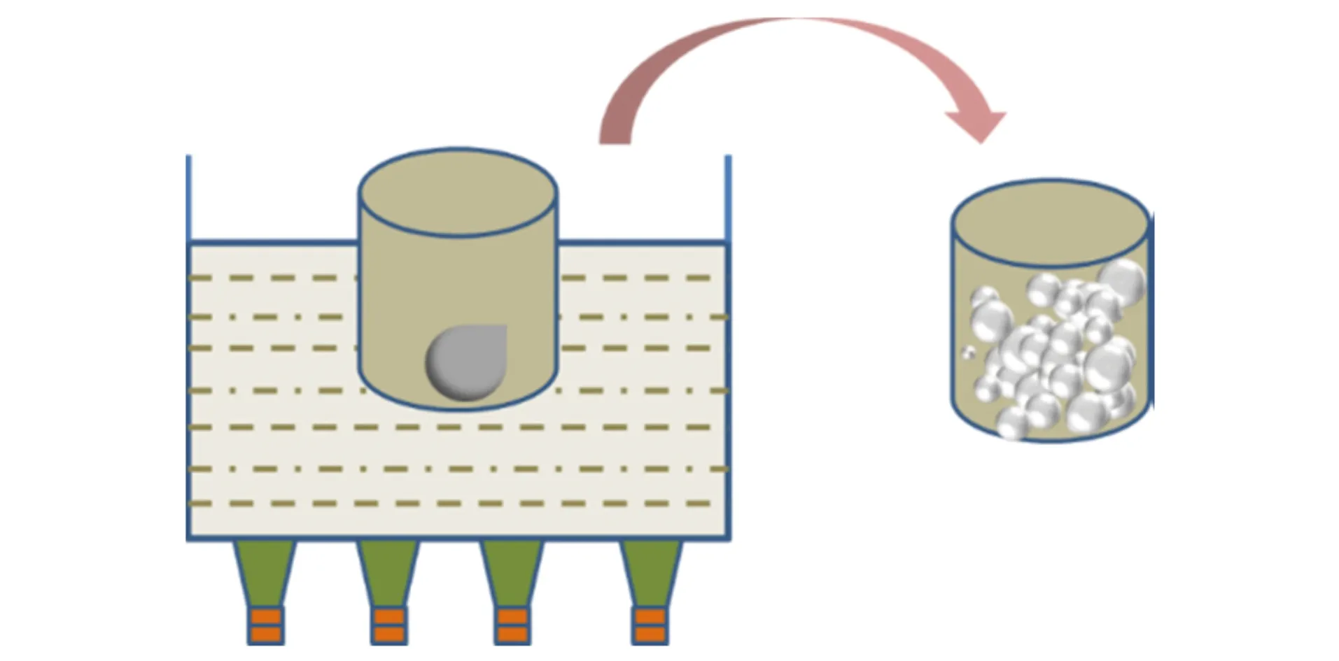
图1 Galinstan微球制备过程示意图Fig.1 The scheme for fabricating Galinstan microspheres
1.2.3 分布均匀的亚微米级Galinstan微球的制备
静置Galinstan微球悬浊液,分别将静置30,60,90,120,150,180 min的悬浊液用吸管吸取上清液,滴在30 mm×30 mm的硅片上。
1.3测试与表征
用Microtrac S3500热场发射扫描电镜(SEM)表征Galinstan微球的形貌;用干湿两用微米级激光粒度仪表征Galinstan微球的粒径分布范围[17]。
2 试验结果与讨论
2.1Galinstan微球粒径与超声功率的关系
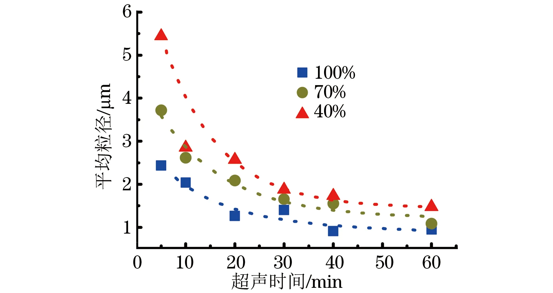
图2 20 ℃超声时Galinstan微球平均粒径与超声功率的关系曲线Fig.2 The relationship curves between mean diameters of Galinstan microspheres and ultrasonic powers at 20 ℃
图2为20 ℃时Galinstan微球平均粒径与超声功率的关系曲线,可以看出平均粒径与超声功率成反比。超声5 min、功率从40%增加到100%时,Galinstan微球平均粒径由5.4 μm减小到2.4 μm;超声60 min、功率从40%增加到100%时,Galinstan微球平均粒径由1.5 μm减小到0.9 μm。试验结果表明在达到最小平均粒径时,随着超声时间的延长超声功率对Galinstan微球平均粒径的影响变小。
由图2还可以看出,Galinstan微球平均粒径随着超声时间的延长而减小;超声功率为100%、超声时间从5 min增加到60 min时,Galinstan微球平均粒径由5.5 μm减小到1.0 μm。图2中的虚线表明超声时间和微球平均粒径的关系可以用单指数函数进行拟合,超声5 min到40 min微球平均粒径的变化比较明显,但从40 min到60 min微球平均粒径的变化比较平缓,最终趋于平衡,也就是说再延长超声时间对微球平均粒径的影响很小。
2.2Galinstan微球粒径与超声温度的关系
超声温度是影响Galinstan微球粒径的另一重要因素[18]。图3a)和图3b)分别是超声功率为100%时在40 ℃和20 ℃下超声60 min制备的Galinstan微球的SEM形貌,可以明显看出在20 ℃超声制备的微球粒径比40 ℃制备的要小。为了定量地表征温度对Galinstan微球平均粒径的影响,对图3a)和图3b)的微球粒径进行统计绘制了图3c),可以看出40 ℃制备微球的平均粒径为2.5 μm,是20 ℃制备微球的近2倍。为了进一步研究温度和时间对Galinstan微球平均粒径的影响,图3d)显示超声5 min时,20 ℃和40 ℃制备微球的平均粒径差是0.5 μm;到超声40 min时,20 ℃和40 ℃制备微球的平均粒径差是1 μm。

图3 100%功率下Galinstan微球粒径与超声温度的关系Fig.3 The relationship between diameters of Galinstan microspheres and ultrasonic temperatures at 100% power: a) SEM morphology of microspheres prepared by ultrasonic at 40 ℃; b) SEM morphology of microspheres prepared by ultrasonic at 20 ℃; c) diameter distribution statistics of microspheres prepared by ultrasonic at different temperatures; d) effects of ultrasonic temperature and time on mean diameters of the microspheres
2.3Galinstan微球粒径与溶剂的关系
超声选择的溶剂是另一个影响Galinstan微球粒径的重要因素。试验中选择表面张力相差比较大的两种溶剂,分别是丙酮(20 ℃时的表面张力为23.7 mN·m-1)和水(20 ℃时的表面张力为72.7 mN·m-1)。图4a)是Galinstan液态金属在去离子水中于20 ℃,100%功率下超声60 min制备的Galinstan微球SEM形貌,与图3b)中的微球粒径相比,可见在水中超声制备的Galinstan微球粒径比在丙酮中制备的要大。为进一步表征溶剂与微球粒径的关系,对图4a)和图3b)中的微球粒径进行统计绘制了图4b),可以看出在水中超声制备的Galinstan微球平均粒径是1.3 μm,是在丙酮中超声制备的1.5倍。试验说明溶剂的表面张力越小,制备的Galinstan微球的平均粒径也越小。
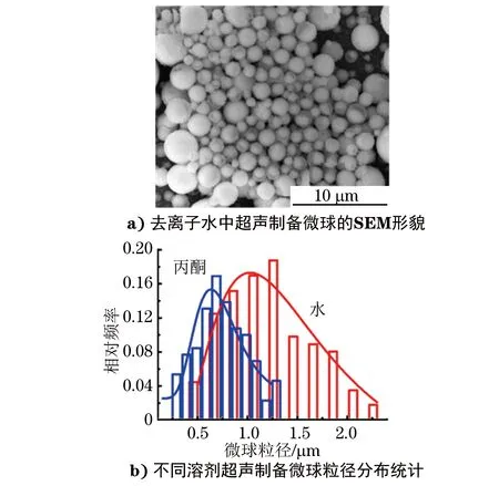
图4 Galinstan微球粒径与溶剂的关系Fig.4 The relationship between diameters of Galinstan microspheres and solvents: a) SEM morphology of microspheres prepared by ultrasonic in deionized water; b) diameter distribution statistics of microspheres prepared by ultrasonic in different solvents
超声波在液体中传播时,液体分子受到周期性交变声场的作用。如果声压达到一定值,液体在波的膨胀阶段产生气泡并迅速成长;在波的压缩阶段这些已经成长到足够大的气泡在液体中瞬间空爆,产生冲击波,向四周辐射,这个过程称为空化效应过程。空爆产生瞬时的局部高温、高压,理论计算和实际测量的结果表明,其加热和冷却的速率>1010K·s-1,温度可达5 000 K以上,压力高达100 MPa[19],因此超声空化能够产生足够的能量作用在Galinstan液态金属表面,制备出Galinstan微球。
Galinstan微球形成过程伴随着Galinstan的融合与分裂,超声空化的形成是超声制备Galinstan微球的前提。增大超声功率会使空化效应增强,空爆时在Galinstan表面产生的能量更强,从而制备出粒径更小的Galinstan微球。
延长超声时间会使超声空爆在Galinstan微球表面产生的能量更多,使比较大的微球变得更小,随着超声时间的延长,较大尺寸的微球逐渐消失,当超声时间达到一定值时Galinstan微球粒径趋于稳定。
一般来说,温度升高,空化阈值下降,其原因是随着温度的升高,蒸汽压增大,表面张力系数σ及黏滞系数η则下降[20],然而由于温度的升高,蒸气压上升,又会导致空化强度或空化效应下降,不利于小尺寸微球的形成。另一方面温度升高不利于Galinstan表面氧化层的形成,从而影响更小尺寸Galinstan微球的形成。
液体的表面张力系数σ增大,意味着空化泡收缩力增大,要求空化阈值增高,在去离子水中更不易形成空化,因而不易于形成更小尺寸的Galinstan微球。
2.4多次沉降法制备均匀分布的亚微米级Galinstan微球
通过以上研究,优化超声条件,在丙酮溶液中制备较小尺寸的Galinstan微球。从图5a)可以看出超声法制备的Galinstan微球粒径基本小于2 μm,但粒径分布范围很宽。根据Stokes定律[21]
式(1)表示半径为r的球体,在黏滞系数为η的流体中以速率v运动时受到的粘滞阻力Fd,以ρp和ρf分别表示球体和流体的密度,则可以推导出下式
式中:g为重力加速度。
在液态金属悬浊液中ρf是一样的,对于每一个液态金属微球来说ρp可以近似相等,因为液态金属微球表面的氧化层很薄对其密度影响很小。因此可以得出大尺寸Galinstan微球具有更大的沉降速率,从而可以将不同尺寸的Galinstan微球分离开来。基于此设计的试验过程如图5b)所示,超声后的Galinstan悬浊液静置一段时间,每隔一段时间取上清液,可以得到粒径分布均匀的Galinstan微球。
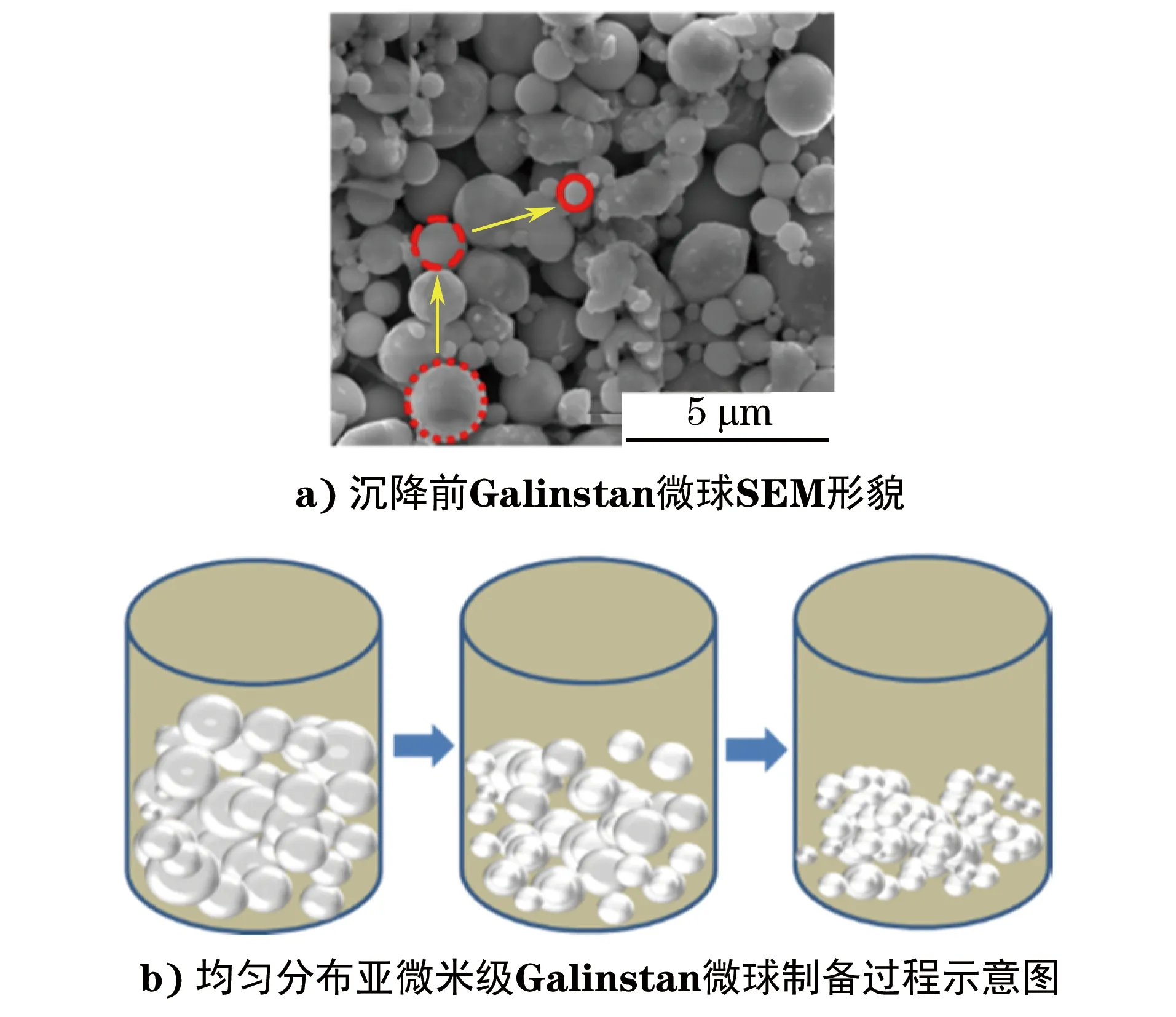
图5 沉降前Galinstan微球SEM形貌及均匀分布亚微米级 Galinstan微球制备过程示意图Fig.5 The a) SEM morphology of Galinstan microspheres before settling and b) schematic diagram of preparation process of uniformly distributed sub-microsized Galinstan microspheres
通过上述过程,随着沉降时间的延长便可获得分布越来越均匀的亚微米级Galinstan微球,如图6a)~c)所示分别是沉降30,60,180 min的Galinstan微球的SEM形貌,沉降30 min时微球平均粒径由1.3 μm减小到1 μm,沉降180 min时微球平均粒径变为0.7 μm;对图6a)~c)中的微球粒径进行统计得到图6d),可见随着沉降时间的延长一方面粒径变小,另一方面粒径分布范围变窄,其中沉降30,60,180 min对应高斯分布的标准差σ分别为0.20,0.15,0.09 μm,且沉降180 min时Galinstan微球粒径分布范围为0.2~0.8 μm。图6e)描述了沉降时间与Galinstan微球平均粒径的关系,可以看出沉降90 min前的平均粒径变化比较明显,说明沉降时间对微球粒径的影响很大;沉降超过90 min后微球平均粒径变化比较小,甚至趋于稳定。
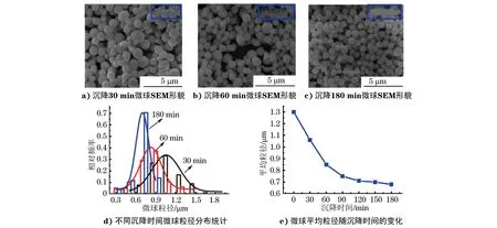
图6 Galinstan微球粒径与沉降时间的关系Fig.6 The relationship between diameters of Galinstan microspheres and settling times: a) SEM morphology of microspheres after settling for 30 min; b) SEM morphology of microspheres after settling for 60 min; c) SEM morphology of microspheres after settling for 180 min; d) diameter distribution statistics of microspheres after settling for different times; e) mean diameters of the microspheres changing with settling times
3 结论
(1) 使用超声法制备Galinstan液态金属微球时,Galinstan微球粒径随超声功率、时间的增加而减小,随超声温度、溶剂表面张力的增加而增大;通过优化超声工艺参数,可以获得粒径小于2 μm的Galinstan微球。
(2) 优化超声条件,将Galinstan微球悬浊液通过多次沉降可获得粒径分布范围为0.2~0.8 μm的Galinstan微球,平均粒径为0.7 μm,且其高斯分布的标准差仅为0.09 μm。
[1] DICKEY M D. Emerging applications of liquid metals featuring surface oxides[J]. ACS Applied Materials & Interfaces,2014,6(21):18369-18379.
[2] DICKEY M D,CHIECHI R C,LARSEN R J,etal.Eutectic gallium-indium (EGaIn):A liquid metal alloy for the formation of stable structures in microchannels at room temperature[J].Advanced Functional Materials,2008,18(7):1097-1104.
[3] LIU T Y,SEN P,KIM C J.Characterization of nontoxic liquid-metal alloy galinstan for applications in microdevices[J]. Journal of Microelectromechanical Systems,2012,21(2):443-450.
[4] ROSSET S,NIKLAUS M,DUBOIS P,etal.Metal ion implantation for the fabrication of stretchable electrodes on elastomers[J].Advanced Functional Materials,2009,19(3):470-478.
[5] ZHANG W,OU J Z,TANG S,etal.Liquid metal/metal oxide frameworks[J].Advanced Functional Materials,2014,24(24):3799-3807.
[6] SEN P,KIM C J. Microscale liquid-metal switches:A review[J].IEEE Transactions on Industrial Electronics,2009,56(4):1314-1330.
[7] LADD C,SO J H,MUTH J,etal. 3D printing of free standing liquid metal microstructures[J].Advanced Materials,2013,25(36):5081-5085.
[8] FASSLER A,MAJIDI C.Liquid-phase metal inclusions for a conductive polymer composite[J].Advanced Materials,2015,27(11):1928-1932.
[9] BOLEY J W,WHITE E L,KRAMER R K.Mechanically sintered gallium-indium nanoparticles[J]. Advanced Materials,2015,27(14):2355-2360.
[10] TANG S Y,ZHU J Y,SIVAN V,etal.Creation of liquid metal 3D microstructures using dielectrophoresis[J]. Advanced Functional Materials,2015,25(28):4445-4452.
[11] ZOU L,WITHAYACHUMNANKUL W,SHAH C M,etal.Dielectric resonator nanoantennas at visible frequencies[J].Optics Express,2013,21(1):1344-1352.
[12] HASHIMOTO M,MAYERS B,GARSTECKI P,etal.Flowing lattices of bubbles as tunable,self-assembled diffraction gratings[J]. Small,2006,2(11):1292-1298.
[13] MOHAMMED M,XENAKIS A,DICKEY M.Production of liquid metal spheres by molding[J].Metals,2014,4(4):465-476.
[14] THELEN J,DICKEY M D,WARD T. A study of the production and reversible stability of EGaIn liquid metal microspheres using flow focusing[J]. Lab on a Chip,2012,12(20):3961-3967.
[15] BROWN E N,KESSLER M R,SOTTOS N R,etal.In situ poly (urea-formaldehyde) microencapsulation of dicyclopentadiene[J]. Journal of Micro-encapsulation,2003,20(6):719-730.
[16] TANG S Y,AYAN B,NAMA N,etal.On-chip production of size-controllable liquid metal microdroplets using acoustic waves[J].Small,2016,12(28):3861-3869.
[17] 杜煜.激光衍射法测定超细氢氧化铝粒度中分散条件的控制[J].理化检验-物理分册,2013,49(1):46-49.
[18] DELMAS T,PIRAUX H,COUFFIN A C,etal.How to prepare and stabilize very small nanoemulsions[J].Langmuir,2011,27(5):1683-1692.
[19] BANG J H,SUSLICK K S. Applications of ultrasound to the synthesis of nanostructured materials[J]. Advanced Materials,2010,22(10):1039-1059.
[20] 叶菁.一种测定液体表面张力系数的新方法[J].理化检验-物理分册,2014,50(12):886-889.
[21] ELOY C,DOARÉ O,DUCHEMIN L,etal. A unified introduction to fluid mechanics of flying and swimming at high reynolds number[J]. Experimental Mechanics,2009,50(9):1361-1366.
UniformlyDistributedSub-microsizedGalinstanLiquidMetalMicrospheresPreparedbyUltrasoicMethod
ZHANGPeitong1,2,LIUYiwei2,GUOQiang1,LIUGang2,LIRunwei2
(1. School of Materials Science and Engineering, Shanghai University, Shanghai 200444, China; 2. Ningbo Institute of Materials Technology and Engineering, Chinese Academy of Sciences, Ningbo 315201, China)
Gallium-indium-tin alloy (Galinstan) liquid metal microspheres were prepared by ultrasonic method, and the effects of ultrasonic process parameters on the diameter of Galinstan microspheres were studied. Then, the uniformly distributed sub-microsized Galinstan microspheres were obtained by controlling the settling time. The results show that the diameter of Galinstan microspheres decreased with the increase of ultrasonic time and power, and increased with the increase of ultrasonic temperature and solvent surface tension. By optimizing the ultrasonic process parameters, Galinstan microspheres with the diameter less than 2 μm could be obtained. Galinstan microspheres with a diameter range of 0.2-0.8 μm could be obtained by multiple sedimentation to remove the large-size microspheres. This method could greatly improve the homogeneity of particle diameter distribution of the microspheres.
ultrosonic method; liquid metal; sub-microsized Galinstan microsphere; diameter; process parameter
TB31
A
1001-4012(2017)10-0701-06
10.11973/lhjy-wl201710002
2017-03-06
国家自然科学基金资助项目(51525103;11474295)
张配同(1990-),男,硕士研究生,主要从事柔性可穿戴设备及传感器研究
刘宜伟(1983-),男,博士,副研究员,主要从事柔性敏感材料与可穿戴器件研究,liuyw@nimte.ac.cn
猜你喜欢
钢铁钒钛(2023年5期)2023-11-17 08:48:34
纺织学报(2023年9期)2023-10-31 08:27:08
酒·饮料技术装备(2018年1期)2018-04-28 09:08:56
资源再生(2017年3期)2017-06-01 12:20:59
小布老虎(2016年4期)2016-12-01 05:46:08
厦门理工学院学报(2016年1期)2016-12-01 04:50:53
上海金属(2016年2期)2016-11-23 05:34:45
水利科技与经济(2016年7期)2016-04-25 13:03:00
核科学与工程(2015年2期)2015-09-26 11:57:24
上海金属(2014年3期)2014-12-19 13:09:06