cN0甲状腺微小乳头状癌多个淋巴结转移的危险因素分析
张磊 杨进宝 孙庆贺 刘跃武 陈革 陈曙光 刘子文 李小毅
·临床研究与应用·
cN0甲状腺微小乳头状癌多个淋巴结转移的危险因素分析
张磊①杨进宝②孙庆贺③刘跃武①陈革①陈曙光①刘子文①李小毅①
目的:术后病理证实的淋巴结转移在临床淋巴结转移阴性(clinical lymph node negative,cN0)的甲状腺乳头状癌(papillary thyroid carcinoma,PTC)中并不罕见,本研究旨在探讨临床淋巴结转移阴性的甲状腺微小乳头状癌(papillary thyroidmicrocarcino⁃ma,PTMC)淋巴结转移的危险因素,特别是多个淋巴结转移(>5枚)的危险因素。方法:回顾性分析中国医学科学院北京协和医院2013年11月至2014年10月行手术的cN0 PTMC患者1 268例,其中男性270例,女性998例。分析患者的临床病理学特征,通过单因素、多因素分析寻找淋巴结转移及多个转移的风险因素。结果:1 268例患者中共出现淋巴结转移416例(32.8%),多个淋巴结转移43例(3.4%),淋巴结转移的危险因素的单因素分析中,男性(42.22%vs.30.26%,P<0.01)、年龄<40岁(<40岁为48.39%,40~59岁为27.62%,年龄≥60岁为22.45%,P<0.03)、多发病灶(41.00%vs.29.03%,P<0.01)、无慢性淋巴细胞性甲状腺炎(36.44%vs.20.62%,P<0.01)、肿瘤直径>0.5 cm(35.77%vs.23.05%,P<0.01)者淋巴结转移比例显著增加。多因素分析中,男性(OR=1.516,P<0.001)、多发病灶(OR=1.743,P<0.001)、肿瘤直径>0.5 cm(OR=1.788,P<0.001)是淋巴结转移的独立危险因素;而年龄较大患者(40~59岁OR=0.388;60岁及以上OR=0.301,P<0.001)、慢性淋巴细胞性甲状腺炎(OR=0.472,P<0.001)是出现淋巴结转移的保护性因素。在多个淋巴结转移危险因素的多因素分析中,男性(OR=2.383,P=0.002)是多个淋巴结转移的独立危险因素;而年龄≥40岁则是保护性因素(OR=0.270,P<0.001)。结论:cN0 PTMC淋巴结转移并不少见,但是多个淋巴结转移少见,男性、年龄<40岁的患者淋巴结转移、多个转移的风险明显增加。
甲状腺微小乳头状癌 cN0 多个淋巴结转移 危险因素
甲状腺乳头状癌(papillary thyroid carcinoma,PTC)是甲状腺最常见的实体肿瘤,约占所有甲状腺恶性肿瘤的90%[1]。近年来,其发病率快速增长,在韩国甚至成为最常见的恶性肿瘤[2]。在所有PTC中,直径<1 cm的甲状腺微小乳头状癌(papillary thyroid microcarcinoma,PTMC)的增加最为明显,约占新发PTC的一半。这部分患者总体预后良好,其长期生存率可达99%以上[3-5]。
尽管PTMC的生存预后较好,但是部分患者在长期生存中依然面临复发的风险。有文献报道,出现少量淋巴结转移的情况下,患者出现复发的比例约为5%,而在出现多个淋巴结转移时,其复发的比例约为20%[6]。因此,2015年美国甲状腺协会(American Thyroid Association,ATA)更新了复发危险分层,把出现多个淋巴结转移(>5枚转移性淋巴结)作为复发的中危因素[7]。
手术后病理发现PTMC中,淋巴结转移并不罕见,其转移比例最高可达约40%[8]。但术前超声预测对于颈部中央区淋巴结转移的敏感性并不理想[9],因此有大量临床淋巴结阴性(clinical lymph node nega⁃tive,cN0)的患者可能存在淋巴结转移[10-11]。而对于这一部分患者,是否行预防性淋巴结清扫一直存在争议。不主张预防性清扫的观点认为,预防性淋巴结清扫不能改善预后,同时增加并发症[6];主张预防性清扫的观点则认为,预防性清扫可能改善局部复发,评估复发风险[12-15]。因此,在cN0 PTMC患者中筛选淋巴结转移的高风险者、特别是可能有潜在多个淋巴结转移患者,给予合理的治疗,对于降低复发风险具有重要的临床价值。
既往有关cN0 PTMC研究中主要关注淋巴结转移的风险因素,缺乏对多个转移比例及风险因素的研究。本研究的主要目的是通过对cN0 PTMC患者临床病理资料进行分析,评估cN0患者出现淋巴结转移的风险因素、并进一步筛选多个淋巴结转移的风险因素。
1 材料与方法
1.1 病例资料
本研究回顾性分析2013年11月至2014年10月中国医学科学院北京协和医院行手术的cN0的初治PTMC患者1 268例,其中男性270例,女性998例。本研究通过超声检查来判断颈部淋巴结转移情况,若无以下征象则判定为cN0:颈部淋巴结横/长径>0.5 cm,皮髓质分界不清或髓质结构消失,与原发灶相似的砂砾样钙化或囊性变,皮质内高回声团块,皮质周围血流丰富或有不规则血流[16]。
1.2 方法
入选标准:1)初治且术后病理证实PTMC患者;2)符合cN0诊断标准;3)手术切除范围至少包含患侧腺体及患侧中央区淋巴结。排除标准:1)前证实为cN1的患者;2)非初治患者;3)未行中央区淋巴结清扫;4)术后病理非PTC患者。
中央区淋巴结清扫范围参照2009年ATA中央区淋巴结定义范围[17]。淋巴结多个转移定义为淋巴结转移数量>5枚。观察的临床病理资料包括:性别、年龄(A组<40岁,B组40~59岁,C组60岁及以上)[8,18]、甲状腺癌家族史、慢甲炎、多发病灶、包膜侵犯、肿瘤大小。对于多发癌灶,取最大肿物直径作为该患者的肿瘤大小进行统计学分析。
1.3 统计学分析
采用SPSS 22.0软件进行统计学分析。影响淋巴结转移及多个转移的潜在危险因素的单因素分析应用χ2检验及Fisher精确概率法检验。应用Logistic回归模型进行多因素分析。以P<0.05为差异具有统计学意义。
2 结果
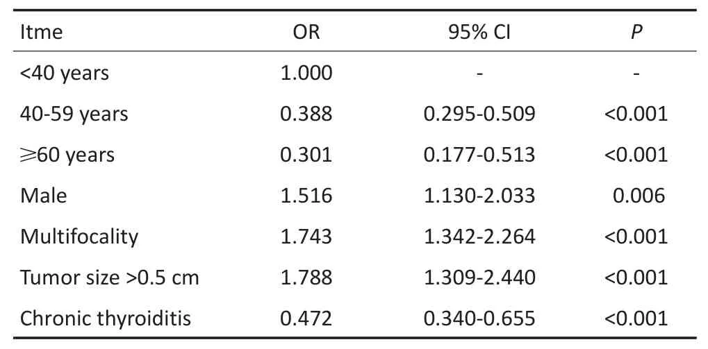
1 268例PTMC患者中,淋巴结转移率为32.8%(416/1 268),多个淋巴结转移率为3.4%(43/1 268)。淋巴结转移的危险因素的单因素分析中,男性的转移比例显著高于女性(42.2%vs.30.3%,P<0.05),合并慢甲炎的患者出现淋巴结转移的比例更低(20.6%vs.36.4%,P<0.05),多发病灶的患者淋巴结转移比例更高(41.0%vs.29.0%,P<0.05),直径>0.5 cm的患者出现转移比例更高(35.8%vs.23.1%,P<0.05),年轻的患者较年长患者出现淋巴结转移的比例更高(A组48.4%,B组27.6%,C组22.5%,P<0.05,表1)。多因素分析显示,男性(OR=1.52,P<0.05)、多发病灶(OR=1.743,P<0.05)、直径>0.5 cm(OR=1.79,P<0.05)是出现淋巴结转移的独立危险因素,慢甲炎(OR=0.47,P<0.05),年龄较大的患者(B组OR=0.39,P<0.05;C组OR=0.30,P<0.05)是淋巴结转移的保护性因素(表2)。
多个淋巴结转移的危险因素的单因素分析中,男性的转移比例显著高于女性(6.30%vs.2.61%,P<0.05),直径>0.5 cm患者出现多个转移比例更高(4.01%vs.1.36%,P<0.05),年轻患者出现多个转移的比例更高(A组7.62%,B组2.05%,C组0,P<0.05),见表1。多因素分析中,男性是出现多个淋巴结转移的独立危险因素(OR=2.383,P<0.05),相比较年轻患者,年龄较大患者出现多个淋巴结转移的风险更低(B组OR=0.27,P<0.05),见表3。

表1 cN0 PTMC患者淋巴结转移、多个转移风险因素的单因素分析Table 1 Univariate analysisof risk factors for LNM and high-volume LNM in cN0 PTMCpatients

表2 cN0 PTMC患者淋巴结转移风险因素的多因素分析Table 2 Multivariate analysis of risk factors for LNM in cN0 PTMC patients

表3 cN0 PTMC患者多个淋巴结转移风险因素的多因素分析Table 3 Multivariate analysis of risk factors for high-volume LNM in cN0PTMCpatients
3 讨论
颈部超声及CT目前被广泛应用于颈部转移性淋巴结的筛查[19-20]。由于超声具有操作简便,速度快的特点,且对于淋巴结微结构识别优于CT,因此被推荐作为术前诊断的主要手段。由于颈部解剖学特点,超声对于中央区淋巴结转移的诊断的准确性并不理想[9,21-22],因此在cN0患者中,仍然会有大量患者术后证实存在淋巴结转移。在本研究中,cN0 PTMC患者的淋巴结转移率为32.8%。既往的研究表明男性、肿瘤大小以及年龄等是PTMC出现淋巴结转移的独立危险因素[23],本研究再次证实了相似的结果:年龄<40岁、男性、多发病灶、肿瘤直径>0.5 cm患者的淋巴结转移率分别为48.39%、42.22%、41.0%、35.77%,均是cN0 PTMC淋巴结转移的独立危险因素。因此,对于cN0 PTMC中接近1/3的患者出现淋巴结转移的情况,尤其对于有淋巴结转移高风险因素的患者,常规采取预防性中央区淋巴结清扫是有合理性的,一些指南也推荐常规的预防性清扫[24-25],其中部分患者可能因此改变疾病分期、治疗决策[26]。
但是,另一方面需要仔细探讨cN0中仅有少量、较小的淋巴结转移(也因此无法被术前超声发现的病变)的临床价值及其对预后(无论是生存预后、还是复发预后)的影响。研究已显示PTMC的整体预后良好,长期生存可达99%以上,局部或区域复发率为2%~6%,远处转移率仅为1%~2%[3-5];少量的颈部淋巴结转移并不增加患者的复发风险以及长期预后,因此淋巴结转移尤其是少量转移对PTMC患者的预后影响可能有限[27]。事实上,多项尸检研究发现,隐匿存在于甲状腺中的乳头状癌的比例为1%~35.6%(总体为11.5%),而且这一人群中的10%有隐匿的淋巴结转移病灶[14]。由此可见,在“正常人群中”可以有PTC及其转移病灶存在,有可能不对正常人产生“威胁”。在一项针对1 235例cN0低危PTMC患者的非首选手术治疗的临床观察中,5年、10年新发临床证实淋巴结转移率为1.7%和3.8%[18],与本研究中的多个转移率(3.4%)近似。因此,在临床中重点关注、诊治这些多个淋巴结转移的患者具有积极的临床意义。有研究表明多个转移患者复发较少量转移者显著增加,2015年更新的ATA指南也明确将出现多个淋巴结转移作为复发风险分层中的重要危险因素[6]。
本研究证实,男性cN0 PTMC患者出现多个淋巴结转移的独立危险因素(OR=2.383,P<0.05),相较年轻患者,年龄较大患者出现多个淋巴结转移的风险更低(40~59岁,OR=0.27,P<0.05);其对应的多个转移率分别为:男性为6.3%,年龄<40岁为7.62%,40~59岁为2.05%,60岁及以上为0。这与本研究前期的结论类似[8],尤其值得注意的是在年龄这一危险因素中,将其分为3组更能区分出不同风险的患者,年龄<40岁患者,因为有更长的预期寿命、淋巴结转移风险较大,应该给予更积极的治疗。
综上所述,cN0 PTMC患者中仍有很大比例存在淋巴结转移,尽管部分研究指出预防性清扫可能带来相应的并发症风险[6,28],但对于有经验的临床医生,预防性淋巴结清扫并不增加并发症率,同时有可能减少局部复发及二次手术[29-32];尤其对于男性、年轻患者,其淋巴结转移、多个转移的比例明显增加、风险显著增高,对这部分患者,更应考虑给予积极的预防性淋巴结清扫,以减少复发以及再次手术带来的风险。
[1]Davies L,Welch HG.Current thyroid cancer trends in the United States[J].JAMAOtolaryngolHead Neck Surg,2014,140(4):317-322.
[2]Jung KW,Won YJ,Kong HJ,et al.Cancer statistics in Korea:incidence,mortality,survival,and prevalence in 2011[J].Cancer Res Treat,2014,46(2):109-123.
[3]Hay ID.Managementof patientsw ith low-risk papillary thyroid carcinoma[J].Endocr Pract,2007,13(5):521-533.
[4]MazzaferriEL.Management of low-risk differentiated thyroid cancer[J].Endocr Pract,2007,13(5):498-512.
[5]Londero SC,KrogdahlA,Bastholt L,et al.Papillary thyroid carcinoma in denmark,1996-2008:outcome and evaluation ofestablished prognostic scoring systems in a prospective national cohort.[J].Thyroid,2015,25(1):78-84.
[6]Haugen BR,Alexander EK,Bible KC,etal.2015American thyroid association management guidelines for adult patients w ith thyroid nodulesand differentiated thyroid cancer:the American thyroid association guidelines task force on thyroid nodules and differentiated thyroid cancer[J].Thyroid,2016,26(1):1-133.
[7]Adam MA,Pura J,Goffredo P,et al.Presence and number of lymph nodemetastasesare associated w ith comprom ised survival for patients younger than age 45 yearswith papillary thyroid cancer[J].J Clin Oncol,2015,33(21):2370-2375.
[8]Zhang L,Yang J,Sun Q,et al.Risk factors for lymph nodemetastasis in papillary thyroid m icrocarcinoma:Older patients w ith fewer lymph node metastases[J].Eur JSurg Oncol,2016,42(10):1478-1482.
[9]Zhang L,Liu H,Xie Y,etal.Risk factorsand indication for dissection of right paraesophageal lymph nodemetastasis in papillary thyroid carcinoma[J].Eur JSurgOncol,2016,42(1):81-86.
[10]Koo BS,ChoiEC,Yoon YH,et al.Predictive factors for ipsilateralor contralateral central lymph node metastasis in unilateral papillary thyroid carcinoma[J].Ann Surg,2009,249(5):840-844.
[11]Sugitani I,Fujimoto Y,Yamada K,et al.Prospective outcomesof selective lymph node dissection for papillary thyroid carcinoma based on preoperative ultrasonography[J].World JSurg,2008,32(11):2494-2502.
[12]HartlDM,Mamelle E,Borget I,et al.Influence of prophylactic neck dissection on rate of retreatment for papillary thyroid carcinoma[J].World JSurg,2013,37(8):1951-1958.
[13]Popadich A,Levin O,Lee JC,et al.Amulticenter cohort study of total thyroidectomy and routine central lymph node dissection for cN0 papillary thyroid cancer[J].Surgery,2011,150(6):1048-1057.
[14]BarczynskiM,Konturek A,Stopa M,et al.Prophylactic centralneck dissection for papillary thyroid cancer[J].Br JSurg,2013,100(3):410-418.
[15]Lee YC,Na SY,Park GC,et al.Occult lymph nodemetastasisand risk of regionalrecurrence in papillary thyroid cancerafterbilateralprophylactic centralneck dissection:amulti-institutionalstudy[J].Surgery,2017,161(2):465-471.
[16]Podnos YD,Sm ith D,Wagman LD,et al.The implication of lymph nodemetastasison survivalin patientsw ithwell-differentiated thyroid cancer[J].Am Surg,2005,71(9):731-734.
[17]American thyroid association surgeryworking G,American association of endocrines,American academy of O-H,et al.Consensus statement on the term inology and classification of centralneck dissection for thyroid cancer[J].Thyroid,2009,19(11):1153-1158.
[18]Ito Y,M iyauchiA,Kihara M,et al.Patientage issignificantly related to the progression of papillarym icrocarcinoma of the thyroid under observation[J].Thyroid,2014,24(1):27-34.
[19]KouvarakiMA,Shapiro SE,Fornage BD,et al.Role of preoperative ultrasonography in the surgicalmanagement of patientswith thyroid cancer[J].Surgery,2003,134(6):946-954.
[20]Som PM,Brandwein M,Lidov M,et al.The varied presentations of papillary thyroid carcinoma cervicalnodaldisease:CTand MR findings[J].AJNRAm JNeuroradiol,1994,15(6):1123-1128.
[21]Choi JS,Kim J,Kwak JY,et al.Preoperative staging of papillary thyroid carcinoma:comparison of ultrasound imaging and CT[J].AJR Am JRoentgenol,2009,193(3):871-878.
[22]Hwang HS,Orloff LA.Efficacy of preoperative neck ultrasound in the detection of cervical lymph nodemetastasis from thyroid cancer[J].Laryngoscope,2011,121(3):487-491.
[23]SunW,Lan X,Zhang H,etal.Risk factors for centrallymph nodemetastasis in cN0 papillary thyroid carcinoma:a systematic review andmeta-analysis[J].PLoSOne,2015,10(10):e0139021.
[24]Takam iH,Ito Y,NoguchiH,et al.Treatment of thyroid tumor:Japanese clinicalguidelines[M].2010,Springer,2013:111-113.
[25]Endocrine society of Chinese medical association,et al.Management guidelines of thyroid nodules and differentiated thyroid cancer[J].Chin JEndocrinolMetab,2012,28(10):779-797.[中华医学会内分泌学会,中华医学会外科学分会内分泌学组,等.甲状腺结节和分化型甲状腺癌诊治指南[J].中华内分泌代谢杂志,2012,28(10):779-797.]
[26]Pacini F,Castagna MG,Brilli L,et al.Thyroid cancer:ESMO clinical practice guidelines for diagnosis,treatment and follow-up[J].Ann Oncol,2012,(23Suppl7):vii110-119.
[27]Randolph GW,Duh QY,Heller KS,et al.The prognostic significance ofnodalmetastases from papillary thyroid carcinoma can be stratified based on the size and number ofmetastatic lymph nodes,as wellas the presence of extranodalextension[J].Thyroid,2012,22(11):1144-1152.
[28]Nixon IJ,Wang LY,Ganly I,et al.Outcomes for patientswith papillary thyroid cancerwho do not undergo prophylactic centralneck dissection[J].Br JSurg,2016,103(3):218-225.
[29]Bonnet S,HartlD,Leboulleux S,et al.Prophylactic lymph node dissection for papillary thyroid cancer less than 2 cm:implications for radioiodine treatment[J].J Clin Endocrinol Metab,2009,94(4):1162-1167.
[30]Chisholm EJ,Kulinskaya E,Tolley NS.Systematic review and metaanalysis of the adverse effects of thyroidectomy combined w ith central neck dissection as compared w ith thyroidectomy alone[J].Laryngoscope,2009,119(6):1135-1139.
[31]Sancho JJ,Lennard TW,Paunovic I,et al.Prophylactic central neck disection in papillary thyroid cancer:a consensus report of the European Society of Endocrine Surgeons(ESES)[J].Langenbecks Arch Surg,2014,399(2):155-163.
[32]Zhao W,You L,Hou X,et al.The Effect of prophylactic centralneck dissection on locoregionalrecurrence in papillary thyroid cancerafter total thyroidectomy:a systematic review and meta-analysis :pCND for the locoregional recurrence of papillary thyroid cancer[J].Ann SurgOncol,2016:1-10.
(2017-03-29收稿)
(2017-06-29修回)
(编辑:孙喜佳 校对:郑莉)
Risk facto rs fo r high-vo lum e lym ph nodem etastases in cN 0 pap illary thyroid m icrocarcinom a
LeiZHANG1,Jinbao YANG2,Qinghe SUN3,Yuewu LIU1,Ge CHEN1,Shuguang CHEN1,Ziwen LIU1,XiaoyiLI1
1Department of General Surgery,Peking Union MedicalCollege Hospital,Beijing 100730,China;2Department of General Surgery Second,Bethune International Peace Hospital,Shijiazhuang 050082,China;3Department of General Surgery,Cangzhou People Hospital,Cangzhou 061000,China
Objective:Lymph nodemetastasis(LNM)often occurs in cN0 papillary thyroidm icrocarcinoma(PTMC).The risk factors for lymph nodemetastasis,especially for high-volumemetastasis,were investigated in this study.Methods:Themedical recordsof 1,268 consecutive PTMCpatientsadm itted in the Peking Union MedicalCollege Hospital from 2013 to 2014were reviewed.Their clinicaland pathological featureswere collected.Univariate andmultivariate analyseswere performed to identify the risk factors for LNM/highvolume LNM.Results:Of the 1,268 patients,416 patients(32.8%)and 43(3.4%)had LNM and high-volume LNM,respectively.According to the univariate analysis results for the risk factorsof LNM,male(42.22%vs.30.26%,P<0.01),<40 years(<40 years,48.39%;40-59 years,27.62%;≥60 years22.45%,P<0.03),multifocality(41.00%vs.29.03%,P<0.01),w ithout chronic thyroiditis(36.44%vs.20.62%,P<0.01),tumor size >0.5 cm(35.77%vs.23.05%,P<0.01)were associated w ith LNM.Meanwhile,according to themultivariate analysis results,male,multifocality,and tumor size>0.5 cm are independent risk factors for LNM(OR=1.516,1.743,and 1.788,respectively,all P<0.05).The protective factors for LNM are 40-59 years,≥60 years,and chronic thyroiditis(OR 0.388,0.301,and 0.472,respectively,all P<0.05).In the univariate analysis of risk factors for high-volume LNM,the results indicated that beingmale(6.30%vs.2.61%,P=0.005),<40 years(<40 years,7.62%;40-59 years,2.05%;≥60 years0,P<0.001),and tumor size >0.5 cm(4.01%vs.1.36%,P=0.027)are associated w ith high-volume LNM.Inmultivariate analysis,the results suggest that beingmale is an independent risk factor for LNM(OR=2.383,P=0.002),whereasage of40-59 years isa protective factor for LNM(OR=0.270,P<0.001).Conclusion:Lymph nodemetastasisoften ocucrs in cN0PTMC,whereashigh-volume LNM is rare.Beingmale and<40 yearsold are risk factors forboth LNM and highvolume LNM.
papillary thyroidm icrocarcinoma,cN0,high-volume lymph nodemetastasis,risk factors
XiaoyiLI;E-mail:li.xiaoyi@163.com

10.3969/j.issn.1000-8179.2017.16.352
①中国医学科学院北京协和医院基本外科(北京市100730);②白求恩和平医院普外二科;③沧州市人民医院普外科
李小毅 Li.xiaoyi@263.net
张磊 专业方向为甲状腺微小癌研究。
E-mail:zhanglei_tj@163.com

