Realization of UVB Lasing in High Quality Cubic ZnMgO Films
ZHANG Han, PEI Lei-lei, SU Shi-chen
(Guangdong Provincial Key Laboratory of Nanophotonic Functional Materials and Devices, Institute of Opto-electronic Materials and Technology, South China Normal University, Guangzhou 510631, China)
Realization of UVB Lasing in High Quality Cubic ZnMgO Films
ZHANG Han, PEI Lei-lei, SU Shi-chen*
(Guangdong Provincial Key Laboratory of Nanophotonic Functional Materials and Devices, Institute of Opto-electronic Materials and Technology, South China Normal University, Guangzhou 510631, China)
The optical and structural properties of high-quality epitaxial Zn1-xMgxO films deposited by pulsed-laser deposition (PLD) were studied. Zn1-xMgxO films with ~45% Mg incorporation were measured by EDS (Energy dispersive spectroscopy). XRD (X-ray diffraction) measurement results show that Zn0.55Mg0.45O films have a cubic phase structure without phase separation and are epitaxial grown along thec-axis of Al2O3substrate. In the films, intense UVB optical pumped stimulated emission of this pure cubic-phase ZnMgO can be observed. The lasing threshold is about 22 kW/cm2. Lasing occurs at UVB wavelength of ~310 nm under optical pumping.
ZnMgO; PLD; threshold
1 Introduction
Semiconductor oxide materials based on energy-gap engineering have garnered widespread interest in many aspects, for instance, in catalysts, sensors, electronic devices, UV detectors and solar cells, among others[1-11].Among these semiconductor oxide materials, ZnO as a direct wide band gap semiconductor material, because of its wide band gap energy (3.37 eV at room temperature), large exciton binding energy (60 meV), much higher than the room temperature heat ionization energy 26 meV, and high optical gain (300 cm-1). So that exciton-stimulated ultraviolet radiation at room temperature or higher can be achieved, which makes ZnO has attracted extensive studies in the preparation of UV light-emitting diodes and laser devices.
However, the P-type doping of ZnO is still a significant difficulty[12]. In addition, another key problem in the application of ZnO materials is the regulation of the band. To utilize the optical and electrical properties of ZnO sufficiently, an excellent method is to dope proper transition elements, such as Co, Mn, Fe, Ni,etc.[13-14]. Since the radius of Mg2+(0.057 nm) is very close to the radius of Zn2+(0.060 nm), the lattice mismatch of Mg in the position of substitution of Zn is very small (only 0.1%), therefore, Mg is an appropriate element. By varying the Mg composition, the band gap can be tuned from 3.37 to 7.8 eV for wurtzite and cubic-structured MgxZn1-xO, extending the cutoff wavelength from UV-A (320-400 nm) to UV-B (280-320 nm) and UV-C (200-280 nm) regions[15-17]. Thus, ZnMgO has become a very suitable material for the preparation of ZnO/ZnMgO superlattices, quantum wells and UVB optoelectronic devices.
Due to the high energy and short wavelength of the photon in the UVB band, the UVB laser source based on wide bandgap semiconductor has a broad prospect in high density data storage, laser precision machining, large screen display, biomedicine, food sterilization, water purification and so on, while the resulting economic and environmental benefits will be immeasurable.
Because the lattice structure of MgO and ZnO is cubic rock salt and hexagonal wurtzite structure,when the molar fraction of Mg is in the range of 0.4-0.6, the phase separation phenomenon exists in the ZnMgO material. Despite the existence of phase separation, pulsed laser deposition(PLD)[18-20], molecular beam epitaxy(MBE)[21-23], RF reactive magnetron sputtering[24-25]and metal organic chemical vapor deposition(MOCVD)[26-27], such continuous improvement in epitaxial film deposition techniques has contributed the successful growth of wurtzite ZnMgO with a Mg content up to 0.37(4.28 eV) and cubic ZnMgO for more than 0.62(5.40 eV) Mg content.
Band gap engineering and stimulated emissions of nanostructures with different Mg doping concentrations were demonstrated[28-30], however, the threshold for the ZnMgO nanowire is about 200 kW/cm2. It is noteworthy that few reports have been previously reported on the use of cubic ZnMgO thin films (low thresholds) in UVB lasers. It is widely accepted that high-quality film is fundamentally important for high performance optoelectronic devices. In this letter, we report the demonstration of ultraviolet (UV) laser action in Zn0.55Mg0.45O high quality films grown on sapphire substrates by PLD.
2 Experiments
Using pulsed laser deposition (PLD-450) to grow thec-plane sapphire ZnMgO films with thickness of 300 nm. In order to conduct systematic studies and obtain conclusive results, films are grown with substrate temperatures (Tsub=600 ℃) and oxygen pressures (P(O2)=0.5 Pa). The substrate was continuously rinsed in acetone, ethanol and distilled water and dried by nitrogen gas and then loaded into a growth chamber. In order to ensure uniform ablation without damage, the target is continuously rotated throughout the deposition process.The target used in the present study was made of Mg-doped high purity ZnO powder(99.999%, Kurt J. Lesker Co.) to achieve Mg incorporation of 45%(mole fraction) in ZnO. The background pressure for the growth is 10-4Pa.The 248 nm laser pulse from the Coherent COMPexPro 102 excimer laser with a pulse energy of 300 mJ and a repetition rate of 2 Hz was used for the PLD growth. After the end of the growth and natural cooling to room temperature, remove the sample.
3 Results and Discussion
Fig.1 shows XRD patterns of Zn0.55Mg0.45O film. As shown inθ-2θangular scan in the XRD patterns, only two strong peaks at 36.67° and 41.68° can be observed. The peak at 41.68° is from thec-plane sapphire substrate(006), the appearance of (111) peak at 36.67° indicates that Zn0.55Mg0.45O film is highlyc-axis preferred orientation with pure cubic phase. Obviously, the XRD of MgO (111) peak and ZnO (002) peak are at 2θ=37°and 34.4°, respectively. High-quality cubic ZnMgO(111) without phase separation was prepared in this figure and the diffraction angle is 36.67°. It is clear that the optical band gap increases with increasing Mg doping concentration. In addition, the lattice constantsaandcaxis of ZnMgO films decrease slightly with the increase of Mg content, which is indicated by the lattice geometric equation:
(1)
As a result, XRD results showed that (111)c-axis-preferred orientation peak has a slight shift toward the direction of angle increases with increasing Mg concentration. The above results are consistent with the reasoning by the Bragg’s law:
2dsinθ=nλ.
(2)
Thefullwidthathalfmaximum(FWHM)ofthe(111)diffractionpeakofZnMgOthinfilmasshownininsertofFig.1.TheFWHMisabout0.16°.TheaboveresultsalsoprovethattheZnMgOfilmsarehigh-qualitycubicstructure.

Fig.1XRDθ-2θangular scan of the Zn0.55Mg0.45O films deposited onc-plane sapphire substrates, the insert is theω-rocking curve of the (111) of Zn0.55Mg0.45O film.
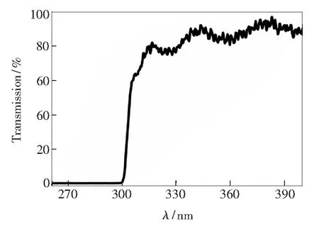
A typical transmission spectrum of the ZnMgO films is shown in Fig.2. The films show a high transmission of over 80% in the ultraviolet spectrum region, while they have a very sharp absorption edge at around 310 nm (4 eV). Therefore, ZnMgO thin film material for the preparation of UVB optoelectronic devices has important significance. The typical feature of phase separation is the appearance of multiphase absorption edges. For this sample, no multiphase absorption edge was observed, which confirms that in our case does not occur in phase separation which is often observed in ZnMgO alloys with high Mg content. Thus, the absence of phase separation largely improves the performance of the devices can be obtained on our films. Similarly, this reveals that this cubic ZnMgO film has a very high crystalline quality.

Fig.2 Transmission spectrum of the cubic ZnMgO films in the UV spectrum
The bright-field transmission electron microscope(TEM)micrograph in cross section from a Zn0.55Mg0.45O sample is shown in Fig.3, with the corresponding diffraction pattern from the ZnMgO film. As shown in Fig.3(a), the Zn0.55Mg0.45O/Al2O3structure clearly appears smooth cross-section, which is a typical image of a single crystalline material. In addition, the Zn0.55Mg0.45O films were found to cohere well to the sapphire substrate and displayed a uniform thickness on a microscopic scale. The diffraction crystal lattice pattern in Fig.3(b) shows the films are crack free, homogeneous, well covered with granular deposits and without any evidence of lattice defects. As a consequence, the micrograph and diffraction patterns clearly show the structure of single cubic phase ZnMgO (111), there is no evidence of any phase separation, which is consistent with previous results.

Fig.3 Cross-sectional TEM and transmission lattice micrographs of a Zn0.55Mg0.45O film
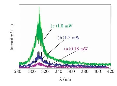
Lasing characteristics of ZnMgO film were investigated by aQ-switch Nd∶YAG laser (266 nm) under pulsed operation (6 ns, 30 Hz). Fig.4 shows the evolution of the emission spectrum as the average pump power (hereinafter referred to as pump power) increases. Comparative analysis of the three color lines in the figure, the central wavelength of the stimulated emission does not change with the pump power, which is always in the UVB about 310 nm. In the pump power from low to high changes in the process, showing a different stimulated emission spectrum, at low excitation intensity, the spectrum consisted of a single wide spontaneous emission peak. When the pump power is increased, the emission peak became narrower due to the preferential amplification at the frequency close to the maximum value of the gain spectrum. When the pump power increases further, more sharp peaks appear, and the line-width of peak drops sharply. When the pump power density is increased above the threshold, the emission intensity of the narrower feature becomes dominant. The narrow and strong emission exhibits a superlinear increase, accompanied by a slight redshift, Which indicates the appearance of stimulated emission. The stimulated emission may be attributed to the stimulated recombination of exciton-exciton scattering[26-27].

Fig.4 Stimulated emission spectra of the Zn0.55Mg0.45O films at different average pump powers ranging from 0.18 mW (a), 1.5 mW (b), 1.8 mW (c), respectively.
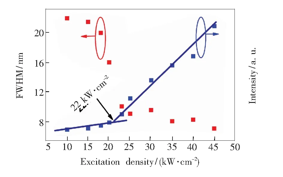
Discrete red dots in Fig.5 show FWHM as the non-linearity reduction function of the excitation density, the FWHM narrowed much more rapidly with the excitation density at the threshold. Another set of blue dots in the figure indicates the excitation density as a non-linear increasing function of the emission intensity. On the blue dot-fitted line, it is observed that the emission intensity increased much more rapidly with the excitation density above the excitation density of multiple sharp peaks emerged in the emission spectrum, which is the threshold behavior. This increased process is superlinear, which the threshold value is 22 kW/cm2. This proves that spontaneous radiation is transformed into stimulated radiation.

Fig.5 FWHM and emission intensity of the Zn0.55Mg0.45O films as a non-linear function of the excitation density
These results indicate that laser action has occurred in the Zn0.55Mg0.45O films. Compared with other literatures, this result of threshold power was lower than several orders of magnitude. This is a momentous progress to improve the work performance of laser devices.
The optical cavity formed by repeated scattering has a different loss. When the pump power increases, the gain first achieves low loss cavity loss. After that, laser oscillation occurs in these cavities, and the laser frequency is determined by the cavity resonance. Laser emission from these resonators results in a small amount of discrete emission spectra narrow peaks. When the pump power is further increased, the gain increases and exceeds the loss in the lossier cavities. Due to the laser oscillations in these cavities, the emission spectrum adds more discrete peaks. These theories are consistent with the experimental data in this paper.
4 Conclusion
In conclusion, pure cubic MgxZn1-xO thin films with Mg content of 45% have been prepared by PLD. High crystalline quality non-phase separation of the Zn0.55Mg0.45O films was observed in the results of XRD and TEM. Under optical pumping, excitation occurs in the UVB wavelength of ~310 nm. As the excitation density increased, the peak intensity increased superlinear, and the linewidth of peak decreased dramatically. The high-quality Zn0.55Mg0.45O alloys films with a thresholds of 22 kW/cm2that we prepared have potential applications in various optoelectronic devices.
[1] LIU Y, YU L, HU Y,etal.. A magnetically separable photocatalyst based on nest-like γ-Fe-O-/ZnO double-shelled hollow structures with enhanced photocatalytic activity [J].Nanoscale, 2012, 4(1):183-187.
[2] ETACHERI V, ROSHAN R, KUMAR V. Mg-doped ZnO nanoparticles for efficient sunlight-driven photocatalysis [J].AcsAppl.Mater.Interf., 2012, 4(5):2717-2725.
[3] WANG X, LIAO M Y, ZHONG Y T,etal.. ZnO hollow spheres with double-yolk egg structure for high-performance photocatalysts and photodetectors [J].Adv.Mater., 2012, 24(25):3421-3425.
[4] LIU D, LI C C, ZHOU F,etal.. Rapid synthesis of monodisperse au nanospheres through a laser irradiation-induced shape conversion, self-assembly and their electromagnetic coupling SERS enhancement [J].Sci.Rep., 2015, 5:7686-7686.
[5] LEE C T, LIN H Y, TSENG C Y. Nanomesh electrode on MgZnO-based metal-semiconductor-metal ultraviolet photodetectors [J].Sci.Rep., 2015, 5:13705.
[6] WANG L K, JU Z G, ZHANG J Y,etal.. Single-crystalline cubic MgZnO films and their application in deep-ultraviolet optoelectronic devices [J].Appl.Phys.Lett., 2009, 95(13):131113-1-3.
[7] CHEN Y Y, WANG C H, CHEN G S,etal.. Self-powered n-MgxZn1-xO/p-Si photodetector improved by alloying-enhanced piezopotential through piezo-phototronic effect [J].NanoEnergy, 2015, 11:533-539.
[8] ZENG H, CAI W P, HU J L,etal.. Violet photoluminescence from shell layer of Zn/ZnO core-shell nanoparticles induced by laser ablation [J].Appl.Phys.Lett., 2006, 88(17):171910-1-3.
[9] ZHENG Q, HUANG F, DING K,etal.. MgZnO-based metal-semiconductor-metal solar-blind photodetectors on ZnO substrates [J].Appl.Phys.Lett., 2011, 98(22):221112-1-3.
[10] KUMAR M H, YANTARA N, DHARANI S,etal.. Flexible, low-temperature, solution processed ZnO-based perovskite solid state solar cells [J].Chem.Commun., 2013, 49(94):11089-11091.
[11] LIANG Z Q, ZHANG Q F, WIRANWETCHAYAN O,etal.. Effects of the morphology of a ZnO buffer layer on the photovoltaic performance of inverted polymer solar cells [J].Adv.Funct.Mater., 2012, 22(10):2194-2201.
[12] TANG K, GU S L, YE J D,etal.. High-quality ZnO growth, doping, and polarization effect [J].J.Semicond., 2016, 37(3):031001.
[13] TIAN F B, DUAN D F, LI D,etal.. Miscibility and ordered structures of MgO-ZnO alloys under high pressure [J].Sci.Rep., 2014, 4:5759-5759.
[14] SARD A. Sol-gel derived Li-Mg co-doped ZnO films: preparation and characterizationviaXRD, XPS, FESEM [J].J.AlloysCompds., 2012, 512(1):171-178.
[15] TENG C W, MUTH J F, ÖZGÜR Ü, etal.. Refractive indices and absorption coefficients of MgxZn1-xO alloys [J].Appl.Phys.Lett., 2000, 76(8):979-981.
[16] OHTOMO A, KAWASAKI M, KOIDA T,etal.. MgxZn1-xO as a Ⅱ-Ⅳ widegap semiconductor alloy [J].Appl.Phys.Lett., 1998, 72(19):2466-2468.
[17] TIAN Y, MA X Y , LI D S,etal.. Electrically pumped ultraviolet random lasing from heterostructures formed by bilayered MgZnO films on silicon [J].Appl.Phys.Lett., 2010, 97(6):061111-1-3.
[18] PARK W I, YI G C, JANG H M. Metalorganic vapor-phase epitaxial growth and photoluminescent properties of Zn1-x-MgxO(0≤x≤0.49) thin films [J].Appl.Phys.Lett., 2001, 79(13):2022-2024.
[19] SHARMA A K, NARAYAN J, MUTH J F,etal.. Optical and structural properties of epitaxial MgxZn1-xO alloys [J].Appl.Phys.Lett., 1999, 75(21):3327-3329.
[20] CHOOPUN S, VISPUTE R D, YANG W,etal.. Realization of band gap above 5.0 eV in metastable cubic-phase MgxZn1-xO alloy films [J].Appl.Phys.Lett., 2002, 80(9):1529-1531.
[21] TANAKA H, FUJITA S, FUJITA S. Fabrication of wide-band-gap MgxZn1-xO quasi-ternary alloys by molecular-beam epitaxy [J].Appl.Phys.Lett., 2005, 86(19):192911-1-3.
[22] YANG W, HULLAVARAD S S, NAGARAJ B,etal.. Compositionally-tuned epitaxial cubic MgxZn1-xO on Si(100) for deep ultraviolet photodetectors [J].Appl.Phys.Lett., 2003, 82(20):3424-3426.
[23] CHEN H X, DING J J, MA S Y. Structural and optical properties of ZnO∶Mg thin films grown under different oxygen partial pressures [J].PhysicaE, 2010, 42(5):1487-1491.
[24] TIAN C, JIANG D Y, TAN Z D,etal.. Effects of thermal treatment on the MgxZn1-xO films and fabrication of visible-blind and solar-blind ultraviolet photodetectors [J].Mater.Res.Bull., 2014, 60:46-50.
[25] JU Z G, SHAN C X, YANG C L,etal.. Phase stability of cubic Mg0.55Zn0.45O thin film studied by continuous thermal annealing method [J].Appl.Phys.Lett., 2009, 94(10):101902-1-3.
[26] WANG L K, JU Z G, ZHANG J Y,etal.. Single-crystalline cubic MgZnO films and their application in deep-ultraviolet optoelectronic devices [J].Appl.Phys.Lett., 2009, 95(13):131113-1-3.
[27] HSU H C, WU C Y, CHENG H M,etal.. Band gap engineering and stimulated emission of ZnMgO nanowires [J].Appl.Phys.Lett., 2006, 89(1):013101-1-3.
[28] MUHAMMAD M M, MOHAMMAD S L, ZHENG Z,etal.. Ultraviolet random lasing from asymmetrically contacted MgZnO metal-semiconductor-metal device [J].Appl.Phys.Lett., 2014,105(21):211107-1-3.
[29] BAGNALL D M, CHEN Y F, ZHU Z,etal.. Optically pumped lasing of ZnO at room temperature [J].Appl.Phys.Lett., 1997, 70(17):2230-2232.
[30] CHEN Y F, TUAN N T, SEGAWA Y,etal.. Stimulated emission and optical gain in ZnO epilayers grown by plasma-assisted molecular-beam epitaxy with buffers [J].Appl.Phys.Lett., 2001, 78(11):1469-1471.

张晗(1991-),女,山东济宁人,硕士研究生,2015年于曲阜师范大学获得学士学位,主要从事半导体光电子材料与器件的研究。
E-mail: 15625138017@163.com

宿世臣(1979-), 男, 黑龙江佳木斯人,博士,副研究员,2009年于中科院长春光机所获得博士学位,主要从事宽禁带半导体的研究。
E-mail: shichensu@126.com
2017-03-07;
2017-04-17
国家自然科学基金(61574063); 广东省科技计划(2016A040403106); 广州市科技计划(2016201604030047)资助项目 Supported by National Natural Science Foundation of China(61574063); Science and Technology Program of Guangdong Province(2016A040403106); Science and Technology Project of Guangzhou City(2016201604030047)
高质量立方相ZnMgO的制备与紫外受激发射特性研究
张 晗, 裴磊磊, 宿世臣*
(广东省光电功能材料与器件重点实验室, 华南师范大学 光电材料与技术研究所, 广东 广州 510631)
利用脉冲激光沉积(PLD)设备在蓝宝石衬底上制备了高质量Zn1-xMgxO单晶薄膜,并对其结构和光学特性进行了深入细致的研究。通过能量衍射谱(EDS)确认Zn1-xMgxO薄膜的Mg组分为45%。在Zn0.55Mg0.45O薄膜的X射线衍射谱(XRD)中观测到了明显的位于36.67°的衍射峰,对应的是(111)晶向的立方相ZnMgO。从透射光谱中可以看出,Zn0.55Mg0.45O具有陡峭的吸收边,没有发生相分离,在透射电镜图谱中也得到了证实。该ZnMgO薄膜还表现出了优异的光学特性,在Zn0.55Mg0.45O材料体系中实现了峰位位于310 nm的紫外光泵浦受激发射,其激光发射的阈值仅为22 kW/cm2。
氧化锌镁; 脉冲激光沉积; 阈值
1000-7032(2017)07-0905-06
O484.4 Document code: A
10.3788/fgxb20173807.0905
*Corresponding Author, E-mail: shichensu@126.com
- 发光学报的其它文章
- 基于碳量子点荧光恢复的三聚氰胺测定方法
- ZnO/Cu2O异质结纳米阵列制备及光催化性能
- C-mount封装激光器热特性分析与热沉结构优化研究
- High-efficiency Flexible Organic Solar Cells with UV-ozone Treated Silver Modification ITO Electrode
- Preparation of Eu2+ Activated Barium Phosphosilicate Phosphors with Two-color Emission by A New Two-step Method
- 铜掺杂氧化锌纳米棒的非线性光学响应竞争特性

