消化道颗粒细胞瘤临床特点及疗效分析
2017-05-22 13:35马豫茜李国华樊淑梅
中国医药科学 2017年6期
马豫茜+李国华+樊淑梅

[摘要] 目的 分析消化道颗粒细胞瘤(GCT)患者的临床特征,对治疗效果进行分析。方法 回顾分析 2012年 12月~2016年10月在我院住院的5 例消化道 GCT患者的临床资料,分析其临床特征,治疗效果。结果5 例消化道 GCT 患者中,食管 GCT4 例(其中1例合并早期食管癌),盲肠 GCT 1例,所有患者均表现为非特异性消化道症状或无消化道症状,均行术前内镜及超声内镜检查诊断为消化道黏膜下隆起性病变,分别疑诊脂肪瘤、 类癌及异位胰腺。1例术前考虑消化道 GCT,5例患者术前均未活检,4例行内镜下黏膜剥离术(ESD),1例行单环套扎+圈套器切除,术后病理及免疫组化分析显示肿瘤细胞均表达 S-100,确诊为消化道 GCT,未见明显恶性特征,术后随访3~48个月,均无复发及转移。结论 消化道GCT 可无特异性消化道症状、发病率低,内镜下亦无特异性表现,因此明确诊断仍依赖于病理结果,对于<2cm 的黏膜下 GCT,内镜下治疗效果好,但需长期随访,排除恶变可能。
[关键词] 消化道;颗粒细胞瘤;特点
[中图分类号] R735 [文献标识码] A [文章编号] 2095-0616(2017)06-154-04
[Abstract] Objective To explore the clinical characteristicas of granular cell tuomr, and to analyze the curative effect. Methods Data of 5 cases of GCT patients cured in our hospital from December 2012 to October 2016 were retrospectively analyzed. The clinical characteristics and therapeutic effect were analyzed. Results Among the 5 cases of GCT patients, there were GCT4 cases of esophagus(1 cases with early esophageal cancer) and GCT of cecum in 1 cases. All patients presented with nonspecific gastrointestinal symptoms or no gastrointestinal symptoms. All cases were diagnosed by endoscopic and endoscopic ultrasonography, and they respectively were lipoma, carcinoid and ectopic pancreas. 1 cases of gastrointestinal tract GCT were considered before operation, and all of the 5 patients had no biopsy before operation, and endoscopic mucosal resection(ESD) was performed in all of the 4 patients, and the other cases were treated with single loop ligation and snare excision(n=1). Postoperative pathological and immunohistochemical analysis showed that the tumor cells all expressed S-100, which was diagnosed as digestive tract GCT, and there was no obvious malignant feature. No recurrence and metastasis were observed after 3-48 months of follow-up. Conclusion There are no specific gastrointestinal symptoms and low incidence of GCT in the digestive tract. Therefore, the definite diagnosis depends on pathological results. For less than 2cm of submucosal GCT, endoscopic treatment effect is good, but need long-term follow-up, exclude the possibility of malignancy.
[Key words] Digestive tract; Granular cell tumor; Characteristic
顆粒细胞瘤(granular cell tumor,GCT)是一种来源于外周神经,具分化的有雪旺细胞(Schwanns cell)分化特点的罕见软组织肿瘤,免疫组化显示颗粒细胞瘤细胞主要表达S-100蛋白,1926年由Abrikossof首次报道[1],临床发生率低,任何年龄均可发病,但以30~60岁多见[2],可发生于身体任何部位,一般好发于口腔、头颈部、躯干和四肢及外阴的皮肤或皮下软组织,而消化道仅占8%,其中食管占2%[3-4],下段>中段>上段,发生于胃、小肠和胆道的少见,发生于盲肠者及食管GCT合并早期食管癌者更加罕见,目前该病多为个案报道。我们对5例消化道GCTs的临床、内镜特点及内镜下治疗效果进行研究、总结,分析其特点及治疗效果,现报道如下。
1 资料与方法
1.1 一般资料
2012年12月~2016年10月在佛山市顺德区第一人民医院住院的5例消化道GCTs患者,男3例,女2例,年龄31~64岁,平均49.5岁。
1.2 方法
回顾性分析5例消化道GCTs患者的临床资料:一般资料、临床表现、内镜表现、超声内镜表现、病理及免疫組化结果、治疗方法及预后,分析临床特点及治疗效果。
2 结果
2.1 5例消化道GCT患者临床表现
消化系统颗粒细胞瘤患者临床症状以腹部不适、吞咽不畅、胸骨后不适、烧心,见表1,图1。
2.2 5例消化道GCT患者超声内镜表现
5例位于黏膜下层,呈低回声改变,均未累及肌层,界限清楚,见表2,图1~2。
2.3 5例消化道GCTs治疗方法
4例行ESD治疗,1例行单环黏膜套扎+圈套器切除术,1例出血术中穿孔,予以钛夹夹闭伤口,2例创面少量渗血,予以电凝止血后无活动性出血,5例均完整切除,送病理学检查,术后恢复良好,见表3,图3。
2.4 病理及免疫组化结果
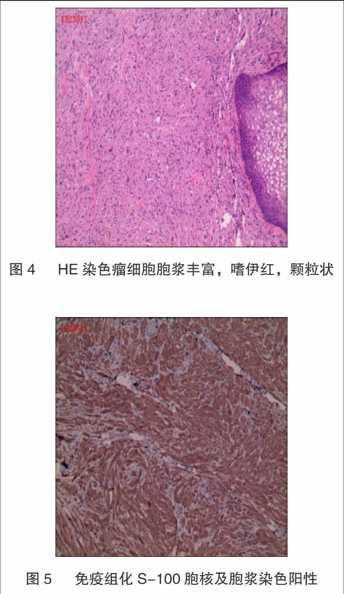
术后病理HE(图4)肿瘤细胞排列紧密,呈巢状或条索状,细胞大小一致,胞质丰富有大量嗜酸性颗粒细胞,核小、圆而居中。免疫组化(图5)肿瘤细胞S-100均呈弥漫性强阳性。
2.5 术后随访
5例患者其中4例随访1~2年,复查胃镜及肠镜未见肿瘤复发。1例颗粒细胞瘤并浅表食管癌患者(内镜治疗)术后3个月随访1次,复查食管黏膜未见复发。
3 讨论
消化道颗粒细胞瘤可发生在食管、胃、小肠、大肠、肛管、甚至胆管、胰腺等部位[5-10]。其中食管发生比例最高,盲肠相对较少,食管颗粒细胞瘤合并覆盖黏膜鳞状细胞癌的,罕见报道。本组5例消化道GCT,1例发生于盲肠,1例食管颗粒细胞瘤合并被覆上皮早期鳞癌。
消化道GCT无特异性症状,多表现为腹部不适、胸骨后不适,吞咽不畅等消化道症状,根据肿瘤大小及部位的不同而症状各异。部分患者为体检时意外发现,本研究5例患者的临床表现列表可以看出,GCT可无任何症状或仅有非特异性的消化道症状。除此之外,消化道GCT可与其他疾病并存,甚至多部位肿瘤。有临床发现,食管GCT与食管鳞状细胞癌并存[11],这与我们研究发现的其中1例病例符合。因此,我们在内镜检查过程中,不但要观察隆起型黏膜下病变,其覆盖表面上皮或临近组织也应仔细观察,避免多发肿瘤的漏诊。
消化道颗粒细胞瘤的内镜表现具有一定特征性[12]。主要为:(1)白光镜下表现为表面光滑的淡黄色或灰白色隆起,边界清晰;(2)超声内镜表现为来源与黏膜下层或黏膜肌层为主的低回声病变,病变边界清晰,内部回声均匀,随着诊断技术的发展,超声内镜对GCT的诊断率有所提高[13],但单纯内镜检查仍无法与间质瘤、类癌及脂肪瘤等鉴别。临床诊断最终依靠病理及免疫组化[14],诊断标准:(1)瘤细胞胞浆丰富,嗜酸性颗粒状,排列成索状或巢状;(2)免疫组化S-100表达阳性。
消化道GCTs以良性居多,且以黏膜下层为主,适合内镜下EMR、ESD黏膜套扎切除等治疗。我组研究5例病例采用ESD及单环套扎+切除术治疗GCTs均未出现出血、穿孔、腹膜炎或纵膈感染等并发症,患者恢复良好,且经复查无复发或转移等并发症,因此,内镜治疗不失为临床值得应用及推广的微创治疗方法,但GCT仍有恶变可能,通过淋巴及血道转移至肝、肺、骨甚至胰腺,导致死亡[15],因此应强调术后复查的重要性。
[参考文献]
[1] Abrikossoff A.Uber Myomeausgehend von der uergestreiften will- kurlichen Muskulatur[J].Virchows Arch Pathol Anat,1926,260:215- 233
[2] Endo S,Hirasaki S,Doi T,et al.Granular cell tumor occurring in the sigmoid colon treated by endoscopic mucosal resection using a transparent cap (EMR-C)[J].J Gastroenterol,2003,38(4):385-389.
[3] 张芳,徐永红,闫领,等.食管颗粒细胞瘤的研究进展[J].世界华人消化杂志,2016,24(17):2647-2653.
[4] Narra SL,Tombazzi C,Datta V,et al.Granular cell tumor of the esophagus: report of five cases and review of the literature[J].Am J Med Sci,2008,335(5):338-341.
[5] An S,Jang J,Min K,et al.Granular cell tumor of the gastrointestinal tract: Histo-logic and immunohistochemical analysis of 98 cases[J].Hum Pathol,2015,46(6):813-819.
[6] Thumallapally N,Ibrahim U,Kesavan M,et al.Esophageal Granular Cell Tumor: A Case Report and Review of Literature[J].Cureus,2016,8(9):e782.
[7] Sevilla Ribota S,Perez-Bedmar Delgado J,Dominguez Canete JJ,et al.Endoscopic resection of rectal granular-cell tumor using elastic band ligation[J].Rev Esp Enferm Dig,2016,108(10):677-680.
[8] Chopade TR,Smith CL,Maley WR,et al.Granular Cell Tumor of the Common Hepatic Duct as an Unusual Cause of Jaundice in a Hepatitis C Patient[J].ACG Case Rep J,2016,3(2):115-117.
[9] Pertile D,Scabini S,Romairone E, et al.Gastric Abrikosoff tumor (granular cell tumor): Case report[J].G Chir,2010,31(10):433-434.
[10] Kanno A,Satoh K,Hirota M,et al.Granular cell tumor of the pancreas: A case report and review of literature[J].World J Gastrointest Oncol,2010,2(2):121-124.
[11] Saito K,Kato H,Fukai Y,et al.Esophageal granular cell tumor covered by intramucosal squamous cell carcinoma: Report of a case[J].Surg Today,2008,38(7):651- 655.
[12] 潘晨,張永宏,杨杰.食管颗粒细胞瘤1例并文献复习[J].国际消化病杂志,2012,50(3):188-189,191.
[13] 钱燕敏,许国强,陈洪潭,等.食管颗粒细胞瘤超声内镜图像的计算机分析研究[J].中华消化杂志,2013,33(4):223-225.
[14] An S,Jang J,Min K,et al.Granular cell tumor of the gastrointestinal tract: histologic and immunohistochemical analysis of 98 cases[J].Hum Pathol,2015,46(6): 813-819.
[15] Fujita F,Eguchi S,Takatsuki M,et al.A recurrent granulosa cell tumor of the ovary 25 years after the initial diagnosis: A case report[J]. Int J Surg Case Rep,2015,12:7-10.
(收稿日期:2017-02-04)
猜你喜欢
保健与生活(2022年5期)2022-03-15
科学导报·学术(2020年47期)2020-11-17
科教导刊·电子版(2020年19期)2020-10-09
中国保健营养(2019年9期)2019-10-20
学习与科普(2019年4期)2019-09-10
中学课程辅导·教师教育(中)(2016年9期)2016-10-20
科技视界(2016年21期)2016-10-17
考试周刊(2016年76期)2016-10-09
中华少年(2009年9期)2009-09-14