青少年特发性脊柱侧凸术后 adding-on 现象病例分析及分类初探
杨长伟 夏士博 杨明园 赵检 朱晓东 李明
青少年特发性脊柱侧凸术后 adding-on 现象病例分析及分类初探
杨长伟 夏士博 杨明园 赵检 朱晓东 李明
目的初步探讨青少年特发性脊柱侧凸 ( adolescent idiopathic scoliosis,AIS ) 患者术后远端adding-on 现象的原因和分类。方法回顾分析在我院行手术矫形、术后随访期发生明显 adding-on 现象的14 例 AIS 患者。根据其影像学、年龄及骨骼成熟度等参数的不同,将所有 adding-on 病例分为 3 种类型:I 型为伴有术后双肩失平衡型;II 型为伴有术后冠状位失平衡型;III 型为不伴有双肩和冠状位失平衡型,并对各类型的临床和影像学特点进行初步总结。结果I 型 6 例 ( 42.9% ),患者术后明显双肩失平衡,平均双肩高度差 ( radiographic shoulder height,RSH ) 为 20 ( 15~25 ) mm。术后 2 年的随访中,RSH 减少到 6 ( 0~12 ) mm,但是其远端 adding-on 角度平均增加 14° ( 5°~23° )。II 型 3 例 ( 21.4% ),术后明显冠状位失平衡,平均冠状面失平衡值为 45 ( 39~54 ) mm。末次随访时,冠状位失衡平均减小 13 ( 8~17 ) mm,但 adding-on 角度平均增加10° ( 5°~14° )。III 型 5 例 ( 35.7% ),所有患者 Risser 征均为 0 度,术后即刻无明显双肩和冠状位失平衡,但是随访期中仍然发生 adding-on,其 adding-on 角度平均增加 10° ( 6°~15° )。长期随访结果表明,I 型和 II 型均无再次手术者,但是 1 例 III 型患者因侧凸进展迅速接受了再次手术。结论 本次研究提出 adding-on 现象的初步分型,描述其相关临床及影像学特点,将有助于 adding-on 的发病机制的探讨。
脊柱侧凸;青少年;肌肉骨骼生理现象;Adding-on 现象
青少年特发性脊柱侧凸 ( adolescent idiopathic scoliosis,AIS ) 是一种原因不明、脊柱在冠状位上偏离中线同时伴有椎体旋转和矢状位曲度改变的畸形,是小儿骨骼肌肉系统中最常见的畸形之一。
手术治疗仍是目前对进展性 AIS 主要的有效治疗方式。椎弓根螺钉因其三柱固定的特点,矫形能力增强,广泛应用于后路侧凸矫形融合。随着临床的广泛应用,很多学者注意到一些病例在术后随访中,远端未融合节段发生侧凸向原主弯方向加重的情况。这一现象最初由 Suk 等[1]最先报道并将其称为 adding-on 现象。其后很多学者也进行了研究,报道其发生率为 2%~21%[2-4]。目前公认的 adding-on诊断标准为:术后即刻至 2 年随访中,下端椎向融合节段远端移动并且冠状面 Cobb’s 角增加 5° 以上;或者下固定椎 ( lowest instrumented vertebra,LIV ) 远端邻近椎间盘成角变化>5°[4]。
虽然大多数术后出现 adding-on 的患者并无明显症状,但这一现象会导致矫正丢失,邻近椎间盘楔形变,严重者甚至需再次手术延长融合节段[5-6],文献报道其再次手术率为 7.3%[7]。目前的研究发现了 adding-on 的一些危险因素,包括 LIV 选择不恰当、患者骨骼发育不成熟、术后双肩不平衡及 LIV到骶骨中垂线距离 ( LIV-CSVL ) 过大等[3,5-6,8-10]。这些研究表明,adding-on 是一个复杂的现象,可能存在不同类型以对应不同原因。因此,笔者对所在单位 2009 年 9 月至 2013 年 11 月接受手术矫形并发生adding-on 的 14 例 AIS 患者进行回顾分析,并予以初步分类探讨。
资料与方法
一、入组与排除标准
1. 入组标准:( 1 ) 明确诊断为 AIS 并接受前路或者后路矫形患者;( 2 ) 有完整的术前、术后1 周及术后 2 年的全脊柱正侧位 X 线片;( 3 ) 满足adding-on 的诊断标准。
2. 排除标准:其它类型脊柱侧凸,如先天性脊柱侧凸、神经肌肉型脊柱侧凸等,以及没有完整的影像学资料者。
二、数据采集
一般资料包括:年龄、性别、Risser 征和 Lenke分型。影像学参数包括:LIV 的位置 [ 与稳定椎( stable vertebrae,SV ) 的关系 ]、双肩高度差 ( radiographic shoulder height,RSH )、冠状面平衡 ( C7铅垂线到骶骨中垂线的距离 )、adding-on 角度 ( LIV 上终板与远端第二椎体下终板夹角 )。adding-on 角度改变为术后 2 年随访时与术后即刻角度差值。根据adding-on 病例特征分为不同类型,总结各种类型的一般特征。
结 果
研究回顾分析 241 例 AIS 病例,最终纳入 14 例( 5.8% )。根据不同特点,将 14 例 adding-on 病例分为 3 个类型,现将各分型及其临床特征介绍如下。
一、I 型:术后双肩失平衡型
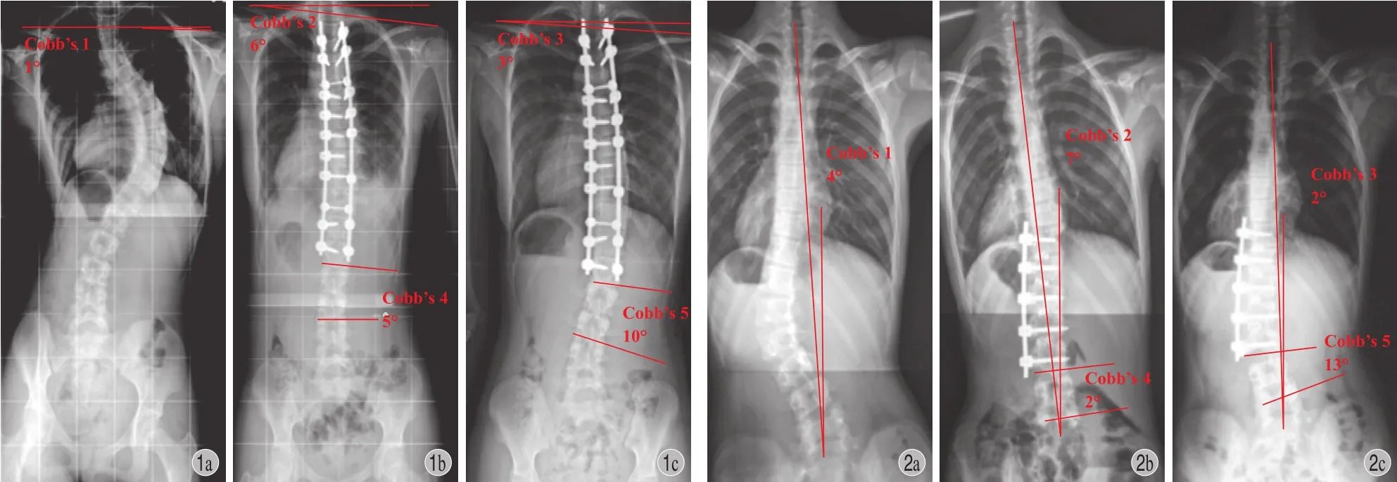
14 例 adding-on 病例中,6 例 ( 42.9% ) 术后伴有明显双肩失平衡。其平均年龄为 14 岁,平均 Risser征为 2.5 度。6 例中 3 例为 Lenke 2 型 ( 2 例 Lenke 2 AN,1 例 Lenke 2 A+ ),2 例为 Lenke 1 BN,1 例Lenke 3 CN (表1)。这些患者术后首次站立摄 X 线片时存在明显的术后双肩不平衡,平均 RSH 为 20 ( 15~25 ) mm,所有患者均为左肩高。术后 2 年随访中,LIV 以下节段向右侧凸,右肩增高,双肩失平衡明显改善,RSH 平均减少至 6 ( 0~12 ) mm,但是远端 adding-on 角度平均增加了 14° ( 5°~23° )。进一步随访表明,这些患者 adding-on 角度没有再进展,均未再次手术干预 (表1、图1)。
二、II 型:术后躯干失平衡型
14 例中,3 例 ( 21.4% ) 术后首次站立摄 X 线片出现明显冠状位失平衡,躯干向左偏移,平均冠状面失平衡值为 45 ( 39~54 ) mm。平均年龄为 14 岁,平均 Risser 征为 II 度。3 例均为 Lenke 5 CN 型,其 LIV 均到稳定椎以上 2 个椎体 ( SV-2 )。2 年随访时,融合椎以下节段向左弯曲以代偿躯干失平衡,冠状位失衡平均减小 13 ( 8~17 ) mm。但 adding-on角度平均增加 10° ( 5°~14° )。进一步随访结果提示,这些患者 adding-on 角度没有再进展,均未再次手术 (表2、图2)。
三、III 型:无术后双肩失平衡和躯干失平衡型
5 例 ( 35.7% ) 术后没有明显术后躯干及双肩失平衡,但在随访中仍然发生 adding-on。平均年龄为 12 ( 11~13 ) 岁,所有患者 Risser 征均为 0 度。5 例均为 Lenke 1 型 ( 4 例为 Lenke 1 AN 型,1 例为Lenke 1 B- 型 )。术后 2 年随访时,adding-on 角度平均增加 10° ( 6°~15° ) (表3、图3)。其中 1 例因角度进展迅速,进行翻修手术延长了融合节段。

表1 I 型 adding-on 患者相关资料Tab.1 General information of the patients with Type I adding-on

表2 II 型 adding-on 患者相关资料Tab.2 General information of the patients with Type II adding-on

图1 I 型 adding-on 典型病例,患者,女,13 岁,Lenke 2 AN 型 a:术前双肩基本平齐;b:术后双肩明显不平衡;c:术后 2 年随访,双肩趋向平衡,远端 adding-on 角度增加 15°图 2 II 型 adding-on 典型病例,患者,男,15 岁,Lenke 5 CN 型 a:术前有冠状位失平衡;b:术后冠状位失平衡加重;c:术后 2 年随访,术后冠状位平衡明显改善,但是远端 adding-on 角度增加 11°Fig.1 A typical case with type I adding-on, female, 13 years old, Lenke 2 AN a: Shoulder balance before surgery; b: Significant shoulder imbalance postoperatively; c: Shoulder rebalance during 2-year follow-up, but distal adding-on angle was increased by 15°Fig.2 A typical case with type II adding-on, male, 15 years old, Lenke 5 CN a: Coronal imbalance before surgery; b: Coronal imbalance was aggravated postoperatively; c: Coronal rebalance during 2-year follow-up, but distal adding-on angle was increased by 11°
讨 论
本研究描述了 3 种不同类型的 adding-on 现象,笔者认为进一步研究 adding-on 的原因及预防方法时,要根据不同类型分组研究,将不同类型混杂无助于明确其真正的原因,这也与目前很多研究揭示其复杂的相关因素一致。
I 型患者,主要是 Lenke 1、2 型,术后即刻存在明显的双肩失平衡,末次随访时患者双肩平衡明显改善,但是融合节段下出现右侧弯曲,以代偿性减少 RSH,达到双肩平衡。这一现象与一些学者发现的术后双肩失平衡可能会加剧 adding-on 相一致[9,11-12]。因此,笔者推测这一类型 adding-on 的发生是对术后双肩失平衡的代偿,融合节段的选择及上胸弯处理不当造成双肩失衡是该类型发生 adding-on现象的主要原因。对此类病例,术前应仔细计划,术中撑开及加压操作时,须密切观察双肩平衡情况。然而进一步随访表明,这些患者 adding-on 角度进展不大,不需要再次手术干预,这表明尽管I 型 adding-on 虽然发生率高,但是对患者临床效果影响相对较小。因此,是采取减少远端融合节段而适度容忍这一现象的发生,还是延长融合避免其发生,不同学者也有不同观点[4]。

表3 III 型 adding-on 患者相关资料Tab.3 General information of the patients with Type III adding-on

图3 III 型 adding-on 典型病例,患者,女,11 岁,Lenke 1 AN型,Risser 征 0 度 a:术前双肩及冠状位平衡良好;b:术后患者无明显双肩失平衡及冠状位失平衡;c:术后 2 年随访,出现adding-on 角度为 15°Fig.3 A typical case with type III adding-on, female, 11 years old, Lenke 1 AN, Risser 0° a: Shoulder balance and coronal balance before surgery; b: No significant shoulder or coronal imbalance postoperatively; c: Distal adding-on angle was increased by 15° during 2-year follow-up
对于 II 型,主要发生在以胸腰 / 腰弯为主弯( Lenke 5 型 ) 的患者,术后即刻均存在明显冠状位失平衡,为了适应胸 / 腰弯的显著变化和重建冠状面平衡,LIV 远端的活动椎间盘发生楔形变化以代偿性重建冠状面平衡[13-14],这一类型 adding-on 现象事实上与很多文献已经报道的 Lenke 5 型术后 LIV 远端椎间盘楔形变是同一现象[14]。预防方法主要是术前评估远端未融合节段的代偿能力,必要时延长节段或术中适度矫正侧凸,以适应远端倾斜,不至于冠状位失平衡。进一步随访结果提示,与 I 型 adding-on患者类似,这些患者 adding-on 角度进展不大,都没有再次手术。
III 型 adding-on 为术后不伴有肩部或者冠状位失平衡的类型,主要发生于低龄、骨骼发育成熟度低的患者,本研究中的 III 型 adding-on 患者平均年龄为 12 岁,所有患者 Risser 征都是 0 度,提示此类型 adding-on 的发生机制可能与侧凸自然进程的加重有关,这在很多其他学者研究中已得到证实[3,8,15]。而且这部分患者可能进展很快,甚至需要再次手术,本研究发现 1 例末次随访时主弯角度增加到45° 以上,从而被迫选择手术延长融合节段。由于本研究纳入病例较少,III 型 adding-on 是否与融合节段的选择有关仍然不能明确,目前能采取的预防手段只有适当延长融合节段。对这部分病例需要更大样本的深入研究,以阐明其危险因素和有效的预防措施。
虽然本研究对 adding-on 现象进行了分类,但是这一分类是初步的,还存在很多缺陷:首先,研究仅为单中心的 14 例病例,样本量很少,需要更多的病例来验证分型的完整性;第二,分类主要依据患者影像学及临床特征,主观性也比较大,其科学性和临床指导的有效性也需进一步探讨。因此,需要更多的大样本调查来进一步完善 adding-on 现象的分类研究。
根据 adding-on 患者的临床及影像学特点,可以分为不同的类型,其对应的危险因素也不同。这种分型将有助于对 adding-on 发生机制的理解,从而采取有效的预防方法以避免其发生。
[1] Suk SI, Kim WJ, Lee CS, et al. Indications of proximal thoraciccurve fusion in thoracic adolescent idiopathic scoliosis: recognition and treatment of double thoracic curve pattern in adolescent idiopathic scoliosis treated with segmental instrumentation[J]. Spine, 2000, 25(18):2342-2349.
[2] Lehman RA Jr, Lenke LG, Keeler KA, et al. Operative treatment of adolescent idiopathic scoliosis with posterior pedicle screw-only constructs: minimum three-year followup of one hundred fourteen cases[J]. Spine, 2008, 33(14): 1598-1604.
[3] Matsumoto M, Watanabe K, Hosogane N, et al. Postoperative distal adding-on and related factors in Lenke type 1A curve[J]. Spine, 2013, 38(9):737-744.
[4] Cho RH, Yaszay B, Bartley CE, et al. Which Lenke 1A curves are at the greatest risk for adding-on... and why[J]? Spine, 2012, 37(16):1384-1390.
[5] Wang Y, Hansen ES, Høy K, et al. Distal adding-on phenomenon in Lenke 1A scoliosis: risk factor identification and treatment strategy comparison[J]. Spine, 2011, 36(14): 1113-1122.
[6] Lakhal W, Loret JE, de Bodman C, et al. The progression of lumbar curves in adolescent Lenke 1 scoliosis and the distal adding-on phenomenon[J]. Orthop Traumatol Surg Res, 2014, 100(Suppl 4):S249-254.
[7] Sponseller PD, Betz R, Newton PO, et al. Differences in curve behavior after fusion in adolescent idiopathic scoliosis patients with open triradiate cartilages[J]. Spine, 2009, 34(8):827-831.
[8] Upasani VV, Hedequist DJ, Hresko MT, et al. Spinal deformity progression after posterior segmental instrumentation and fusion for idiopathic scoliosis[J]. J Child Orthop, 2015, 9(1):29-37.
[9] Cao K, Watanabe K, Hosogane N, et al. Association of postoperative shoulder balance with adding-on in Lenke Type II adolescent idiopathic scoliosis[J]. Spine, 2014, 39(12): E705-712.
[10] 耿翔, 王孝宾, 吕国华, 等. Lenke 1A 型青少年特发性脊柱侧凸患者胸弯融合术后附加现象的原因分析[J]. 中国脊柱脊髓杂志, 2013, 23(7):622-627.
[11] Matsumoto M, Watanabe K, Kawakami N, et al. Postoperative shoulder imbalance in Lenke Type 1A adolescent idiopathic scoliosis and related factors[J]. BMC Musculoskelet Disord, 2014, 15:366.
[12] Cao K, Watanabe K, Kawakami N, et al. Selection of lower instrumented vertebra in treating Lenke type 2A adolescent idiopathic scoliosis[J]. Spine, 2014, 39(4):E253-261.
[13] Wang T, Zeng B, Xu J, et al. Radiographic evaluation of selective anterior thoracolumbar or lumbar fusion for adolescent idiopathic scoliosis[J]. Eur Spine J, 2008, 17(8):1012-1018.
[14] Sun Z, Qiu G, Zhao Y, et al. The effect of unfused segments in coronal balance reconstitution after posterior selective thoracolumbar / lumbar fusion in adolescent idiopathic scoliosis[J]. Spine, 2014, 39(24):2042-2048.
[15] 丁旗, 邱勇, 孙旭, 等. 主胸腰弯或腰弯型青少年特发性脊柱侧凸行前路选择性融合术后胸弯失代偿的危险因素[J]. 中华外科杂志, 2012, 50(6):518-523.
( 本文编辑:王萌 )
Cases report of postoperative adding-on phenomenon in patients with adolescent idiopathic scoliosis and a preliminary classification
YANG Chang-wei, XIA Shi-bo, YANG Ming-yuan, ZHAO Jian, ZHU Xiao-dong, LI Ming. Department of Spine Surgery, Changhai Hospital of the second Military Medical University, Shanghai, 200433, China Corresponding author: LI Ming, Email: limingch@21cn.com
ObjectiveTo explore the factors and classification of distal adding-on phenomenon in the patients with adolescent idiopathic scoliosis ( AIS ) after correction surgery.MethodsMedical records of 14 AIS patients suffering from adding-on phenomenon after surgical therapy in our hospital were retrospectively reviewed. Three different types of adding-on phenomenon were classifed according to their various parameters such as image, age, skeletal maturity and so on, and clinical and imaging characteristics of each type were summarized. Addingon phenomenon was divided into 3 subtypes: postoperative shoulder imbalance ( Type I ), postoperative trunk imbalance ( Type II ) and neither shoulder nor trunk imbalance after surgery ( Type III ).ResultsSignifcant shoulder imbalance was observed in 6 patients ( 42.9% ) immediately after surgery who met the criteria of Type I, with the mean radiographic shoulder height ( RSH ) of 20 mm ( range: 15 - 25 mm ). During the 2-year follow-up, the mean RSH was decreased to 6 mm ( range: 0 - 12 mm ). The mean distal adding-on angle was increased by 14° ( range: 5° - 23° ). Three patients ( 21.4% ) met the criteria of Type II, and the trunk shift was increased from 39 mm to 54 mm after surgery ( average: 45 mm ). At the fnal follow-up, the trunk shift was decreased to 13 mm ( range: 8 - 17 mm ), while the mean adding-on angle was increased by 10° ( range: 5° - 14° ). Five patients ( 35.7% ) who met the criteria of Type III were found well-balanced in both shoulder and trunk after surgery. Risser sign was 0 in all the patients, while their mean adding-on angle was still increased by 10° ( range: 6° - 15° ). Furthermore, no tendency of progressionor re-operation was noticed in the patients with Type I and Type II during the long-term follow-up. However, 1 patient with Type III received re-operation due to rapid progression of scoliosis. Conclusions Three subtypes of adding-on phenomenon are introduced, as well as their clinical and imaging characteristics, which may make us understand the pathogenetic mechanisms more profoundly.
Scoliosis; Adolescent; Musculoskeletal physiological phenomena; Adding-on phenomenon
10.3969/j.issn.2095-252X.2017.01.003
R682, R619
上海市自然基金 ( 16ZR1449100 )
200433 上海,第二军医大学长海医院骨科脊柱外科 ( 杨长伟、杨明园、赵检、朱晓东、李明 );200433 上海,第二军医大学 ( 夏士博 )
李明,Email: limingch@21cn.com
2016-10-10 )

