胸椎及胸腰椎后凸型休门病患者术后近端交界性后凸对比分析
杜长志 孙旭 李松 徐亮 王斌 朱泽章 钱邦平 邱勇
胸椎及胸腰椎后凸型休门病患者术后近端交界性后凸对比分析
杜长志 孙旭 李松 徐亮 王斌 朱泽章 钱邦平 邱勇
目的比较胸椎及胸腰椎后凸型休门病患者术后近端交界性后凸 ( proximal junctional kyphosis,PJK ) 的发生率,并探讨其相关危险因素。方法回顾性分析 2006 年 4 月至 2014 年 7 月,于我院接受后路内固定矫形融合手术治疗、术后随访 2 年以上的 60 例休门病后凸畸形患者,手术年龄为 13~24 岁,平均( 17.6±4.1 ) 岁。根据后凸顶椎位置不同,将患者分为胸椎组 ( 顶椎位于 T9及以上 ) 和胸腰椎组 ( 顶椎位于T10及以下 ),其中胸椎组 25 例,胸腰椎组 35 例。于站立位全脊柱 X 线片上测量全脊柱最大后凸 Cobb’s 角( global kyphosis,GK )、近端交界角 ( proximal junctional angle,PJA )、胸椎后凸角 ( thoracic kyphosis,TK )、腰椎前凸角 ( lumbar lordorsis,LL )、矢状面轴向垂直距离 ( sagittal vertical axis,SVA ) 和最上端固定椎 ( upper instrumented vertebra,UIV ) -C7矢状面距离参数,对比两组间 PJK 发生率和矢状面形态的影像学参数。结果本研究平均随访 ( 31.1±11.9 ) 个月。胸椎组和胸腰椎组 GK 矫正率分别为 ( 39.8±10.5 ) % 和 ( 48.6±14.4 ) % ( P=0.012 ),末次随访时,后凸矫正丢失分别为 ( 4.2±7.1 ) % 和 ( 9.3%±8.6 ) % ( P=0.018 )。术后随访中,19 例发生了术后 PJK,发生率为 31.7%,最常见的原因为 PJK I 型 ( 后方韧带破坏型 )。其中胸腰椎组的 PJK发生率显著高于胸椎组 ( 42.9% vs. 16.0%,P=0.027 )。2 例因进展性 PJK 接受支具治疗,1 例因 PJK 伴顽固性背痛进行翻修手术。自术前至术后,胸腰椎组 UIV-C7矢状面距离绝对值始终大于胸椎组。胸腰椎组的 UIV 位置和融合节段均显著低于胸椎组 [ T(6.0±1.9)vs. T(2.7±1.1),( 10.2±1.8 ) vs. ( 12.4±0.9 ),P<0.001 ]。UIV 使用钩的PJK 发生率显著高于使用椎弓根螺钉患者 ( 80% vs. 27.3%,P<0.001 ),使用卫星棒者无一例发生 PJK。结论休门病后凸畸形术后 PJK 的发生率为 31.7%,主要发生于胸腰段后凸畸形患者。UIV 位置低及使用钩、过大的后凸矫正率和 UIV-C7矢状面距离等可能是胸腰椎后凸患者并发 PJK 的主要危险因素。
脊柱后凸;Scheuermann 病;脊柱融合术;危险因素;近端交界性后凸
休门病 ( Scheuermann disease ) 又称休门病后凸畸形,是由多节椎体楔形变导致的脊柱后凸畸形,也是青少年结构性后凸畸形最常见病因[1],后凸顶椎通常位于胸椎或胸腰椎,其发病率为 0.4%~10%[2]。对于有明显后凸畸形外观、伴有顽固性腰背痛或神经功能障碍,以及保守治疗无效的进展性后凸,手术是惟一有效的治疗手段[3-4]。近年来,在脊柱畸形内固定矫形手术取得满意效果的同时,近端交界性后凸 ( proximal junctional kyphosis,PJK ) 作为一种术后并发症也逐渐受到关注。PJK 通常因手术近端内固定交界区的应力改变引起,可导致局部内固定松动、断裂和患者胸背痛,严重时甚至需要手术翻修[5]。既往研究提示,PJK 的发生和发展是多因素共同作用的结果[6],与患者年龄、近端融合椎和融合节段选择不当、后凸过度矫正及脊柱骨盆矢状面序列改变等密切相关[7-9]。然而,文献中较少有对休门病后凸畸形患者术后并发 PJK 及其危险因素的报道,且现有文献中尚缺乏对胸椎和胸腰椎休门病后凸畸形术后 PJK 发生情况的对比分析。回顾性分析 2006 年 4 月至 2014 年 7 月,在我科接受后路内固定矫形融合手术的 60 例休门病后凸畸形患者的病例资料,比较胸椎和胸腰椎后凸型休门病患者术后 PJK 的发生率,并探讨其相关危险因素。
资料与方法
一、入组与排除标准
1. 入组标准:( 1 ) 年龄 12~25 岁;( 2 ) 满足休门病诊断:至少 3 个相邻椎体的楔形变>5°[10];( 3 ) 所有患者术前常规行站立位全脊柱 X 线片、全脊柱 CT 平扫+三维重建及全脊柱 MRI,评估患者脊柱及脊髓畸形情况;( 4 ) 行后路多节段 Ponte 截骨矫形内固定融合手术;( 5 ) 最上端固定椎 ( upper instrumented vertebra,UIV ) 位于 T1~12之间;( 6 ) 随访≥24 个月且有完整的临床和影像学资料。
2. 排除标准:( 1 ) 既往手术史;( 2 ) 术后即刻CT 显示 UIV 或 UIV-1 置钉不良[11]。
二、一般资料
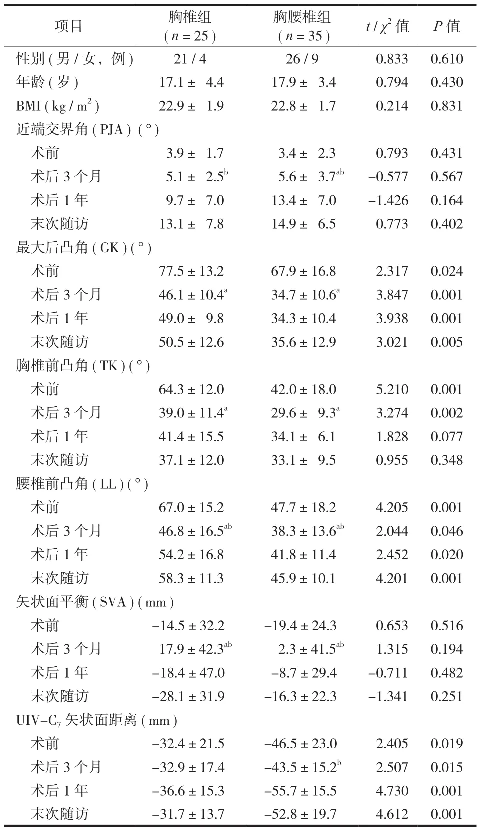
本组 60 例,其中男 47 例,女 13 例;手术年龄13~24 岁,平均 ( 17.6±4.1 ) 岁。根据后凸顶椎位置不同,将休门病后凸畸形患者分为两组:胸椎组( 顶椎位于 T9及以上 ) 和胸腰椎组 ( 顶椎位于 T10及以下 )[12]。其中胸椎组 25 例,胸腰椎组 35 例,两组患者性别、手术年龄、体质量指数 ( body mass index,BMI ) 差异无统计学意义 ( 表1 )。
三、手术方法
患者于全麻下取俯卧位,取后路正中纵向切口,根据术前制定的内固定范围暴露需要融合的节段,其中近端融合至上端椎,远端融合至后凸第一个前凸椎间盘下方的相邻椎体或是矢状面稳定椎。预先植入椎弓根螺钉或椎板钩,然后按照从尾侧向头侧,在预定节段逐个行后路 Ponte 截骨。从远端开始双侧植入预弯棒。首先将连接棒插入远端椎弓根螺钉内,旋紧螺栓,再以连接棒作为杠杆将其逐一压入其它螺钉钉槽内。棒放置和畸形矫正后,紧固螺钉。剪除的棘突制成植骨条,制备椎板植骨床。置管引流,逐层关闭切口。
四、影像学测量方法和评估参数
在术前、术后 3 个月、术后 1 年和末次随访时均摄站立位全脊柱 X 线片,用 Surgimap Spine 软件 ( Nemaris,美国 ) 测量以下参数:( 1 ) 近端交界角 ( proximal junctional angle,PJA ):UIV 的下终板与 UIV+2 的上终板之间的夹角[13-14];( 2 ) 全脊柱最大后凸 Cobb’s 角 ( global kyphosis,GK ):矢状面上端椎上终板和下端椎下终板之间的夹角;( 3 ) 胸椎后凸角 ( thoracic kyphosis,TK ):T5椎体上终板与T12椎体下终板之间的夹角;( 4 ) 腰椎前凸角 ( lumbar lordorsis,LL ):L1上终板与 S1上终板之间的夹角,前凸时记为正值;( 5 ) 以矢状面轴向垂直距离( sagittal vertical axis,SVA ) 代表矢状面平衡:C7铅垂线与骶骨后上角之间的水平距离;当 C7铅垂线位于 S1后上角前方时记为正值;( 6 ) UIV-C7矢状面距离:UIV 中心与 C7铅垂线之间的水平距离,当 UIV位于 C7铅垂线前方时记为正值。以上所有指标由同一位脊柱外科医师独立完成,连续测量 2 次后取其平均值。
PJK 定义为同时满足以下标准:( 1 ) PJA≥10°;( 2 ) 与术前测量相比,PJA 增加至少 10°[15]。参照 Yagi 等[16]分类标准,PJK 分为 3 种类型:( 1 ) I 型:后方韧带破坏;( 2 ) II 型:近端交界区骨折;( 3 ) III 型:内固定拔出或失败。
五、危险因素分析
对 PJK 发生的潜在危险因素进行评估,包括患者因素 ( 手术年龄、性别、BMI、GK、TK、LL 等 )和手术因素 ( UIV 位置、UIV-C7矢状面距离、融合节段、内固定材料、GK 矫正率等 ) 。
六、统计学处理
应用 SPSS 13.0 软件进行统计学分析。计量资料服从正态分布,采用 t 检验;计数资料采用 χ2检验。对胸椎组和胸腰椎组组内术前、术后 3 个月与末次随访时的各参数进行对比,P<0.05 为差异有统计学意义。
结 果
本组术后随访 24~46 个月,平均 ( 31.1±11.9 )个月。胸椎组术前和术后 GK 分别为 ( 77.5±13.2 ) °和 ( 46.1±10.4 ) °,胸腰椎组则为 ( 67.9±16.8 ) ° 和( 34.7±10.6 ) °。胸腰椎组的 GK 矫正率大于胸椎组,差异有统计学意义 [ ( 48.6±14.4 ) % vs. ( 39.8± 10.5 ) %,P=0.012 ]。末次随访时,胸腰椎组较胸椎组有明显的后凸矫正丢失 [ ( 9.3±8.6 ) % vs. ( 4.2± 7.1 ) %,P=0.018 ]。
胸椎组和胸腰椎组术前 PJA 分别为 ( 3.9±1.7 ) °和 ( 3.4±2.3 ) °,术后均进行性加重,至末次随访时分别为 ( 13.1±7.8 ) ° 和 ( 14.9±6.5 ) °。最终,19 例发生了术后 PJK ( 图 1 ),发生率为 31.7% ( 19 / 60 ),发生时间平均为术后 ( 8.4±4.2 ) ( 2~14 ) 个月,其中 84.2% ( 16 / 19 ) 发生于术后 1 年内。根据 Yagi 分类标准,I 型 12 例 ( 63.2% ),II 型 4 例 ( 21.1% ),III 型 3 例 ( 15.7% )。在 PJK 患者中,胸椎组 4 例( 16.0% ),胸腰椎组 15 例 ( 42.9% ),胸腰椎组的PJK 发生率高于胸椎组,差异有统计学意义 ( P=0.027 )。随访过程中,2 例 PJK 患者 ( I 型 1 例,III型 1 例 ) 因进展性 PJK 接受支具治疗,1 例 ( III 型 )因内固定拔出、顽固性背痛进行翻修手术,且这3 例均为胸腰椎后凸畸形患者。
如表1 所示,两组患者术后 3 个月时 GK、TK均较术前明显减小,后凸畸形得到了良好的矫正。相应地,LL 也较术前显著减小,但在随访时逐渐增大。两组患者术后 3 个月 SVA 较术前显著增大,躯干呈正平衡,但随访时两组患者 SVA 均进行性减小,渐趋稳定于负平衡状态。术前胸椎组 GK、TK 和 LL 均显著大于胸腰椎组,而胸腰椎组 UIV-C7矢状面距离绝对值却显著大于胸椎组,这种差异在手术后持续存在。胸腰椎组的 UIV 位置主要选择在T(6.0±1.9)节段,显著低于胸椎组的 T(2.7±1.1)节段。同时,其手术融合节段也短于胸椎组 ( 10.2±1.8 vs. 12.4±0.9 ),差异均有统计学意义 ( P<0.001 )。在5 例 UIV 内固定使用钩的患者中 ( 均为胸腰椎后凸畸形患者 ),4 例 ( 80% ) 出现了术后 PJK,PJK 发生率显著高于 UIV 使用椎弓根螺钉患者 ( 15 / 55,27.3% )。而手术使用卫星棒的 12 例 ( 胸椎后凸 5 例,胸腰椎后凸 7 例 ) 无一例发生 PJK,PJK 均发生于使用传统双棒者 ( 19 / 48,39.6% )。典型病例见图 1。
讨 论
一、休门病患者 PJK 的发生率
目前,一般认为 PJK 是邻近节段疾病的一种特定影像学表现,其在休门病中并不少见。但由于随访时间、病例数、内固定材料及手术方案等差异,文献报道中 PJK 的发生率各不相同。Lowe 等[17]最先报道了休门病患者的 PJK,认为 Luque 内固定器械会增加术后 PJK 发生的风险;随后,Lowe 等[13]又对 32 例使用 CD 内固定器械的休门病患者进行分析,在 42 个月的随访中,PJK 的发生率高达 31%。近年来,尽管后路三维矫形技术已广泛应用于休门病后凸畸形的治疗,但仍未能有效防止术后 PJK 的发生。单纯后路矫形手术,固定初期效果较好,但易出现矫形不足、假关节形成及矫形丢失等情况,大大增加了 PJK 发生的风险[14]。Denis 等[18]对 67 例随访 5 年,发现 PJK 的发生率高达 30%。而一期前后路联合手术,先对椎体前柱松解、撑开,再行融合矫形,有效规避了单纯后路手术的弊端,脊柱矢状位力线得到了更好的恢复。Lim 等[14]对 20 例行该手术的休门病患者平均随访 38 个月,其 PJK 的发生率仅为 6%。但该术式容易导致肋间神经、膈肌及血管的损伤,且要求对开胸技巧熟练掌握,须谨慎使用[19]。对于严重后凸畸形的休门病患者,只有通过短缩截骨并伸直脊柱,才能达到真正的畸形矫正。Yanik 等[20]对 29 例行 Ponte 截骨的休门病患者随访 24.9 个月,17% 的患者发生了 PJK。本研究的60 例行后路多节段 Ponte 截骨矫形的休门病患者,在平均 31 个月的随访中,PJK 的发生率为 31.7%,且主要发生于胸腰段后凸畸形。由此看出,休门病患者术后有较高的 PJK 发生率,且其发生可能是多因素共同作用的结果,其发生率仍需多中心、长期的随访。

图1 患者,男,14 岁,休门病胸腰椎后凸畸形,行脊柱后路多节段 Ponte 截骨矫形内固定融合术,融合节段 T7~L4a~b:术前,GK 为 71°,PJA 为 3°;c~d:术后 3 个月,GK 为 32°,PJA为 7°;e~f:末次随访,GK 为 35°,PJA 为 24°,PJK I 型 ( 后方韧带破坏型 )Fig.1 A 14-year-old male with Scheuermann’s thoracolumbar kyphosis, who received posterior spinal instrumented correction and fusion combined with multi-level Ponte osteotomies. Fusion level: T7- L4 a - b: Before operation, GK: 71°, PJA: 3°; c - d: At 3 months after operation, GK: 32°, PJA: 7°; e - f: At the latest follow-up, GK: 35°, PJA: 24°. Type I of PJK: posterior ligamentous failure
表1 胸椎组和胸腰椎组一般资料及术前、术后影像学参数比较(±s)Tab.1 Comparison of the general information and radiographic parameters between Scheuermann’s patients with thoracic kyphosis versus thoracolumbar kyphosis before and after surgery (±s)

表1 胸椎组和胸腰椎组一般资料及术前、术后影像学参数比较(±s)Tab.1 Comparison of the general information and radiographic parameters between Scheuermann’s patients with thoracic kyphosis versus thoracolumbar kyphosis before and after surgery (±s)
注:a与术前比较,差异有统计学意义 ( P<0.05 );b与末次随访比较,差异有统计学意义 ( P<0.05 )Notice:aWhen compared with the preoperative value, there were statistically significant differences ( P < 0.05 );bWhen compared with the value at the latest follow-up, there were statistically significant differences ( P < 0.05 )
项目 胸椎组( n = 25 )胸腰椎组( n = 35 ) t / χ2值 P 值性别 ( 男 / 女,例 ) 21 / 4 26 / 9 0.833 0.610年龄 ( 岁 ) 17.1± 4.4 17.9± 3.4 0.794 0.430 BMI ( kg / m2) 22.9± 1.9 22.8± 1.7 0.214 0.831近端交界角 ( PJA ) ( ° )术前 3.9± 1.7 3.4± 2.3 0.793 0.431术后 3 个月 5.1± 2.5b 5.6± 3.7ab -0.577 0.567术后 1 年 9.7± 7.0 13.4± 7.0 -1.426 0.164末次随访 13.1± 7.8 14.9± 6.5 0.773 0.402最大后凸角 ( GK ) ( ° )术前 77.5±13.2 67.9±16.8 2.317 0.024术后 3 个月 46.1±10.4a 34.7±10.6a 3.847 0.001术后 1 年 49.0± 9.8 34.3±10.4 3.938 0.001末次随访 50.5±12.6 35.6±12.9 3.021 0.005胸椎前凸角 ( TK ) ( ° )术前 64.3±12.0 42.0±18.0 5.210 0.001术后 3 个月 39.0±11.4a 29.6± 9.3a 3.274 0.002术后 1 年 41.4±15.5 34.1± 6.1 1.828 0.077末次随访 37.1±12.0 33.1± 9.5 0.955 0.348腰椎前凸角 ( LL ) ( ° )术前 67.0±15.2 47.7±18.2 4.205 0.001术后 3 个月 46.8±16.5ab 38.3±13.6ab 2.044 0.046术后 1 年 54.2±16.8 41.8±11.4 2.452 0.020末次随访 58.3±11.3 45.9±10.1 4.201 0.001矢状面平衡 ( SVA ) ( mm )术前 -14.5±32.2 -19.4±24.3 0.653 0.516术后 3 个月 17.9±42.3ab 2.3±41.5ab 1.315 0.194术后 1 年 -18.4±47.0 -8.7±29.4 -0.711 0.482末次随访 -28.1±31.9 -16.3±22.3 -1.341 0.251 UIV-C7矢状面距离 ( mm )术前 -32.4±21.5 -46.5±23.0 2.405 0.019术后 3 个月 -32.9±17.4 -43.5±15.2b 2.507 0.015术后 1 年 -36.6±15.3 -55.7±15.5 4.730 0.001末次随访 -31.7±13.7 -52.8±19.7 4.612 0.001
二、不同后凸顶椎位置对术后并发 PJK 的影响
本研究中,胸腰椎后凸患者的术后 PJK 发生率高达 42.9%,且主要为 I 型即后方韧带破坏型,因此,胸腰椎后凸患者的高 PJK 发生率必须引起临床医生的足够重视。近期的研究表明,近端交界区韧带破坏是导致 PJK 不可忽略的因素。最初研究认为,Luque 技术所引起的 PJK 主要是由于后路手术及椎板下钢丝对脊柱后方肌肉及韧带组织的破坏[17]。Cammatata 等[21]的生物力学研究同样表明,后路双侧关节突关节、棘上韧带和棘间韧带切除后将显著降低后方张力,使 PJK 发生率显著增加。该结论在本次研究中也得到了印证,鉴于此,术中注意对融合节段两端棘上韧带、棘间韧带以及关节囊等结构的保护是预防 PJK 的必要手段。
对休门病患者行手术矫形时,融合节段的选择一直是学术界争论的焦点。目前,多数学者认为近端应融合至上端椎,远端应融合至矢状面稳定椎。如上端融合节段过短,UIV 位置过低,未能包括整个后凸,极有可能导致术后 PJK 的发生[22]。Denis等[18]发现,融合至上端椎组的 PJK 发生率为 8% ( 3 / 40 ),而未融合至上端椎组的 PJK 的发生率高达63% ( 17 / 27 )。本研究中胸腰椎后凸组的高 PJK 发生率,可能也与其 UIV 位置较低,且融合节段较短有关。对此,Lim 等[14]认为,为避免术后 PJK 的发生,应将休门病患者的 UIV 都融合到 T2。但这种手术策略不仅使手术费用增多,还会带来高神经损伤及运动功能受限的风险,而且该决策仍缺乏长期随访结果的支持,须慎重选择。笔者认为,针对胸腰椎后凸畸形的休门病患者,在常规融合节段的基础上,适当延长上端融合节段,可能会降低术后 PJK的发生风险。
Lowe 等[13]认为,休门病患者 GK 的过度矫正也是术后并发 PJK 的一个重要危险因素。本研究中,虽然胸腰椎后凸患者术前和术后的 GK 均显著小于胸椎组,但其 GK 矫正率却显著大于胸椎组。胸腰椎组的高矫正率可能与其后凸畸形略小以及柔软度较好有关。对此,Hosman 等[23]认为,为防止 PJK 发生,休门病患者矫形术后的 GK 应维持在正常值的上限,以 40°~50° 为佳。本研究中的胸腰椎后凸患者的术后 GK 均<40°,且随访时存在明显的后凸矫正丢失现象,极可能是机体对 GK 过度矫正的适应性代偿反应。这些发现均为 GK 过度矫正是术后并发 PJK 的危险因素提供了有力佐证。同时,本研究还首次发现,UIV-C7矢状面距离过大可能也是术后PJK 发生的危险因素,当 UIV 距离 C7铅垂线后方越远时,上端固定椎体倾斜角相对越大,UIV 以上躯干重心也相对越靠前,此时较大的应力作用于 UIV局部椎体前柱,加速了椎间盘前缘的退变,使得UIV 局部后方椎间隙张开,最终导致 PJK 的出现。
Papagelopoulos 等[24]研究发现,如 UIV 单独使用钩,将使固定不充分的近端交界区出现轻度后凸,进而导致 PJK 的发生。但新型内固定出现后,刚性过强的后路内固定系统又使得应力过分集中在UIV 局部,也未能有效避免 PJK 的发生[25]。脊柱融合术后近端交界区的应力集中是并发 PJK 的主要原因。因此,有学者通过预留 2 个螺纹在骨皮质外,用椎弓根螺钉不完全植入 UIV 及其下方椎体的方法降低局部应力,有效避免了 PJK 的发生[20]。截骨区作为后凸畸形的主要矫正区,其周围的应力最强,卫星棒通过四棒交替加压抱紧的方法,极大限度地闭合截骨面,有效分散了围截骨区应力。本研究中,笔者使用该技术,通过预留近端应力过渡区,也显著降低了术后 PJK 的发生。
三、PJK 的处理
虽然 PJK 在休门病患者中高发,但这常常仅是一种影像学表现,对患者生活质量并无严重影响[16]。目前,尚无对 PJK 的严重程度评估的统一标准。对于已经发生 PJK 的患者,是否需要进行手术干预,以及何时进行手术治疗,仍没有定论。为了明确翻修手术的适应证,国际脊柱研究学会提出了PJK 严重程度评分量表[26],即当神经损伤、局部疼痛、内固定并发症、后凸角度、交界区骨折情况以及 UIV 位置这 6 项总评分达到 7 分以上时,建议行手术治疗。该评分能较全面地评估 PJK 进展和对患者功能的影响,拥有良好的可信度和可重复性,可为 PJK 的临床治疗提供有效的参考[27]。本研究中,仅 2 例 PJK 患者接受支具治疗,1 例因内固定脱出、持续背痛而行翻修手术,其他 PJK 患者未进行任何特殊处理。因此,当出现无症状 PJK 时,一般不需要特别治疗,嘱其定期随访观察;而如果患者存在 PJK 持续进展或有比较明显临床症状时,需手术翻修,以提高患者的生活质量。
综上所述,经过 2 年以上的随访,休门病后凸畸形患者矫形术后 PJK 的发生率为 31.7%,且主要发生于胸腰段后凸畸形患者。PJK 的发生是 PJA 逐渐增加的动态累积过程,且是多因素共同作用的结果。UIV 位置过低及使用钩、过大的后凸矫正率和UIV-C7矢状面距离等可能是胸腰椎患者并发 PJK 的主要危险因素。合理延长上端融合节段,联合使用卫星棒等可在一定程度上降低 PJK 发生的风险。
[1] Axenovich TI, Zaidman AM, Zorkoltseva IV, et al. Segregation analysis of Scheuermann disease in ninety families from Siberia[J]. Am J Med Genet, 2001, 100(4):275-279.
[2] Makurthou AA, Oei L, El Saddy S, et al. Scheuermann disease: evaluation of radiological criteria and population prevalence[J]. Spine, 2013, 38(19):1690-1694.
[3] Lamartina C. Posterior surgery in Scheuermann’s kyphosis[J]. Eur Spine J, 2010, 19(3):515-516.
[4] Arlet V, Schlenzka D. Scheuermann’s kyphosis: surgical management[J]. Eur Spine J, 2005, 14(9):817-827.
[5] Hart RA, McCarthy I, Ames CP, Proximal junctional kyphosis and proximal junctional failure[J]. Neurosurg Clin N Am, 2013, 24(2):213-218.
[6] Arlet V, Aebi M. Junctional spinal disorders in operated adult spinal deformities: present understanding and future perspectives[J]. Eur Spine J, 2013, 22(Suppl 2):S276-295.
[7] Bridwell KH, Lenke LG, Cho SK, et al. Proximal junctional kyphosis in primary adult deformity surgery: evaluation of 20 degrees as a critical angle[J]. Neurosurgery, 2013, 72(6): 899-906.
[8] Lee JH, Kim JU, Jang JS, et al. Analysis of the incidence and risk factors for the progression of proximal junctional kyphosis following surgical treatment for lumbar degenerative kyphosis: minimum 2-year follow-up[J]. Br J Neurosurg, 2014, 28(2): 252-258.
[9] Yang SH, Chen PQ. Proximal kyphosis after short posterior fusion for thoracolumbar scoliosis[J]. Clin Orthop Relat Res, 2003, 411(411):152-158.
[10] Sørensen KH. Scheuermann’s juvenile kyphosis: clinical appearances, radiography, aetiology, and prognosis[M]. Copenhagen: Munksgaard. 1964: 273.
[11] Chen Z, Chen X, Zhu Z, et al. Does addition of crosslink to pedicle-screw-based instrumentation impact the development of the spinal canal in children younger than 5 years of age[J]? Eur Spine J, 2015, 24(7):1391-1398.
[12] Nasto L, Perez-Romera AB, Shalabi ST, et al. Sagittal alignment of the cervical spine following correction of Scheuermann kyphosis[J]. Spine J, 2016, 16(4):S102.
[13] Lowe TG, Kasten MD. An analysis of sagittal curves and balance after cotrel-dubousset instrumentation for kyphosis secondary to Scheuermann’s disease: a review of 32 patients[J]. Spine, 1994, 19(15):1680-1685.
[14] Lim M, Green DW, Billinghurst JE, et al. Scheuermann kyphosis: safe and effective surgical treatment using multisegmental instrumentation[J]. Spine, 2004, 29(16):1789-1794.
[15] 邱勇. 重视成人脊柱畸形术后的近端交界性后凸[J]. 中国脊柱脊髓杂志, 2014, 24(8):677-679.
[16] Yagi M, King AB, Boachie-Adjei O. Incidence, risk factors, and natural course of proximal junctional kyphosis: surgical outcomes review of adult idiopathic scoliosis. Minimum 5 years of follow-up[J]. Spine, 2012, 37(17):1479-1489.
[17] Lowe TG. Double L-rod instrumentation in the treatment of severe kyphosis secondary to Scheuermann’s disease[J]. Spine, 1987, 12(4):336-341.
[18] Denis F, Sun EC, Winter RB. Incidence and risk factors for proximal and distal junctional kyphosis following surgical treatment for Scheuermann kyphosis: minimum five-year follow-up[J]. Spine, 2009, 34(20):E729-734.
[19] Riaz S, Lakdawalla RH. Neurologic compression by thoracic disc in a case of scheuermann kyphosis-an infrequent combination[J]. J Coll Physicians Surg Pak, 2005, 15(9):573-575.
[20] Yanik HS, Ketenci IE, Polat A, et al. Prevention of proximal junctional kyphosis after posterior surgery of Scheuermann kyphosis: an operative technique[J]. Clin Spine Surg, 2015, 28(2):E101-105.
[21] Cammarata M, Aubin CÉ, Wang X, et al. Biomechanical risk factors for proximal junctional kyphosis: a detailed numerical analysis of surgical instrumentation variables[J]. Spine, 2014, 39(8):E500-507.
[22] Geck MJ, Macagno A, Ponte A, et al. The Ponte procedure: posterior only treatment of Scheuermann’s kyphosis using segmental posterior shortening and pedicle screw instrumentation[J]. J Spinal Disord Tech, 2007, 20(8):586-593.
[23] Hosman AJ, Langeloo DD, de Kleuver M, et al. Analysis of the sagittal plane after surgical management for Scheuermann’s disease: a view on overcorrection and the use of an anterior release[J]. Spine, 2002, 27(2):167-175.
[24] Papagelopoulos PJ, Klassen RA, Peterson HA, et al. Surgical treatment of Scheuermann’s disease with segmental compression instrumentation[J]. Clin Orthop Relat Res, 2001, (386):139-149.
[25] Weinstein SL. Adolescent idiopathic scoliosis: prevalence and natural history[J]. Instr Course Lect, 1989, 38:115-128.
[26] Hart R, Burton DC, Shaffrey CI. Proximal junctional failure (PJF) classification and severity scale: development and validation of a standardized system[R]//Annual Meeting of the AANS/CNS Section on Disorders of the Spine and Peripheral Nerves. 2013.
[27] Lau D, Clark AJ, Scheer JK, et al. Proximal junctional kyphosis and failure after spinal deformity surgery: a systematic review of the literature as a background to classification development[J]. Spine, 2014, 39(25):2093-2102.
( 本文编辑:王萌 )
Comparison of proximal junctional kyphosis after surgery between Scheuermann’s patients with thoracic kyphosis versus thoracolumbar kyphosis
DU Chang-zhi, SUN Xu, LI Song, XU Liang, WANG Bin, ZHU Ze-zhang, QIAN Bang-ping, QIU Yong. Department of Orthopedics, Nanjing Drum Tower Hospital, Clinical College of Nanjing Medical University, Nanjing, Jiangsu, 210008, China Corresponding author: SUN Xu, Email: drsunxu@163.com
Objective To investigate the incidence and risk factors of proximal junctional kyphosis ( PJK ) after posterior spinal instrumented correction and fusion between Scheuermann’s patients with thoracic kyphosis versus thoracolumbar kyphosis.MethodsSixty patients with Scheuermann’s kyphosis, whose mean age was ( 17.6 ± 4.1 ) years old ( range: 13 - 24 years old ) were recruited in this retrospective study. All the patients received posterior spinal instrumented correction and fusion from April 2006 to July 2014 in our hospital and were followed up for at least 2 years. The patients were divided into 2 groups according to the kyphosis apex level: the thoracic group ( apex at or above T9) ( n = 25 ) and the thoracolumbar group ( apex at or below T10) ( n = 35 ). Radiographic measurements including global kyphosis ( GK ), proximal junctional angle ( PJA ), thoracic kyphosis ( TK ), lumbar lordorsis ( LL ), sagittal vertical axis ( SVA ) and upper instrumented vertebra ( UIV ) -C7sagittal distance were performed on the standing upright lateral radiographs of the spine before and after surgery and at the latest follow-up. The incidenceof PJK and sagittal parameters were compared between the 2 groups.ResultsThe average follow-up was ( 31.1 ± 11.9 ) months. While the correction rates of GK in the thoracic group and the thoracolumbar group were ( 39.8 ± 10.5 ) % and ( 48.6 ± 14.4 ) % ( P = 0.012 ). The correction loss rates were ( 4.2 ± 7.1 ) % and ( 9.3 ± 8.6 ) % ( P = 0.018 ) at the latest follow up respectively. PJK occurred in 19 patients during the follow-up, thus the incidence was 31.7%. The most common type of PJK was posterior ligamentous failure ( type I ). The incidence of PJK in the thoracolumbar group was signifcantly higher than that in the thoracic group ( 42.9% vs. 16.0%, P = 0.027 ). During the follow-up, 2 patients with progressive PJK received brace treatment and one with PJK underwent revision surgery for intractable back pain. UIV-C7sagittal distance in the thoracolumbar group was signifcantly higher than that in the thoracic group from preoperatively to postoperatively. The thoracolumbar group had lower location of UIV and shorter fusion span than the thoracic group [ T(6.0±1.9)vs. T(2.7±1.1), ( 10.2 ± 1.8 ) vs. ( 12.4 ± 0.9 ), P < 0.001 ]. PJK tended to occur in the patients who were placed with hooks at UIV rather than pedicle screws ( 80% vs. 27.3%, P < 0.001 ). No PJK was found in the patients who were added with satellite rods.ConclusionsThe incidence of PJK after posterior spinal instrumented correction and fusion in the patients with Scheuermann’s kyphosis is approximately 31.7%. The patients with thoracolumbar kyphosis tend to have higher incidence of PJK. Lower location of UIV and internal fixation material, over-correction of GK and larger UIV-C7sagittal distance may be the main risk factors for PJK.
Kyphosis; Scheuermann disease; Spinal fusion; Risk factors; Proximal junctional kyphosis
10.3969/j.issn.2095-252X.2017.01.006
R682, R687.3
青年科学基金项目 ( 81401848 )
210008 南京医科大学鼓楼临床医学院骨科 ( 杜长志、孙旭、邱勇 );210008 南京大学医学院附属鼓楼医院骨科 ( 李松、徐亮、王斌、朱泽章、钱邦平 )
孙旭,Email: drsunxu@163.com
2016-09-28 )

