褪黑素通过HIF—1α/VEGF信号通路减轻急性肾损伤的研究
冯杰+++++佟小雅++++++俞佳丽++++++潘祉谕+++++查艳


[摘要]目的 探讨脂多糖(LPS)诱导的急性肾损伤中低氧诱导因子1α(HIF-1α)和血管内皮生长因子(VEGF)的表达及褪黑素(Mel)对其表达的影响。方法 选取3~5周龄的30只Sprague-Dawley(SD)大鼠作为研究对象,随机分为对照组(n=10)、LPS组(n=10,静脉注射500 μg/kg LPS)、Mel+LPS组(n=10,注射LPS之前15 min给予Mel)。48 h后采用TBA法检测肾组织的丙二醛(MDA)含量和超氧化物歧化酶(SOD)活性,分别用RT-qPCR和免疫组化检测HIF-1α、VEGF mRNA和蛋白表达水平。结果 LPS组肾小管上皮细胞肿胀、肾间质水肿,有炎细胞浸润;Mel+LPS组的上述表现明显减轻。LPS组血清BUN和Scr含量[(35.56±1.83)mmol/L和(46.23±2.87)μmol/L]显著高于对照组[(12.69±0.97)mmol/L和(16.73±1.26)μmol/L]以及Mel+LPS组[(23.17±1.33)mmol/L和(35.67±2.25)μmol/L](P<0.05)。LPS组小鼠肾组织的MDA水平[(697.45±43.13)nmol/(mg·prot)]与对照组[(354.35±28.12)nmol/(mg·prot)]比较急剧增高(P<0.05),Mel+LPS组的MDA[(455.37±30.79)nmol/(mg·prot)]与LPS组比较则明显降低(P<0.05)。LPS组小鼠肾组织的SOD水平[(3.75±0.23)U/(mg·prot)]与对照组[(2.93±0.29)U/(mg·prot)]比较急剧降低(P<0.05),Mel+LPS组的SOD水平[(3.20±0.27)U/(mg·prot)]与LPS组比较明显增高(P<0.05)。LPS组小鼠肾组织的HIF-1α、VEGF mRNA和蛋白水平与对照组相比急剧增高(P<0.05),Mel+LPS组与LPS组比较明显降低(P<0.05)。结论 Mel通过抑制MDA的产生和上调SOD的活性,调控HIF-1α和VEGF的表达,起到了保护LPS诱导的急性肾损伤(AKI)肾脏功能的作用。
[关键词]褪黑素;肾损伤低氧诱导因子1α;血管内皮生长因子
[中图分类号] R-332 [文献标识码] A [文章编号] 1674-4721(2016)10(b)-0012-04
[Abstract]Objective To investigate the expression and influence of melatonin on hypoxia-inducible factor (HIF-1α) and vascular endothelial growth factor (VEGF) in acute kidney injury induced by lipopolysaccharide (LPS).Methods 30 Sprague-Dawly(3-5 weeks) rats were selected and randomly divided into the control group (n=10),the LPS group (n=10,500 μg/kg LPS was injected intravenously) and the Mel+LPS group (n=10,melatonin was administrated orally,15 min later,LPS was given),followed by 48 hours,the rats were killed.The thiobarbituric acid (TBA) test was given to detect the level of MDA and the activity of superoxide dismutase (SOD).The mRNAs and proteins of HIF-1α and VEGF were determined by RT-qPCR and immunohistochemistry.Results There was renal tubular epithelial cells swelling,renal interstitial edema and inflammation in the LPS group,those symptoms was attenuant in Mel+LSP group.The level of Scr and BUN in the LPS group [(35.56±1.83)mmol/L and (46.23±2.87)μmol/L] was higher than that in the control group [(12.69±0.97)mmol/L and (16.73±1.26)μmol/L] and the Mel+LPS group [(23.17±1.33)mmol/L and (35.67±2.25)μmol/L] (P<0.05).The level of MDA in the LPS group [(697.45±43.13)nmol/(mg·prot)] was higher than that in the control group [(354.35±28.12)nmol/(mg·prot)] and the level of MDA in the Mel+LPS group [(455.37±30.79)nmol/(mg·prot)] was lower than that in the LPS group (P<0.05).The level of SOD in the LPS group [(3.75±0.23)U/(mg·prot)] was lower than that in the control group [(2.93±0.29)U/(mg·prot)] and Mel+LPS group[(3.20±0.27)U/(mg·prot)] (P<0.05).The level of HIF-1α and VEGF mRNA and protein in the LPS group was higher than that in the control group,and less than that in the Mel+LPS group.Conclusion By inhibiting the production of MDA and up regulating the activity of SOD,Mel can regulate the expression of VEGF and HIF-1α,and plays a role in protecting LPS induced AKI renal function.
[Key words]Melatonin;Renal injury hypoxia inducible factor-1α;Vascular endothelial growth factor
内毒素血症(endotoxemia,ETM)可出现在儿科多系统的多种疾病中,一旦并发肾损伤常难以救治。缺氧诱导因子-1(hypoxia inducible factor-1α,HIF-1α)在多种肾损伤的动物模型中具有肾脏保护作用,其在肾损伤发病过程中的作用在临床研究中也备受关注。血管内皮生长因子(vascular endothelial growth factor,VEGF)是HIF-1α最重要的靶基因之一,在缺氧情况下可被HIF-1α迅速诱导活化。褪黑素(melatonin,Mel)是松果体分泌的吲哚类神经内分泌激素,目前临床上主要用于内分泌和肿瘤等其他疾病的治疗。近年来的研究表明,Mel对ETM肾损伤具有保护作用。本实验通过观察ETM幼年大鼠肾组织中HIF-1α、VEGF的表达,探讨Mel干预对ETM肾损伤的作用及其对HIF-1α介导的VEGF表达的影响。
1材料与方法
1.1材料
血清肌酐(serum creatinine,Scr)、血尿素氮(blood urea nitrogen,BUN)、超氧化物歧化酶(superoxide dismutase,SOD)活性、丙二醛(malondialdehyde,MDA)检测试剂购自Cell Signaling Technology,脂多糖(lipopo-lysaccharide,LPS)购至Sigma, Mel购自Sigma-Aldrich,兔抗HIF-1α抗体、兔抗VEGF抗体和兔抗β-actin抗体购自Abcam,HRP标记的山羊抗兔IgG购自博兴生物,DAB辣根过氧化物酶显色试剂盒购自碧云天,RT-qPCR试剂盒购自Thermo Fisher Scientific。清洁级3~5周龄雄性Sprague-Dawley(SD)大鼠30只,购自贵州医科大学动物实验中心。
1.2动物模型建立
将3~5周龄清洁级SD大鼠随机分为对照组、LPS组、Mel+LPS组,每组10只。LPS组静脉注射500 μg/kg LPS,Mel+LPS组注射LPS之前15 min给予Mel,然后500 μg/kg LPS静脉注射,48 h后,麻醉下处死幼年大鼠,采血1 ml,取双侧肾脏。
1.3大鼠肾功能相关指标检测及病理学检测
全自动生化仪测Scr、BUN。处死大鼠,取肾组织用于病理检测。石蜡切片常规过碘酸希夫反应(periodic acid-Schiff reaction,PAS)染色,光镜下双盲法观察肾小管间质的变化情况。
1.4大鼠肾脏氧化应激相关指标检测
肾组织匀浆(0.01 mol/L PBS缓冲液以产生10%组织裂解液),按照试剂盒说明书操作,用硫代巴比妥酸(TBA)法测定肾脏的MDA水平,检测535 nm处吸光度,结果用nmol/(mg·prot)表示。用超氧化物歧化酶SOD测定试剂盒检测肾脏SOD活性,按照试剂盒说明书操作,检测550 nm处吸光度,结果用U/(mg·prot)表示。
1.5免疫组织化学法检测肾组织HIF-1α和VEGF的蛋白表达
石蜡切片2 μm,常规脱蜡至水,灭活内源性酶,热修复抗原。加1∶150稀释的兔抗HIF-1α抗体、兔抗VEGF抗体,4℃过夜;生物素标记抗兔IgG(1∶1000),室温孵育2 h;洗涤后,加辣根过氧化物酶标记的链霉亲和素工作液孵育,37℃,30 min,DAB显色,封片、显微镜观察。
1.6 RT-qPCR检测肾组织HIF-1α和VEGF mRNA的表达
Trizol裂解细胞提取总RNA,用紫外可见分光光度仪测定其纯度和含量。反转录得到cDNA。热循环的条件:94℃变性30 s,54℃退火1 min,72℃延伸1 min,循环30次。mRNA的表达变化用2ΔΔCt表示。
1.7统计学处理
采用SPSS 19.0统计学软件对数据进行分析,计量资料以x±s表示,多组间比较采用单因素方差分析,以P<0.05为差异有统计学意义。
2结果
2.1各组肾组织HE染色的病理学变化
LPS组的肾小管上皮细胞肿胀、肾间质水肿,有炎细胞浸润。Mel+LPS组与LPS组比较,上述表现明显减轻(图1)。
2.2各组肾功能变化和氧化应激相关指标的比较
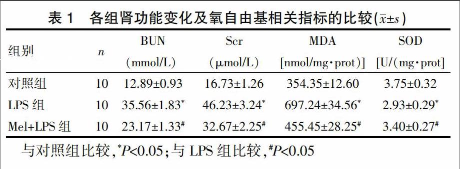
LPS组的血清BUN、Scr含量显著高于对照组,Mel+LPS组的BUN、Scr含量显著低于LPS组,差异有统计学意义(P<0.05)。LPS组小鼠肾组织的MDA水平显著高于对照组,Mel+LPS组小鼠肾组织的MDA水平显著低于LPS组,差异有统计学意义(P<0.05)。LPS组小鼠肾组织的SOD水平显著低于对照组,Mel+LPS组小鼠肾组织的SOD水平显著高于LPS组,差异有统计学意义(P<0.05)(表1)。
2.3 Mel对HIF-1α mRNA和蛋白表达的影响
LPS组小鼠肾组织的HIF-1α mRNA(4.35±1.87)和蛋白水平(6.82±1.97)与对照组(1.00±0.00)和(3.29±0.57)相比急剧增高,差异有统计学意义(P<0.05)。Mel+LPS组的HIF-1α mRNA(2.35±0.76)和蛋白水平(3.95±1.02)与LPS组比较明显降低,差异有统计学意义(P<0.05)(图2、图3)。
2.4 Mel对VEGF mRNA和蛋白表达的影响
LPS组小鼠肾组织的VEGF mRNA(2.01±1.32)和蛋白水平(4.34±1.26)与对照组(1.00±0.00)和(2.53±0.74)相比明显上调,差异有统计学意义(P<0.05)。Mel+LPS组的VEGF mRNA(1.35±0.18)和蛋白水平(2.55±0.75)与LPS组比较明显下调,差异有统计学意义(P<0.05)(图4、图5)。
3讨论
ETM可出现在儿科多系统的多种疾病中,是儿科ICU中重症感染患儿最常见的死亡原因之一。肾脏是其重要靶器官,一旦并发急性肾损伤(AKI)常难以救治[1]。近年来,HIF-1在某些肾脏疾病中的作用逐渐受到关注,然而,HIF-1活性及其对下游基因VEGF的表达调控在ETM幼年大鼠肾损伤过程中扮演的角色尚不清楚[2]。Mel是松果体分泌的吲哚类神经内分泌激素,是目前最为强效的自由基清除剂,在肾脏疾病中发挥着抗氧化、抗凋亡和免疫调节作用[3]。
3.1 Mel对LPS诱导的肾功能的保护作用
Mel主要作为治疗失眠的睡眠调节剂。Mehta等[4]证实,血液透析终末期肾病(end stage renal disease,ESRD)患者体内的Mel水平明显低于正常人,血液透析患者可以通过摄入Mel改善其睡眠质量。日本学者发现,其可延缓肾脏肥大的发生、发展。本研究结果显示,LPS诱导的大鼠肾功能受损,Scr和BUN增高,Mel+LPS使大鼠肾功能明显改善,Scr和BUN明显降低。这一结果验证了Mel能够改善肾功能的结论。
3.2 Mel对氧化应激相关指标的影响
在正常情况下,活性氧被少量释放且被内生的抗氧化剂所拮抗,如过氧化氢酶[5-7]。高水平和低水平的氧分压都会增强氧化应激,这使得维持组织的含氧量处在生理水平,能够避免氧化应激给组织带来的损伤。组织MDA水平可代表组织的氧化应激水平,而SOD为组织清除氧自由基的主要酶[8-11]。Sasabe等[12]发现Mel能够调节MDA和SOD的表达,从而防止细胞的DNA损伤。
在乳腺癌组织内,Mel能够通过SOD抑制HIF-1α,同时抑制HIF-1α诱导的VEGF的表达[13-15]。本研究结果显示,Mel能够保护肾功能,且Mel能够下调HIF-1α和VEGF。
Mel能逆转LPS诱导的肾组织的MDA增高而使SOD减少。LPS诱导的氧化应激加重了AKI小鼠的肾脏功能障碍。Mel通过抑制MDA的产生、上调SOD的表达、活性调控HIF-1α和VEGF的表达起到了抗氧化应激保护LPS诱导的AKI肾脏功能的作用,这为研究Mel在肾脏中的保护作用提供了一定的实验依据。
[参考文献]
[1]Mei S,Livingston M,Hao J,et al.Autophagy is activated to protect against endotoxic acute kidney injury[J].Sci Rep,2016,26(6):22171.
[2]Ogura Y,Jesmin S,Yamaguchi N,et al.Potential amelioration of upregulated renal HIF-1 alpha-endothelin-1 system by landiolol hydrochloride in a rat model of endotoxemia[J].Life Sci,2014,118(2):347-356.
[3]Shiroma ME,Botelho NM,Damous LL,et al.Melatonin influence in ovary transplantation:systematic review[J].J Ovarian Res,2016,9(1):33.
[4]Mehta R,Drawz PE.Is nocturnal blood pressure reduction the secret to reducing the rate of progression of hypertensive chronic kidney disease?[J].Curr Hypertens Rep,2011,13(5):378-385.
[5]Li S,Xu M,Niu Q,et al.Efficacy of procyanidins against in vivo cellular oxidative damage:a systematic review and Meta-analysis[J].PLoS One,2015,10(10):e0139455.
[6]Manquián-Cerda K,Escudey M,ZúnigaG,et al.Effect of cadmium on phenolic compounds,antioxidant enzyme activity and oxidative stress in blueberry Vaccinium corymbosum L. plantlets grown in vitro[J].Ecotoxicol Environ Saf,2016,133:316-326.
[7]Xuan RR,Niu TT,Chen HM.Astaxanthin blocks preeclampsia progression by suppressing oxidative stress and inflammation[J].Mol Med Rep,2016.[Epub ahead of print]
[8]Ma J,Liu B.The effect of microwave radiation on the levels of MDA and the activity of SOD of nasopharyngeal carcinoma cells[J].Lin Chung Er Bi Yan Hou Tou Jing Wai Ke Za Zhi,2008,22(4):160-162.
[9]Flint A,Stintzi A,Saraiva LM.Oxidative and nitrosative stress defences of Helicobacter and Campylobacter species that counteract mammalian immunity[J].FEMS Microbiol Rev,2016.[Epub ahead of print]
[10]Martini D,Del Bo′ C,Tassotti M,et al.Coffee consumption and oxidative stress:a review of human intervention studies[J].Molecules,2016,21(8):pii:E979.
[11]Lalkoviová M,Danielisová V.Neuroprotection and antioxidants[J].Neural Regen Res,2016,11(6):865-874.
[12]Sasabe E,Yang Z,Ohno S,et al.Reactive oxygen species produced by the knockdown of manganese-superoxide dismutase up-regulatehypoxia-inducible factor-1 alpha expression in oral squamous cell carcinoma cells[J].Free Radic Biol Med,2010,48(10):1321-1319.
[13]Vriend J,Reiter RJ.Melatonin and the von Hippel-Lindau/HIF-1 oxygen sensing mechanism:a review[J].Biochim Biophys Acta,2016,1865(2):176-183.
[14]Ozdemir G,Ergün Y,Bakariu S,et al.Melatonin prevents retinal oxidative stress and vascular changes in diabetic rats[J].Eye (Lond),2014,28(8):1020-1027.
[15]Sanchez-Sanchez AM,Antolin I,Puente-Moncada N,et al.Melatonin cytotoxicity is associated to warburg effect inhibition in ewing sarcoma cells[J].PLoS One,2015,10(8):e0135420.

