TIMP-1、MMP-9在乳腺癌患者术前血清中的表达与临床病理特征的关系
马 婕,邱 涵,张 倩,徐正丰
(荆州市中心医院乳腺科,荆州 434020)
TIMP-1、MMP-9在乳腺癌患者术前血清中的表达与临床病理特征的关系
马 婕,邱 涵,张 倩,徐正丰
(荆州市中心医院乳腺科,荆州 434020)
目的:探讨血清基质金属蛋白酶MMP-9及其抑制剂TIMP-1在乳腺癌患者术前血清中的表达与临床病理特征的关系。方法:收集52例乳腺肿瘤患者术前血清,通过ELISA方法检测9例良性肿瘤患者与43例浸润性乳腺癌患者血清中MMP-9和TIMP-1的含量并计算二者比值,用免疫组化技术检病变组织中MMP-9和TIMP-1的表达。结果:与良性肿瘤相比,MMP-9与TIMP-1蛋白表达在术前乳腺癌患者血清中的表达均有显著性升高;对于乳腺癌患者而言,术前血清中MMP-9、TIMP-1的浓度与MMP-9/TIMP-1的比值与患者的TNM分期、肿瘤长度、淋巴结累及之间具有统计学差异,但与患者的年龄、分级无关;MMP-9和TIMP-1二者的浓度之间并无相关性;血清中MMP-9和TIMP-1的浓度与组织蛋白的表达呈正相关。结论:检测术前血清中MMP-9与TIMP的表达有潜力作为良恶性肿瘤鉴别的指标之一;监测乳腺癌患者血清MMP-9和TIMP-1的表达有助于确定肿瘤的转移情况,并对预后的判断具有参考意义。
乳腺癌;基质金属蛋白酶;基质金属蛋白酶抑制剂;术前检测
乳腺癌是女性中最常见的恶性肿瘤,也是女性致死率第二的疾病[1]。然而,在肿瘤的检测中,关键的肿瘤标志物敏感度较低,导致肿瘤很难在早期被检测到。所以在肿瘤的早期进行诊断是十分重要的[2]。常规的检测方法,如超声、CT、MRI等技术在检测微小病灶时并不是很有效,所以,使用新的早期诊断方法以实现肿瘤早期检测是很必要的。目前,一系列的乳腺癌肿瘤标志物已经被发现,包括癌抗原(CA)15-3、癌胚抗原、细胞角蛋白19片段(CYFRA 21.1)[3-4]。这些标志物在早期肿瘤中的敏感性和特异性都较低,所以不适合作为检测靶标[5]。很多种肿瘤细胞都可以分泌基质金属蛋白酶-9(MMP-9),在乳腺癌中,MMP-9与疾病的进展密切相关[6]。金属蛋白酶组织抑制剂-1(TIMP-1)是一种表达
于肿瘤中的糖蛋白,在乳腺癌中也有表达,是MMP-9的天然抑制剂[7]。MMP-9和TIMP-1的表达与乳腺肿瘤的相关性已有很多报道[8],两者在肿瘤组织中的表达已被证实与肿瘤的临床病理特征有关。本研究旨在探讨乳腺癌患者术前血清中MMP-9与TIMP-1的表达与患者临床病理特征的关系,丰富乳腺癌早期诊断的证据。
1 资料与方法
1.1 一般临床资料 收集我院2015年1月~2016年2月间因乳腺肿瘤而收治的52例患者的血清;患者均为女性,年龄区间为35到70,中位年龄为58岁,术前没有任何治疗历史。收集患者术后病变组织,组织病理学诊断显示,所有患者中,良性病变有9例(中位年龄为36岁),恶性乳腺肿瘤43例(中位年龄为55岁)。依照病理分型,浸润性导管癌31例,浸润性小叶癌8例,乳头状癌13例。所有患者均签署知情同意书。
1.2 检测血清MMP-9与TIMP-1的表达 所有患者术前采血2ml,在室温下静置半小时凝固,1000g下离心5min,收集血清。通过双抗夹心酶联免疫分析法检测血清中MMP-9和TIMP-1的含量。MMP-9和TIMP-1定量检测试剂盒均购买于上海生工生物工程有限公司。将MMP-9和TIMP-1系列标准品与样品同时检测,通过对浓度和吸光度进行线性拟合,计算出样本中待测蛋白的浓度,操作严格按照说明书进行。
1.3 检测病变组织中MMP-9和TIMP-1的表达 使用免疫组化方法检测43例乳腺癌患者病变组织中石蜡切片标本中MMP-9和TIMP-1的表达。试剂:鼠抗人MMP-9以及TIMP-1单克隆抗体和SP试剂盒购买于南京凯基生物技术公司。操作按照标准SP法进行,使用MMP-9和TIMP-1单抗为一抗,DAB显色后平行观察三次,采用高倍视野保证结果的准确。
1.4 统计学分析 应用SPASS 17.0软件包对数据进行统计学分析。计量资料表示为“平均值±标准差”,使用t检验和方差分析检验组间差异。采用皮尔森相关系数表示资料的相关性。
2 结果
2.1 乳腺良性肿瘤和乳腺癌患者术前血清MMP-9和TIMP-1的浓度 乳腺癌患者术前血清中MMP-9和TIMP-1的浓度、MMP-9和TIMP-1浓度的比值均高于乳腺良性肿瘤患者(P<0.01,P<0.01,P<0.05)。良性肿瘤和乳腺癌患者血清MMP-9和TIMP-1的测定结果如表1所示:

表1 乳腺良性肿瘤和乳腺癌患者术前血清中MMP-9和TIMP-1浓度
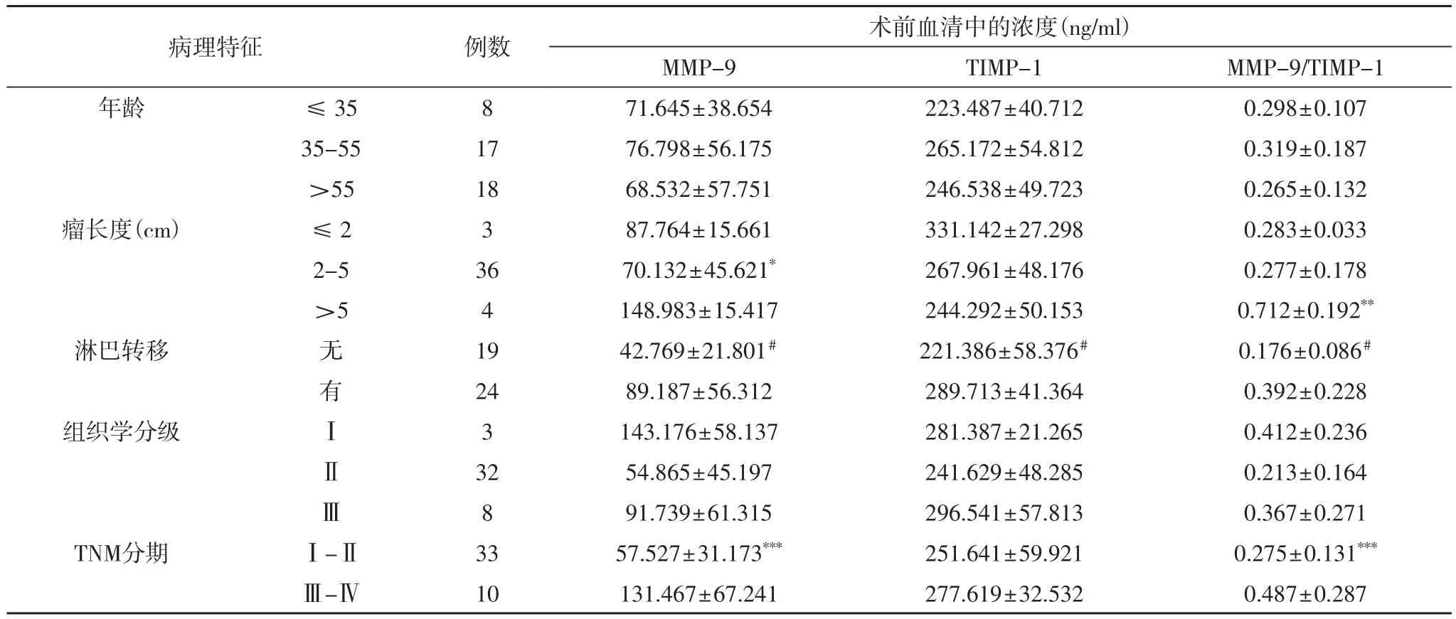
2.2 TIMP-1、MMP-9在乳腺癌患者术前血清中的表达与临床病理特征的关系 乳腺癌患者手术前血清中MMP-9和TIMP-1的浓度以及两者浓度的比值与TNM分期、淋巴结转移有关(P<0.05,P<0.01);TIMP-1的浓度与淋巴结累及相关(P<0.05);MMP-9、TIMP-1与MMP-9/TIMP-1比值与乳腺癌患者的年龄和组织学分级无关;对于肿块大于5cm的患者,其血清MMP-9的浓度和MMP-9/TIMP-1的比值均较瘤块小于5cm的患者高(P<0.05,P<0.01)。三者与临床病理特征的关系见表2。

表2 TIMP-1、MMP-9在乳腺癌患者术前血清中的表达与临床病理特征的关系
2.3 乳腺癌患者术前血清中MMP-9和TIMP-1的浓度之间的相关性 在所有乳腺癌患者中,术前MMP-9和TIMP-1的浓度之间并没有发现显著的相关性(r=0.269,P>0.05)
2.4 乳腺癌患者术前血清中MMP-9与TIMP-1与病变组织中表达的相关性 血清中MMP-9与病变组织中MMP-9的表达呈显著正相关(r=0.476,P<0.05);血清中TIMP-1与病变组织中TIMP-1的表达呈显著正相关(r=0.401,P<0.05); 乳腺癌患者血清中MMP-9与TIMP-1的比值与组织中相应的比值呈显著正相关(r=0.411,P<0.05)。
3 讨论
MMP是一种锌依赖型的蛋白水解酶。其中,MMP-9与肿瘤的侵袭、迁移和不良预后预后有关[9]。MMP的催化活性可被金属蛋白酶组织抑制剂(TIMPs)所抑制,TIMP,尤其是TIMP-1高表达,已在包括乳腺癌在内的多种肿瘤中发现,如鼻咽癌[10]、胰腺癌[11]和胃癌[12]。很多研究已经证实,乳腺癌患者血清中,有较高水平的MMP-9和TIMP-1表达量[13]、[14]。在本研究中,乳腺癌患者血清中MMP-9、TIMP-1和两者的比值都教良性乳腺肿瘤患者显著升高,MMP-9和TIMP-1的比值也具有显著性差异。国外课题组已经得到了类似的数据,然而,研究是以乳腺癌患者的血清与健康受试者相比。本研究将良恶性肿瘤患者术前血清指标进行比较,并发现了其中的显著性差异,这对于区分肿瘤的性质具有一定的意义。近期,Slawomir等人[15]的研究报道了乳腺癌患者血液样本中VEGF、MMP-9和TIMP-1的表达对肿瘤诊断的意义,这与我们的研究结果相一致。我们在后续研究中还将对出现转移和非转移的乳腺癌患者血清MMP-9和TIMP-1的水平进行比较,以期获得预测肿瘤转移的指导数据。将MMP-9和TIMP-1的血清水平与乳腺癌患者的临床病理特征比较,发现两者的浓度和比值与患者的淋巴结累及、肿瘤的长度和TNM分期有关。肿瘤直径大于5 cm的患者血清中MMP-9、TIMP-1和MMP-9/TIMP-1比小于5cm的患者相比显著升高。然而也有研究显示,MMP-9的水平低,患者复发率高[16]。这些争议提示,对MMP-9和TIMP-1进一步加大样本量的长期随访研究十分重要。
[1] Alvarado R, Lari S A, Roses R E, et al. Biology, treatment, and outcome in very young and older women with DCIS[J]. ANN SURG ONCOL, 2012, 19(12): 3777-3784.
[2] Perez-Rivas L G, Jerez J M, Sousa F D, et al. Serum protein levels following surgery in breast cancer patients: a protein microarray approach[J]. INT J ONCOL, 2012, 41(6): 2200-2206.
[3] Bièche I, Lerebours F, Tozlu S, et al. Molecular profiling of inflammatory breast cancer identification of a poor-prognosis gene expression signature[J]. CLIN CANCER RES, 2004, 10(20): 6789-6795.
[4] Kufe D W. MUC1-C oncoprotein as a target in breast cancer: activation of signaling pathways and therapeutic approaches[J]. ONCOGENE, 2013, 32(9): 1073-1081.
[5] Harris L, Fritsche H, Mennel R, et al. American Society of Clinical Oncology 2007 update of recommendations for the use of tumor markers in breast cancer[J]. J CLIN ONCOL, 2007, 25(33): 5287-5312.
[6] García M F, González-Reyes S, González L O, et al. Comparative study of the expression of metalloproteases and their inhibitors in different localizations within primary tumours and in metastatic lymph nodes of breast cancer[J]. INT J CLIN EXP PATHO, 2010, 91(4): 324-334.
[7] Thorsen S B, Christensen S L, Würtz SØ, et al. Plasma levels of the MMP-9: TIMP-1 complex as prognostic biomarker in breast cancer: a retrospective study[J]. BMC CANCER2013, 13(12): 3016-3017.
[8] Shay G, Lynch C C, Fingleton B. Moving targets: Emerging roles for MMPs in cancer progression and metastasis[J]. MATRIX BIOL, 2015, 44(3): 200-206.
[9] Ławicki S, Będkowska G E, Gacuta-Szumarska E, et al. Pretreatment plasma levels and diagnostic utility of hematopoietic cytokines in cervical cancer or cervical intraepithelial neoplasia patients[J]. FOLIA HISTOCHEM CYTO, 2012, 50(2): 213-219.
[10] Kozłowski M, Laudański W, Mroczko B, et al. Serum tissue inhibitor of metalloproteinase 1(TIMP-1)and vascular endothelial growth factor A(VEGF-A)are associated with prognosis in esophageal cancer patients[J]. ADV MED SCI-POLAN, 2013, 58(2): 227-234.
[11] Poruk K E, Firpo M A, Scaife C L, et al. Serum osteopontin and tissue inhibitor of metalloproteinase 1 as diagnostic and prognostic biomarkers for pancreatic adenocarcinoma. [J]. PANCREAS, 2013, 42(2): 193-197.
[12] Grunnet M, Mau-Sørensen M, Brünner N. Tissue inhibitor of metalloproteinase 1(TIMP-1)as a biomarker in gastric cancer: a review[J]. SCAND J GASTROENTERO, 2013, 48(8): 899-905.
[13] Berezov T T, Ovchinnikova L K, Kuznetsova O M, et al. Vascular endothelial growth factor in the serum of breast cancer patients[J]. B EXP BIOL MED+, 2009, 148(3): 419-424.
[14] Grunnet M, Mau-Sørensen M, Brünner N. Tissue inhibitor of metalloproteinase 1(TIMP-1)as a biomarker in gastric cancer: a review[J]. SCAND J GASTROENTERO, 2013, 48(8): 899-905.
[15] Ławicki S, Zajkowska M, Głażewska E K, et al. Plasma levels and diagnostic utility of VEGF, MMP-9, and TIMP-1 in the diagnosis of patients with breast cancer[J]. ONCOTARGETS THER, 2016, 9(1): 911-919.
[16] Poulsom R, Pignatelli M, Stetlerstevenson W G, et al. Stromal expression of 72 kda type IV collagenase(MMP-2)and TIMP-2 mRNAs in colorectal neoplasia. [J]. AM J PATHOL, 1992, 141(2): 389-396.
Clinical significance of the expression of TIMP-1 and MMP-9 in the serum of breast cancer patients before surgery
Ma Jie, Qiu Han, Zhang Qian, Xu Zheng-feng
(Department of Breast Oncology, Jingzhou Central Hospital; Jingzhou 434020, China)
Objective To investigate the clinical significance of the expression of TIMP-1 and MMP-9 in the serum of breast cancer patients before surgery. Methods The serum 52 patients with breast tumor patients were collected before surgery. ELISA and Immunohistochemistry were used respectively to determine the expression of MMP-9 and TIMP-1 in serum and tumor tissues of 9 patients with benign tumor and 43 breast cancer patients. The ratio between MMP-9 and TIMP-1 was also calculated. Results Compared with patients with benign tumor, breast cancer patients showed significantly elevated serum levels of MMP-9, TIMP-1 and MMP-9/TIMP-1; statistical difference of MMP-9, TIMP-1 and MMP-9/TIMP-1 was found in the TNM stage, tumor length and Lymph node involvement, but not in histological grading and age; the serum concentrations of MMP-9 and TIMP-1 showed no relevance; the serum concentration of MMP-9 and TIMP-1 was positively correlated with that in tumor tissues. Conclusion The detection of MMP-9 and TIMP-1 in serum of breast tumor patients before surgery has potential to identify cancer from benign tumor; monitoring the concentration of MMP-9 and TIMP-1 can help to confirm tumor metastasis and judge prognosis.
breast cancer; matrix metalloproteinases; matrix metalloproteinase inhibitor; preoperative examination
R737.9
A
1673-016X(2016)05-0017-03
2016-05-20
荆州市科技局项目(NO.2015031)
马婕,E-mail:majie4506@sina.com

