骨肉瘤皮肤转移一例
樊俊威 阿克拜尔·苏来曼 边毅 董进成 侯巍 万学峰 帕丽达·阿布力孜
830054乌鲁木齐,新疆医科大学第一附属医院皮肤科
骨肉瘤皮肤转移一例
樊俊威 阿克拜尔·苏来曼 边毅 董进成 侯巍 万学峰 帕丽达·阿布力孜
830054乌鲁木齐,新疆医科大学第一附属医院皮肤科
患者女,68岁,发现口腔、手背及肺部多处肿物20余天,无明显自觉症状。体检:各系统检查无异常。皮肤科检查:上颚可见2个蚕豆大小暗红色结节,左手背可见一鸽蛋大小暗红色结节,表面光滑,质硬,活动度差。正电子发射计算机断层显像(PetCT):全身多处转移癌(脑、淋巴结、肺、胃肠道、双肾、多处骨及肌肉间转移)。口腔内上颚结节病理结果:肿瘤细胞分化较差,局部区域可见骨样基质、软骨样结构及肿瘤骨,免疫组化角蛋白阴性,S100局灶性阳性,波形蛋白阳性,CD99阳性,P63阳性,Ki67阳性(60%),考虑右侧上颌骨骨肉瘤。手背皮损组织病理检查:表皮未见明显异常,真皮全层弥漫性中等至较大的组织样细胞浸润,细胞有异形性,局部区域可见红染不规则的骨样基质及肿瘤骨;免疫组化Ki67阳性(60%),CD3、CD10、CD20、Bcl⁃2、BCl⁃6均阴性。诊断:骨肉瘤皮肤转移。患者放弃治疗,发病6个月后死亡。
骨肉瘤;肿瘤转移;皮肤表现;病理过程
骨肉瘤是较常见的发生在20岁以下青少年或儿童的一种恶性骨肿瘤。骨肉瘤皮肤转移罕见,对《中国生物医学文献数据库》、《中国期刊全文数据库》、《万方数据知识服务平台》、《维普期刊资源整合平台》等12个中文数据库1978—2015年12月检索表明,目前中国大陆地区尚没有文献报道。我们报道1例骨肉瘤皮肤转移并复习相关文献。
病历资料
患者女,68岁,以口腔、手背及肺部多处肿物20 d为主诉入住我院。患者于2014年1月初发现左手背出现豆粒大小红色丘疹,无明显自觉症状,后逐渐增大至鸽蛋大小。同时口腔内多发肿物,疼痛明显,不能进食,在当地诊所按炎症给予抗炎治疗无效,后在当地医院住院治疗,头部CT显示颅内占位,胸部CT检查示左肺肿块性质待定,由当地医院转入我院。病程中患者一般情况尚可,神志清,无发热、咳嗽、咳痰、胸闷、气短、胸痛、低热、盗汗、头晕、头痛、恶心、呕吐、腹痛、腹泻等症状,饮食、睡眠差,近1个月体重减轻约5 kg。患者既往体健,否认高血压、糖尿病等慢性病病史。
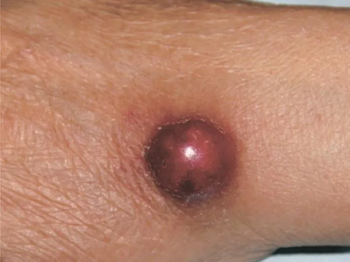
体检:体温37.6 ℃,血压100/60 mmHg(1 mmHg=0.133 kPa),脉搏80次,呼吸21次。浅表淋巴结未触及肿大,各系统检查无异常。皮肤科检查:右侧上颚可见2个蚕豆大小暗红色结节;左手背可见一鸽蛋大小暗红色结节,表面光滑,质硬,活动度差(图1),其余部位未见明显相关皮疹。
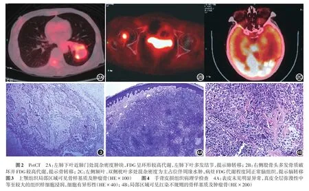
实验室检查:血常规红细胞3.09×10¹²/L,血红蛋白97 g/L,其余未见明显异常。尿粪常规未见异常。血液生化:钾3.07 mmol/L,白蛋白32 g/L,其余未见明显异常。血细胞沉降率68 mm/1 h。肿瘤标记物全套、乙肝、丙肝、梅毒、人免疫缺陷病毒抗体检测未见异常;结核抗体及甲状腺功能检测未见异常。正电子发射计算机断层显像(PetCT):全身多处(脑、淋巴结、肺、胃肠道、双肾、多处骨及肌肉)转移癌(图2)。口腔内右侧上颚结节组织病理检查:肿瘤细胞分化较差,局部区域可见骨样基质、软骨样结构及肿瘤骨(图3),免疫组化:角蛋白阴性,S100局灶性阳性,波形蛋白、P63、CD99、Ki67阳性(60%),结合肿瘤形态及免疫组化,考虑右侧上颌骨骨肉瘤。手背皮损组织病理检查:表皮未见明显异常,真皮全层弥漫性中等至较大的组织样细胞浸润,细胞有异形性,局部区域可见红染不规则的骨样基质及肿瘤骨(图4);免疫组化:角蛋白阴性,S100局灶性阳性,Ki67阳性(60%),CD3、CD10、CD20、Bcl⁃2、Bcl⁃6均阴性。
诊断:结合PetCT结果,上颚和皮肤HE切片及免疫组化,诊断为右侧上颌骨骨肉瘤伴脑、淋巴结、肺、胃肠道、双肾、全身多处骨、肌肉间及皮肤转移。
患者放弃治疗,自动出院。发病6个月后因多器官功能衰竭死于当地医院。

图1 左手背一鸽蛋大小的暗红色结节,表面光滑,质硬,活动度差

讨 论
骨肉瘤好发于儿童及青少年,典型的骨肉瘤好发于远心端股骨、近心端胫骨和肱骨[1],少数也可发生在头骨、上下颌骨和脊椎骨。骨肉瘤是最常见的原发性骨恶性肿瘤,高达90%的患者会发生肺部转移[1⁃3]。
骨肉瘤的皮肤转移极为罕见,1924年Finnerud报道1例原发于右侧肱骨的骨肉瘤患者,在头皮及左上颌部皮肤发生转移[4]。骨肉瘤皮肤转移临床表现通常是1个或多个隆起性的坚硬结节,偶有疼痛感,骨肉瘤皮肤转移与原发性骨肉瘤的组织形态基本相同,在真皮或皮下有多形性的间质增生细胞,
1细胞形态多为梭形,细胞核淡染,尤其大多有类骨质形成。本文患者病理表现与此相符合,但转移性骨肉瘤的组织病理需要与淋巴瘤、黑素瘤、Merkel细胞癌、肺小细胞癌等鉴别,鉴别的关键是能否在切片中找到骨样基质或肿瘤骨,鉴别困难时免疫组化角蛋白阴性、S100阴性、波形蛋白阳性、CD99阳性、CD3阴性、CD10阴性、CD2阴性、Bcl⁃2阴性、Bcl⁃6阴性、Ki67增殖指数等有助于进一步协助鉴别。

表1 1995—2015年全世界报道的骨肉瘤皮肤转移患者16例
1995—2015年的20年间,全世界报道的骨肉瘤皮肤转移病例16例(表1),这些原发于不同部位的骨肉瘤患者年龄5~83岁不等,男8例,女8例,11例发生肺部转移,9例头皮转移,5例腰腹部皮肤转移,2例肩胛部皮肤转移,1例右锁骨上皮肤转移,1例右太阳穴处皮肤转移。头皮转移在骨肉瘤皮肤转移中占相当大的比重,16例患者中9例发生了头皮转移,其原因可能是因为头皮的静脉系统是由无瓣膜的颈椎静脉系统构成,这种逆行静脉构成是骨肉瘤容易发生头皮转移的原因之一[11]。Schwartz[19]认为,肿瘤的皮肤转移往往比原发病灶更容易发现,这种通过无瓣膜的静脉循环传播,比肿瘤在快速成长和复发时速度更快,故骨肉瘤皮肤转移患者往往已经发生了全身转移,提示预后较差。1997年ten Harkel等[7]报道1例14岁男性骨肉瘤患者合并肺部转移,也出现了头皮转移,他认为骨肉瘤头皮转移发生于肺部转移后,是来自肺部的次发转移,头皮病灶的出现,表明肿瘤已经广泛扩散。因此骨肉瘤的皮肤转移,可视为广泛性转移的一个先兆,也可以作为预后不良的判定指标。
[1] Huvos AG.Osteogenic sarcoma.Bone tumors:diag⁃nosis,trement and prognosis[M].Philadelphia:WB Sanuder,1991:85⁃155.
[2] Jeffree GM,Price CH,Sissons HA.The metastatic patterns of osteosarcoma[J].Br J Cancer,1975,32(1):87⁃107.
[3] Webster VJ,Arons I.Intussusception secondary to osteogenic sarcoma metastasis[J].Br J Clin Pract,1987,41(2):628⁃629.
[4] Finnerud CW.Ossifying sarcoma of the skin metastatic from ossifying sarcoma of the humerus[J].Arch Derm Syphilol,1924,10(1):56⁃62.
[5] Myhand RC,Hung PH,Caldwell JB,et al.Osteogenic sarcomawith skin metastases[J].JAm Acad Dermatol,1995,32(5 Pt 1):803⁃805.
[6]Setoyama M,Kanda A,KanzakiT.Cutaneous metastasis of an osteosarcoma.A case report[J].Am J Dermatopathol,1996,18(6):629⁃632.
[7] ten Harkel AD,Hogendoorn PC,Beckers RC,et al.Skin metastases of osteogenic sarcoma:a case report with review of the literature[J].J Pediatr Hematol Oncol,1997,19(3):266⁃267.
[8] Stavrakakis J,Toumbis⁃Ioannou E,Alexopoulos A,et al.Subcutaneous nodules as initial meta⁃static sites inosteosarcoma[J].Int J Dermatol,1997,36(8):606⁃609.
[9] Vaidya S,Jones KP,Fisher C.Letter to the editor:osteogenic sarcoma:cutaneous metastases[J].Med PediatrOncol,2002,38(6):453⁃454.DOI:10.1002/mpo.1364.
[10] Covello SP,Humphreys TR,Lee JB.A case of extraskeletal osteosarcoma with metastasis to the skin[J].J Am Acad Dermatol,2003,49(1):124⁃127.DOI:10.1067/mjd.2003.297.
[11] Collier DA,Busam K,Salob S.Cutaneous meta⁃stasis of osteosar⁃coma[J].J Am Acad Dermatol,2003,49(4):757⁃760.
[12]何秋燕,楊國材,張戊暉,等.頭皮骨肉瘤皮膚轉移[J].中華皮誌,2006,24:123⁃130.Ho CY,Yang KC,Chang WH,et al.Cutaneous metastases of osteosarcomaonthescalp[J].DermatolSinica,2006,24:123⁃130.
[13] Delépine F,Leccia N,Schlatterer B,et al.Inaugural cutaneous metastases of an osteo⁃sarcoma:a case report[J].Rev Chir Orthop Reparatrice Appar Mot,2006,92(7):719⁃723.
[14] Lee WJ,Lee DW,Chang SE,et al.Cutaneous metastasis of extraskeletal osteosarcoma arising in the mediastinum[J].Am J Dermatopathol,2008,30(6):629 ⁃631.DOI:10.1097/DAD.0b013e3181812751.
[15]Larsen S,Davis DM,Comfere NI,et al.Osteosarcoma of the skin[J].Int J Dermatol,2010,49(5):532⁃540.DOI:10.1111/j.1365⁃4632.2010.04315.x.
[16]Ragsdale MI,Lehmer LM,Ragsdale BD,et al.Cutaneous metastasis of osteosarcoma in the scalp[J].Am J Dermatopathol,2011,33(6):e70⁃e73.DOI:10.1097/DAD.0b013e318214a7ea.
[17]Park SG,Song JY,Song IG,et al.Cutaneous extraskeletal osteo⁃sarcoma on the scar of a previous bone graft[J].Ann Dermatol,2011,23(Suppl 2):S160 ⁃S164.DOI:10.5021/ad.2011.23.S2.S160.
[18] Fernandez⁃Pineda I,Bahrami A,Green JF,et al.Isolated subcu⁃taneous metastasis of osteosarcoma 5 years after initial diagnosis[J].J Pediatr Surg,2011,46(10):2029⁃2031.DOI:10.1016/j.jpedsurg.2011.06.011.
[19] SchwartzRA.Cutaneousmetastaticdisease[J].JAmAcadDermatol,1995,33(2 Pt 1):161⁃182.
A case of cutaneous metastasis of osteosarcoma
Fan Junwei,Akebaier.Sulaiman,Bian Yi,Dong Jincheng,Hou Wei,Wan Xuefeng,Palida.Abulize
Department of Dermatology,First Affiliated Hospital of Xinjiang Medical University,Urumqi 830054,China
A 68⁃year⁃old female patient was admitted to the hospital for multiple masses in the mouth and lungs as well as on dorsal hands for more than 20 days without obvious subjective symptoms.No abnormalities were found by physical examination.Dermatological examination showed two bean⁃sized dark⁃red nodules on the upper jaw as well as one pigeon egg⁃sized dark⁃red nodule on the left dorsal hand,and all the nodules were hard with smooth surfaces and limited mobility.Positron emission tomography⁃computed tomography(PetCT)revealed multiple metastases to the brain,lymph nodes,lungs,gastrointestinal tract,both kidneys,multiple bones and intermuscular tissues.Pathology of nodules from the upper jaw showed lowly differentiated tumor cells with osteoid matrix,chondroid structures and tumor bone in local areas,and immunohistochemical examination of tumor cells found positive staining for S100(focally),vimentin,CD99,P63 and Ki⁃67(60%),but negative staining for keratin.A diagnosis of osteosarcoma of the right side of the upper jaw was considered.Pathology of nodules from the dorsal hand revealed no obvious abnormalities in the epidermis,while there was a diffuse infiltration of medium⁃to large⁃sized histiocyte⁃like cells in the whole dermis with cell atypia and irregularly red⁃stained bone matrix and tumor bone in some regions.Immunopathology showed positive staining for Ki67(60%),and negative staining for CD3,CD10,CD20,Bcl⁃2,and Bcl⁃6.A diagnosis of cutaneous metastasis of osteosarcoma was made.The patient refused further treatment and died 6 months after the onset of lesions.
Osteosarcoma;Neoplasm metastasis;Skin manifestations;Pathological processes
Palida.Abulize,Email:palidae@aliyun.com
帕丽达·阿布力孜,Email:palidae@aliyun.com
10.3760/cma.j.issn.0412⁃4030.2016.07.008
2015⁃11⁃16)
(本文编辑:颜艳)

