磁共振对BMI正常人群髓核体积的测量与临床应用研究
文 兵 杜 瑛 胡良波 张 翱 梁学恒 余 洁
磁共振对BMI正常人群髓核体积的测量与临床应用研究
文 兵 杜 瑛 胡良波 张 翱 梁学恒 余 洁
目的:定量研究身高体重指数(BMI)正常的人群椎间盘髓核体积,并探讨其临床应用价值。方法:分析310例BMI正常人群的腰椎磁共振图像,运用磁共振图像处理系统测量FSE-T2WI上腰椎各节段椎间盘髓核的体积。根据年龄不同分为青年组(22~44岁),中年组(45~59岁),老年组(60岁以上)三个组,对不同年龄组男女性的L1/2~L5/S1节段椎间盘髓核进行体积测量和统计学分析。结果:青年组L1/2~L5/S1椎间盘髓核体积男性分别为:2523.50±627.55mm3,3178.89±803.77mm3,3686.88±897.43mm3,3913.35±749.00mm3,3086.26±928.92mm3,女性分别为1922.42±659.36mm3,2248.56±847.48mm3,3066.38±968.56mm3,3124.99±917.92mm3,2818.81±1022.87mm3。中年组L1/2~L5/S1椎间盘髓核体积男性分别为:2502.27±821.06mm3,3056.98±965.97mm3,3629.82±1219.14mm3,3858.20±1035.57mm3,3037.07±834.51mm3,女性分别为1991.65±638.32mm3,2383.49±723.42mm3,2989.30±851.11mm3,3165.12±885.84mm3,2787.01±762.72mm3;老年组L1/2~L5/S1椎间盘髓核体积男性分别为:2455.86±776.34mm3,3019.00±676.15mm3,3515.96±809.14mm3,3802.23±721.39mm3,2984.83±706.73mm3,女性分别为:2030.78±604.28mm3,2246.92±622.53mm3,2812.35±833.55mm3,3020.11±899.33mm3,2750.87±852.667mm3。同年龄组L1/2~L5/S1节段男女之间正常髓核体积差异均有显著性统计学意义(P<0.01),同节段不同年龄组之间正常髓核体积差异均无统计学意义(P>0.05),统计节段中相邻两髓核体积存在相关关系,各节段髓核体积与BMI指数相关性不大,而分别与身高和体重具有一定的相关性。结论:本研究测量了正常成人腰椎间盘髓核体积正常范围,为椎间盘髓核假体体积以及其他科学实验提供方法和参考标准。
椎间盘;髓核;体积;磁共振
【Abstract】Purpose: To quantitative study the volume of nucleus pulposus of lumbar intervertebral disc in population with normal body mass index (BMI), and to explore its clinical application value. Methods: Three hundred and ten patients who were investigated by lumbar MRI were analyzed retrospectively. The measurement was carried out based on sagittal FSE-T2WI images, and then the volumes of nucleus pulposus were calculated.The cases were divided into the young group (22 to 45 years old), the middle age group (46-60 years old), and the elder group (>60 years) according to their age, and the volume of L1/2-L5/S1 nucleus pulposus of the males and females in each group were measured and statistically analyzed respectively. Results: The young group: the volume of L1/2-L5/S1 nucleus pulposus of males was 2523.50±627.55mm³,3178.89±803.77mm³,3686.88±897.43mm³,3913.35±749.00mm³,3086.26±928.92mm³, respectively, and the volume of L1/2-L5/S1 nucleus pulposus of females was 1922.42±659.36mm³,2248.56±847.48mm³,3066.38±968.56mm³,3124.99±917.92mm³,2818.81±1022.87mm³, respectively; The middle age group: the volume of L1/2-L5/S1 nucleus pulposus of males was 2502.27±821.06mm³,3056.98±965.97mm³,3629.82±1219.14mm³,3858.20±1035.57mm³,3037.07±834.51mm³,respectively, and the volume of L1/2-L5/S1 nucleus pulposus of females was 1991.65±638.32mm³,2383.49±723.42mm³,2989.30±851.11mm³,3165.12±885.84mm³,2787.01±762.72mm³, respectively; The elder group: the volume of L1/2-L5/S1 nucleus pulposus of males was 2455.86±776.34mm³,3019.00±676.15mm³,3515.96±809.14mm³,3802.23±721.39mm³,2984.83±706.73mm³, respectively, and the volume of L1/2-L5/ S1 nucleus pulposus of females was 2030.78±604.28mm³,2246.92±622.53mm³,2812.35±833.55mm³,3020.11±899.33mm³,2750.87±852.667mm³, respectively. There was statistical significant difference in the volumes of L1/2-L5/S1 nucleus pulposus between the males and females in the same group (P<0.01). No statistical significant difference was found in the volumes of the same segment between different groups. The volumes of the adjacent nucleus pulposus were statistically correlated. Segmental volume of nucleus pulposus had little correlation with BMI,and had certain correlation between height and weight. Conclusion: This study measured the normal range of the normal adult lumbar nucleus pulposus volume, which can provide reference standard for intervertebral disc nucleus pulposus prosthesis and other scientific experiments.
【Key words】Intervertebral disc; Nucleus pulposus;Volume;Magnetic resonance imaging
腰痛是中老年人常见的临床症状,而由腰椎间盘退变导致的一系列病变是主要原因[1],随着生活及工作方式的变化,椎间盘退变引起症状越来越趋向年轻化[2]。这类患者经保守治疗疗效不佳后一般需手术治疗[3],目前最好的手术方式是髓核置换术[4-5],包括预成型假体和原位注射型假体,均需要对拟置换髓核的体积有一个准确估算,随着3D打印技术在医学领域的应用[6],要求髓核体积的估算越来越精确、需要正常人群的参考值范围。而对于髓核体积的精确测量方法及正常人群髓核体积参考值文献报道较少。本研究以身高体重指数(body mass index,BMI)正常人群为基础,运用磁共振的图像处理系统,对MRI成像的正常腰椎间盘髓核体积进行测量,并探讨方法的可行性及其临床应用的价值。
方 法
1.一般资料
对2013年-2015年在我院进行腰椎MRI检查者4500例,根据两位高年资的影像科医生对腰椎MRI图像的进行评价,独自筛选出Pfirrmann分级[5]Ⅰ级或者Ⅱ级髓核为研究对象,定义其髓核为正常髓核。选取两位医师筛选结果一致认为的正常髓核,并统计被检查者一般信息,同时计算BMI,共统计出BMI范围为18.5~24.99的患者310例纳入研究对象,年龄22~70岁,平均年龄44.3±11.6岁。其中男121例,平均年龄44.3±13.5岁,平均BMI为21.89±1.92;女179例,平均年龄44.5±10.7平均BMI为21.34±1.56。根据WHO对年龄划分的新规定,将所有对象分为青年组(22~44岁)、中年组(45~59岁)和老年组(60岁以上),共选出男性正常髓核L1/2~L5/S1分别为101例、94例、90例、52例、59例,女性正常髓核L1/2~L5/S1分别为157例、149例、145例、88例、104例。
2.测量方法
椎间盘髓核体积模型的建立:用椎间盘髓核体积相当的水注入到小气球里,排净空气后系好,并用将装满水的小气球置于椎间隙模型里,并给予一定的压力让充满水的气球呈扁平椭圆状,再将髓核体积模型进行MRI扫描,利用磁共振自带图像处理系统在FSE-T2WI图像上勾勒出气球的边缘,每个模型通过软件测量3次并取平均值,得出该髓核模型的体积,再与实际注入水的体积进行对比。
对研究对象的腰椎MRI矢状位FSE-T2WI的图像进行分析,利用磁共振图像处理系统,在图像上勾勒出髓核的边界,同样利用软件测量3次取平均值计算得到每个节段髓核的体积。
3.统计学方法
采用SPSS 17.0软件进行统计学分析。不同性别、不同年龄组及不同节段进行对比分析,方差齐性采用单因素方差分析(One-way ANOVA),P<0.05有统计学意义,然后采用LSD法均数间两两比较;方差不齐,采用非参数秩和检验(Krustal-wallistest)进行比较分析,P<0.05有统计学意义,然后采用多重比较Dunnett`s T3法进行两两组间比较。
结 果
椎间盘髓核体积模型:3个髓核体积模型每个运用软件测得的体积大小分别为:4786mm3、 7328mm3、10564mm3,由量筒测量的实际体积分别为3000mm3、7000mm3、11000mm3,误差率依次分别为4.28%、4.69%、5.13%,采用配对设计t检验,P>0.05,两者体积之间无统计学差异。
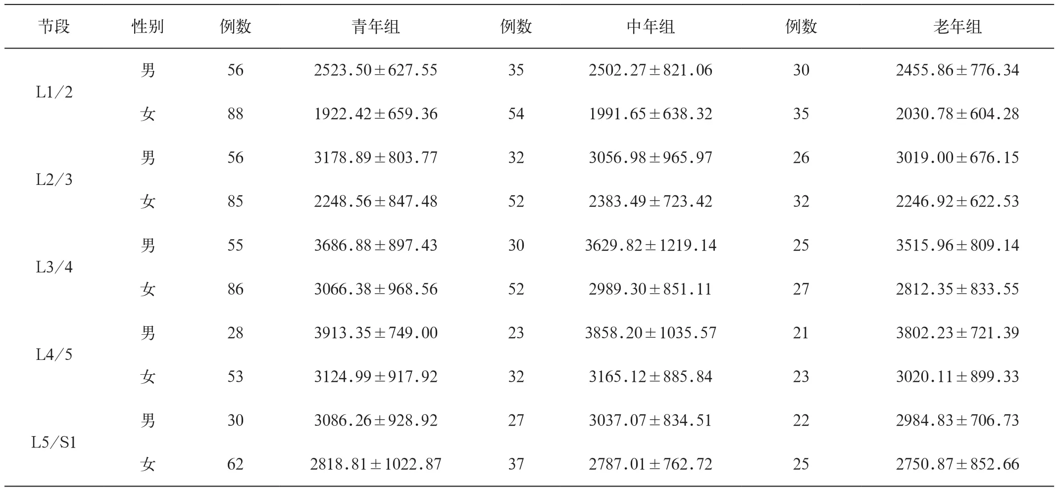
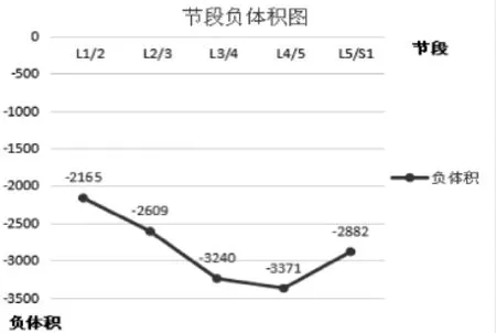
运用软件测量正常髓核体积,见表1。L1/ L2~L5/S1同节段男女之间正常髓核体积差异均有显著统计学意义(P<0.01),见表2。同节段不同年龄组之间正常髓核体积差异均无统计学意义(P>0.05),见表3。L1/2~L4/5体积增大,L4/5~L5/S1体积逐渐减小,采用负体积在数轴上描述图像类似于正常腰椎曲线,见表4和图1。统计不同节段中相邻两髓核体积呈线性相关关系,见表5。不同节段髓核体积与BMI指数、身高及体重的相关性,见表6。
表1 各年龄组各节段男女正常髓核体积(±s ,mm3)

表1 各年龄组各节段男女正常髓核体积(±s ,mm3)
节段性别例数青年组例数中年组例数老年组男L1/2 562523.50±627.55352502.27±821.06302455.86±776.34女881922.42±659.36541991.65±638.32352030.78±604.28 L2/3 L3/4 L4/5男563178.89±803.77323056.98±965.97263019.00±676.15女852248.56±847.48522383.49±723.42322246.92±622.53男553686.88±897.43303629.82±1219.14253515.96±809.14女863066.38±968.56522989.30±851.11272812.35±833.55男283913.35±749.00233858.20±1035.57213802.23±721.39女533124.99±917.92323165.12±885.84233020.11±899.33男L5/S1 303086.26±928.92273037.07±834.51222984.83±706.73女622818.81±1022.87372787.01±762.72252750.87±852.66
表2 同节段不同性别正常髓核体积比较(±s ,mm3)

表2 同节段不同性别正常髓核体积比较(±s ,mm3)
节段例数男例数女P L1/21212505.43±734.501771963.37±630.05P<0.01 L2/31143121.27±817.631692295.07±776.55P<0.01 L3/41103640.32±982.111653011.97±914.57P<0.01 L4/5723864.21±815.521083114.75±845.18P<0.01 L5/S1793044.36±830.541242786.34±912.63P<0.01
表3 同节段不同年龄组正常髓核体积比较(±s ,mm3)

表3 同节段不同年龄组正常髓核体积比较(±s ,mm3)
组别L1/2L2/3L3/4L4/5L5/S1青年组2148.63±756.602602.24±943.843290.83±961.573358.59±979.562890.66±913.37中年组2176.15±821.882615.63±852.643193.05±927.473229.87±1030.062881.22±805.36老年组2219.37±777.682569.90±719.993130.04±894.473323.97±949.872850.19±810.51 P P>0.05P>0.05P>0.05P>0.05P>0.05
表4 各节段正常髓核体积(±s ,mm3)

表4 各节段正常髓核体积(±s ,mm3)
L1/2L2/3L3/4L4/5L5/S1例数298283275180203 X±S2165.57±797.352609.71±880.293240.66±931.013371.46±981.222882.11±907.12

表5 正常髓核体积相邻节段相关性分析

表6 不同节段髓核体积与BMI指数、身高及体重的相关性

图1 各节段正常髓核负体积折线图
讨 论
髓核位于椎间盘中心位置,是人体重要的组织结构,是一种具有良好弹性的胶状物质,被分层的纤维环和软骨紧紧包绕,髓核中含有较丰富的硫酸软骨素、黏多糖蛋白复合体以及大量的水分。一般髓核的含水量会随着年龄的增长而减低,因此而发生髓核的变性,髓核的体积和形态容易发生相应的变化[8],从而引发一系列的临床症状。中老年人是病变的主要人群,在我们这样一个正在步入人口老龄化的国家,对于髓核的进一步研究显得尤为重要。随着科技的发展,医学也在逐步向大数据时代迈进,现代医学影像学的应用就能客观地提供广泛的数据,也能为影像的定量诊断及治疗的精准性提供基础。
对于椎间盘病变的治疗,传统的单纯髓核摘除术会改变生物力学结构;手术融合则容易导致该阶段的运动功能障碍;近年来椎间盘髓核假体[9]的建立及置换[10-11]成为临床手术治疗的重要手段,然而髓核假体如何定量就显得非常重要。对于椎间盘突出的患者进行射频消融术时,也可以通过患者术前术后的磁共振影像定量评估髓核体积的变化。如果采取体位注射的方式来改善或者恢复髓核的力学性能[12],那么注射的体积也是一个考虑的因素。本研究主要研究BMI正常人群的髓核体积的数据范围,建立不同节段、不同年龄、不同性别之间腰椎间盘髓核体积的正常值范围,为腰椎间盘髓核假体的建立和置换、注射以及定量方式,提供可靠的参考值及体积测量方法。本研究结果表明,同年龄组同节段、不同性别,同年龄组同性别、不同节段的髓核体积有统计学差异,需要采取不同型号的髓核假体,因此术前要进行个体化的评估。但是在同节段不同的年龄组中,正常髓核体积的变化不大,说明了成人正常髓核体积具有稳定性,对椎间盘髓核的组织工程学有一定的参考价值[13-14]。同时,本研究用节段-负体积数轴描述了体积变化的趋势,并研究了相邻髓核体积的相关性,其相关性的变化趋势和髓核体积的变化趋势基本一致,但是是否与生物力学具有内在的联系,需要进一步研究。人工髓核置换的患者大多椎间盘发生了退行性改变,置入前已无法获知正常髓核的大小,根据本研究,既可以考虑按照患者成年后的任意年龄正常髓核影像资料设计髓核的大小,也可以考虑根据不同节段髓核的相关性来计算正常髓核体积作为一个参考的指标,具有一定的临床意义。
另外,本研究发现BMI正常范围内人群的正常髓核体积与BMI指数并没有相关性,而是分别与身高和体重存在一定的正相关性。
总之,本研究介绍了定量研究腰椎间盘髓核体积方法,为以后对正常和各种退行性疾病的椎间盘髓核体积改变提供了研究方法,同时也为BMI正常人群腰椎间盘髓核体积提供了正常参考值范围。
[ 1 ]俎金燕, 王晨光, 贾宁阳, 等. 腰椎间盘退行性变的 MR 扩散张量成像初步研究. 中华放射学杂志, 2012, 46: 1002-1005.
[ 2 ]Nazari J, Pope M H, Graveling R A. Feasibility of Magnetic resonance imaging (MRI) in obtaining nucleus pulposus (NP) water content with changing postures . Magnetic Resonance Imaging, 2015,33 : 459-464.
[ 3 ]Fritzell P,Hagg O,Wessberg P,et al.2001 Volvo award winner in clinical studies;lumbar fusion versus nonsurgical treatment for chronic low back pain;a multicenter randomized controlled trial from the Swedish Lumbar Spine Study Group. Spine,2001,26:2521-2534.
[ 4 ]Borges A C, Eyholzer C, Duc F, et al. Nanofibrillated cellulose composite hydrogel for the replacement of the nucleus pulposus . Acta Biomaterialia, 2011, 7 : 3412-3421.
[ 5 ]Schmocker A, Khoushabi A, Bourban P E, et al. Photopolymerization device for minimally invasive implants: application to nucleus pulposus replacement //World Congress on Medical Physics and Biomedical Engineering, June 7-12, 2015, Toronto, Canada. Springer International Publishing, 2015: 1333-1337.
[ 6 ]谷守欣,李克.3D打印技术在骨骼病变的应用进展.中国医学计算机成像杂志,2015,21:387-392.
[ 7 ]Pfirrmann CW,Metzdorf A,Zanetti M,et al.Magnetic resonance classification of lumbar intervertebral disc degeneration. Spine,2001,26 :1873-1878.
[ 8 ]O'Connell GD,Jacobs NT,Sen S,et al.Axial creep loading and unloaded recovery of the human intervertebral disc and the effect of degeneration.J Mech Behav Biomed Mater,2011,4:933.
[ 9 ]Bergknut N1, Smolders LA, Koole LH, et al. Performance of a hydrogel nucleus pulposus prosthesis in an ex vivo canine model. . Biomaterials,2010,31 :6782-6788.
[10]Lewis G. Nucleus pulposus replacement and regeneration/repair technologies: present status and future prospects. J Biomed Mater Res B Appl Biomater, 2012 ,100 :1702-1720.
[11]Borges AC1, Eyholzer C, Duc F, Bourban PE, et al. Nanofibrillated cellulose composite hydrogel for the replacement of the nucleus pulposus. Acta Biomater, 2011 ,7 :3412-3421.
[12]Foss BL, Maxwell TW, Deng Y. Chondroprotective supplementation promotes the mechanical properties of injectable scaffold for human nucleus pulposus tissue engineering. J Mech Behav Biomed Mater,2014,29:56-67.
[13]Mercuri J, Addington C, Pascal R 3rd, , et al. Development and initial characterization of a chemicallys tabilized elastin-glycosaminoglycancollagen composite shape-memory hydrogel for nucleus pulposus regeneration. J Biomed Mater Res A, 2014 ,102 :4380-4393.
[14]Strange DG, Oyen ML. Composite hydrogels for nucleus pulposus tissue engineering. J Mech Behav Biomed Mater,2012,11:16-26.
Measurement of Nucleus Pulposus Volume by Magnetic Resonance Imaging in Population with Normal BMI
WEN Bing, DU Ying, HU Liang-bo, ZHANG Ao, Liang Xue-heng, Yu Jie
Medical Science Foundation of Chongqing Health Bureau No. 2013-2-075
R445.2
A
1006-5741(2016)-04-0346-05
2015.12.28 ;修回时间:2016.07.04)
中国医学计算机成像杂志,2016,22:346-350
重庆医科大学附属永川医院放射科
通信地址:重庆市永川区萱花路439号, 重庆市 402160
胡良波(电子邮箱:hlb123_001@163.com)
重庆市卫生局面上资助项目No. 2013-2-075
Chin Comput Med Imag,2016,22:346-350
Department of Radiology,Yongchuan Hospital Chongqing Medical University
Address:No.239 Dong xuanhua Rd,Chongqing 402160,P.R.C.
Address Correspondence to HU Liang-bo(E-mail: hlb123_001@163.com)

