胃癌淋巴结包膜外侵犯与预后的关系研究
赵玉国,张建文△,许瑞庭(.郴州市第一人民医院普外科,湖南43000;.宁夏回族自治区人民医院微创外科,宁夏银川7500)
胃癌淋巴结包膜外侵犯与预后的关系研究
赵玉国1,张建文1△,许瑞庭2
(1.郴州市第一人民医院普外科,湖南423000;2.宁夏回族自治区人民医院微创外科,宁夏银川750021)
目的初步探讨胃癌淋巴结包膜外侵犯与预后的关系。方法收集郴州市第一人民医院2007年1~12月符合入选标准的患者80例,以常规HE染色法观察淋巴结转移癌包膜外侵犯情况,标本经常规病理检查为阳性则认为淋巴结有癌转移。80例患者均采用电话预约后门诊复查登记形式进行至少5年随访,截至2012年1月或患者死亡、中途失访。结果胃癌淋巴结包膜外侵犯阳性47例,阴性33例;淋巴结包膜外侵犯阳性例数在T分期、N分期、TNM分期方面比较,差异有统计学意义(P<0.01);胃癌预后单因素分析表明,患者平均生存时间与TNM分期、肿瘤大小、术后化疗、CEA水平、包膜外侵犯相关(P<0.01或0.05);多因素分析表明,包膜侵犯为胃癌预后的独立因素(P<0.01)。结论胃癌淋巴结包膜外侵犯对判断胃癌预后有重要作用,淋巴结包膜外侵犯阳性患者预后较差。
胃肿瘤;淋巴结;肿瘤分期;肿瘤侵润;预后
胃癌是胃肠外科常见的消化道恶性肿瘤之一,胃癌的预后和淋巴结转移与否有重要关联,作为胃癌主要转移途径的淋巴转移目前研究较多,但对转移的淋巴结包膜外侵犯(ECS,是指癌细胞突破淋巴结外膜而侵犯其周围的血管、神经、肌肉或脂肪等组织)的机制及始发因素与胃癌预后的关系临床上尚存在争议。本研究通过对郴州市第一人民医院胃肠外科2007年1~12月收治的胃癌根治术患者的资料进行回顾性分析,初步研究胃癌ECS与患者预后的关系,期望对胃癌的诊疗提供较好的理论指导。
1 资料与方法
1.1一般资料收集符合入选标准的患者80例,其中男53例,女27例;年龄22~75岁;肿瘤大于或等于5 cm 35例,<5 cm 45例;肿瘤分期Ⅰ期0例,Ⅱ期21例,Ⅲ期55例,Ⅳ期4例;肿瘤位置:胃窦53例,胃体22例,胃底5例;低分化69例,高中分化11例;术后化疗者60例,未化疗者20例;癌胚抗原(CEA)正常(<5 ng/mL)73例,升高(≥5 ng/mL)7例;ECS阳性47例,阴性33例。入选标准:(1)术后病理证实为胃癌者,有完善的术前检查资料;(2)术前评估分期为cT1~4N1~2M0。剔除标准:(1)术后病理证实为胃其他类型恶性肿瘤,如胃间质瘤、肉瘤、淋巴瘤等;(2)曾行胃切除术后再次手术者,如残胃癌、胃癌复发;(3)其他系统、器官原发恶性肿瘤5年内胃癌患者;(4)同时有2个以上系统、器官恶性肿瘤者。
1.2方法
1.2.1病理检查方法(1)切除的新鲜胃癌标本立即将可疑转移淋巴结分离,连同淋巴结周边的部分正常组织一并分离。标本即刻送病理实验室,以10%福尔马林溶液常规固定,再于脱水机内处理12 h,浸蜡、包埋、风干。切片常规HE染色,切片由经验丰富的同一名病理科主任医师在光学显微镜下阅片,明确是否有淋巴结转移,若淋巴结发生ECS,则特殊报告。(2)分离的淋巴结标本行常规病理检查。本研究以淋巴结包膜的完整性破坏,伴结缔组织增生反应,或包膜外见肿瘤细胞为标准,判断淋巴结是否发生ECS[1]。
1.2.2随访手术当天即为随访起始时间,80例患者均采用电话预约后门诊复查登记形式进行至少5年随访,截至2012年1月或患者死亡、中途失访截止。
1.3统计学处理应用SPSS18.0统计软件进行数据处理,根据不同类型的资料采用相应统计学方法,计数资料以率或构成比表示,采用χ2检验;计量资料以x±s表示,采用t检验;Cox比例风险回归模型采用双侧检验。P<0.05为差异有统计学意义。
2 结 果
2.1各指标ECS发生情况比较结果ECS阳性例数在不同年龄、性别、分化级别、肿瘤位置、肿瘤大小、血CEA水平方面比较,差异均无统计学意义(P>0.05);而在T分期、N分期、TNM分期方面比较,差异均有统计学意义(P<0.01)。见表1。

表1 各指标ECS发生情况比较
2.2胃癌预后单因素分析患者平均生存时间在不同肿瘤TNM分期、不同肿瘤大小、术后化疗与否、不同CEA水平、ECS发生与否等方面比较,差异均有统计学意义(P<0.05或0.01);而在性别、年龄、分化类型方面比较,差异均无统计学意义(P>0.05)。见表2。
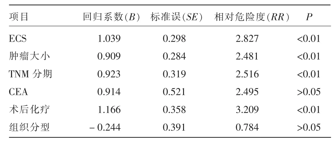
2.3胃癌预后多因素分析单因素分析有统计学意义的结果引入Cox回归模型,结果显示,ECS为胃癌预后的独立因素(P<0.01)。见表3。
表2 胃癌预后单因素分析(±s,月)

表2 胃癌预后单因素分析(±s,月)
项目年龄(岁)n 生存时间t P ≥5 0 <5 0 0 . 0 0 7 >0 . 0 5性别男女0 . 6 0 3 >0 . 0 5分化级别1 . 6 2 8 >0 . 0 5肿瘤位置低分化中分化胃窦胃体胃底4 9 3 1 5 3 2 7 6 9 1 1 5 3 2 2 3 . 0 2 9 >0 . 0 5 T分期5 1 1 4 2 8 . 1 8 3 <0 . 0 1 N分期2 1 . 6 6 3 <0 . 0 1 T N M分期T 1 T 2 T 3 T 4 N 1 N 2 N 3Ⅱ期3 8 . 7 1 7 <0 . 0 1 E C S 1 6 . 3 5 9 <0 . 0 1 C E A (n g / m L)Ⅲ期Ⅳ期阴性阳性<5 ≥5 1 0 . 3 4 9 <0 . 0 1术后化疗5 . 0 2 4 <0 . 0 5肿瘤大小(c m)是否<5 ≥5 3 4 3 1 2 0 2 9 3 1 2 1 5 5 4 3 3 4 7 7 3 7 6 0 2 0 4 5 3 5 4 2 . 5 2 9 ± 3 . 0 2 3 4 2 . 3 3 5 ± 3 . 5 9 9 4 1 . 9 5 5 ± 2 . 9 1 6 4 3 . 2 5 8 ± 3 . 7 6 2 4 1 . 9 6 2 ± 2 . 4 3 1 4 5 . 5 1 5 ± 6 . 8 4 3 4 5 . 0 1 2 ± 2 . 6 4 5 3 3 . 9 7 7 ± 4 . 7 4 3 5 3 . 7 3 3 ± 6 . 4 3 5 6 0 . 0 0 0 ± 0 . 0 0 0 6 1 . 0 0 0 ± 0 . 0 0 0 4 7 . 8 7 2 ± 2 . 6 1 7 2 7 . 4 5 2 ± 3 . 6 3 6 5 7 . 8 0 0 ± 2 . 7 6 4 4 1 . 5 4 6 ± 3 . 8 3 4 3 3 . 2 1 2 ± 3 . 4 8 0 6 0 . 6 3 1 ± 1 . 1 0 7 3 7 . 6 7 5 ± 2 . 6 3 8 1 1 . 7 5 0 ± 4 . 8 0 2 5 3 . 1 5 7 ± 2 . 7 7 6 3 5 . 0 0 8 ± 2 . 9 6 5 4 4 . 1 4 6 ± 2 . 3 6 0 2 5 . 0 0 0 ± 6 . 7 8 6 4 8 . 8 1 3 ± 1 . 9 6 7 2 3 . 3 0 0 ± 5 . 1 3 0 4 8 . 3 8 3 ± 2 . 8 5 6 3 4 . 6 7 9 ± 3 . 3 6 8 1 9 . 7 2 4 <0 . 0 1

表3 胃癌预后多因素分析
3 讨 论
胃癌是消化道常见的恶性肿瘤,严重威胁着人们的生命。在我国,因对胃癌的筛查未普及,多数胃癌患者确诊时已是中期或中晚期,虽经以手术为主的综合治疗,但总体预后仍差,而淋巴转移是影响胃癌预后的主要因素之一。在国际抗癌联盟(UICC)的胃癌N分期标准是依据转移淋巴结的个数,并未考虑转移淋巴结本身的受侵范围,对胃癌预后的判断可能欠准确、合理[2]。本研究结果表明,ECS与胃癌N分期密切相关,是影响胃癌预后的独立危险因素。Wreesmann等[3]在对头颈部鳞状细胞癌疾病特异性生存率和多因素分析中发现,无ECS的患者预后明显较好,而有明显ECS患者较微小ECS患者的生存率明显降低,微小ECS患者与无ECS患者生存率比较无显著差异,时间依赖的受试者工作特征曲线(ROC曲线)分析将ECS 1.7 mm作为判断预后的标准。本组研究结果也表明,胃癌ECS患者预后明显较差,但是本研究未对ECS程度的不同进行进一步分析和讨论,今后的研究应逐步完善。有报道显示,在其他系统和消化器官恶性肿瘤患者中也有ECS的现象,如乳腺癌、口腔癌、结直肠癌、前列腺癌等[4-9]。可以推测,ECS可能是恶性肿瘤的一种特殊转移方式,与传统的淋巴结转移机制可能存在不同。即便如此,当前的胃癌N分期系统中还是缺少对ECS的考虑。作者认为应该将ECS纳入胃癌分期系统,同时建议UICC分期系统将ECS纳入肿瘤分期体系[10-12]。
Jiang等[13]认为,ECS与胃癌的侵略性存在密切相关,是一个重要的独立预测因素,明显降低了胃癌患者无病生存率和总体存活率,也是远期复发的独立危险因素,尤其是腹膜复发,选择全身术后辅助治疗(静脉或动脉)和腹腔区域治疗可能是一个合理的方法。Kumagai等[14]研究表明,结外淋巴结侵犯患者有更高的肝转移发生率,多因素分析显示,ECS与肝转移明显相关;淋巴结转移是肝转移的下一个最重要的风险。本研究结果表明,胃癌预后与ECS、TNM分期、肿瘤大小、术后化疗、CEA水平相关,胃癌患者ECS阳性者预后明显低于阴性者,表明ECS阳性组较ECS阴性组术后肿瘤复发时间明显更早,而肿瘤术后复发是胃癌患者死亡的重要因素;多因素分析结果表明,ECS为胃癌预后的独立因素之一。因此,胃癌ECS能够初步判断胃癌患者预后是否不良。但本组研究结果表明,胃癌预后与肿瘤分化程度无相关性(P>0.05),可能与本组标本数量较少有关,应进一步扩大样本对此进行研究。
综上所述,作者认为,胃癌ECS对判断胃癌预后有重要作用,能够指导术前、术后制订更加合理的治疗方案:(1)针对术前包括影像学检查在内的评估高度怀疑淋巴结转移甚至包膜外侵犯的患者,制订合理的术前新辅助放、化疗方案,使肿瘤降期和减少术后ECS的阳性率;术中如病理能够证实ECS,则可对淋巴结外软组织实行更广泛的切除,力求肿瘤达到R0切除。(2)对于术后病理证实ECS的患者,则可尽早制订术后以放、化疗为主的综合治疗方案,以提高术后生存率。但目前临床上对胃癌ECS的研究报道较少,对于胃癌ECS的确切机制尚不完全清楚。如果对胃癌患者的治疗能充分考虑ECS这一因素,能够对胃癌患者术前分期、术后分期、术后治疗和预后判断提供更多的指导,对患者术后生存时间做出更加准确的评估,使胃癌患者受益。
[1]任振虎.口腔鳞癌颈部淋巴结包膜外侵犯相关因素研究[D].长沙:中南大学,2012.
[2]季加孚,张成海,姚学清,等.膜外淋巴结转移-影响胃癌预后的新因素[J].循证医学,2011,11(2):87-90.
[3]Wreesmann VB,Katabi N,Palmer FL,et al.Influence of extracapsular nodal spread extent on prognosis of oral squamous cell carcinoma[J].Head Neck,2015-10-30[2015-11-09].http://www.ncbi.nlm.nih.gov/pubmed/ ?term=Wrees mann+VB%2C+Katabi+N%2C+Palmer+FL%2Cet+al.+ Influence+of+extracapsular+nodal+spread+extent+on++prognosis+of+ oral+squamous+cell+carcinomaWreesmann+VB%2C+Katabi+N%2C+ Palmer+FL%2Cet+al.+Influence+of+extracapsular+nodal+spread+extent +on++prognosis+of+oral+squamous+cell+carcinoma.
[4]Gooch J,King TA,Eaton A,et al.The extent of extracapsular extension may influence the need for axillary lymph node dissection in patients with T1-T2 breast cancer[J].Ann Surg Oncol,2014,21(9):2897-2903.
[5]Yajima R,Fujii T,Yanagita Y,et al.Prognostic value of extracapsular invasion of axillary lymph nodes combined with peritumoral vascular invasion in patients with breast cancer[J].Ann Surg Oncol,2015,22(1):52-58.
[6]Tay KJ,Gupta RT,Brown AF,et al.Defining the incremental utility of prostate multiparametric magnetic resonance imaging at standard and specialized read in predicting extracapsular extension of prostate cancer[J]. Eur Urol,2015-11-06[2015-11-09].http://www.ncbi.nlm.nih.gov/pubmed/ ?term=Tay+KJ+AND+Gupta+RT+AND+Brown+AF.
[7]Kojima Y,Yoneyama T,Hatakeyama S,et al.Detection of core2 β-1,6-N-acetylglucosaminyltransferase in post-digital rectal examination urine is a reliable indicator for extracapsular extension of prostate cancer[J]. PLoS One,2015,10(9):e0138520.
[8]Song B,Yang Y,Wang YL,et al.Adenovirus expressing IFN-λ(Ad/hIFN-λ)produced anti-tumor effects through inducing apoptosis in humantongue squamous cell carcinoma cell[J].Int J Clin Exp Med,2015,8(8):12509-12518.
[9]Simonen P,Lehtonen J,Kandolin R,et al.F-18-fluorodeoxyglucose positron emission tomography-guided sampling of mediastinal lymph nodes in the diagnosis of cardiac sarcoidosis[J].Am J Cardiol,2015,116(10):1581-1585.
[10]Hu X,Cao L,Yu Y.Prognostic prediction in gastric cancer patients without serosal invasion:comparative study between UICC 7(th)edition and JCGS 13(th)edition N-classification systems[J].Chin J Cancer Res,2014,26(5):596-601.
[11]Tang X,Lan Z,Chen Y,et al.The 7th AJCC/UICC TNM staging system may be not suitable in predicting prognosis of synchronous multiplegastric carcinoma patients with D2 gastrectomy[J].Tumour Biol,2015,36(5):1-7.
[12]Röcken C,Behrens HM.Validating the prognostic and discriminating value of the TNM-classification for gastric cancer-a critical appraisal[J]. Eur J Cancer,2015,51(5):577-586.
[13]Jiang N,Deng JY,Ding XW,et al.Node-extranodal soft tissue stage based on extranodal metastasis is associated with poor prognosis of patients with gastric cancer[J].J Surg Res,2014,192(1):90-97.
[14]Kumagai K,Tanaka T,Yamagata K,et al.Liver metastasis in gastric cancer with particular reference to lymphatic advancement[J].Gastric Cancer,2001,4(3):150-155.
Study on relationship between extracapsular spread of lymph node with prognosis in gastric cancer
Zhao Yuguo1,ZhangJianwen1△,Xu Ruiting2(1.Department of General Surgery,Chenzhou Municipal First People′s Hospital,Chenzhou,Hunan 423000,China;2.Department of Minimally Invasive Surgery,Ningxia Hui Autonomous Region People′s Hospital,Yinchuan,Ningxia 750021,China)
ObjectiveTo investigate the relationship between the extracapsular spread of lymph node with prognosis in gastric cancer.MethodsTotally 80 cases conformed to the inclusion criterion were collected from Chenzhou Municipal First People′s Hospital.The routine HE staining method was used to observe the extracapsular spread situation of lymph nodes in gastric cancer,the samples of the positive routine pathological examination were considered to be lymph nodes metastasis.All 80 cases were followed up for at least 5 years up until January 2012 or death of patient and loss to follow up at halfway by the pattern of outpatient department reexamination and registration after telephone appointment.ResultsThe extracapsular spread of lymph nodes was found to be positive in 47 cases and negative in 33 cases.The cases number of extracapsular spread of lymph nodes had statistical difference in the aspects of the T staging,N staging and TNM staging,and the differences were statistically significant (P<0.01);the single factor analysis of gastric cancer prognosis showed that the average survival time was correlated with the TNM stage,tumor size,postoperative chemotherapy,CEA concentration and extracapsular spread(P<0.01 or 0.05);the multivariate analysis showed that the extracapsular spread of lymph node was an independent prognostic factor for gastric cancer.Conclusion
The extracapsular spread of lymph node has an important role for judging the prognosis of gastric cancer,and the prognosis in the patients with extracapsular spread of lymph node is poor.
Stomach neoplasms;Lymph nodes;Neoplasm staging;Neoplasm invasiveness;Prognosis
10.3969/j.issn.1009-5519.2016.05.014
A
1009-5519(2016)05-0678-03
赵玉国(1974-),硕士研究生,副主任医师,主要从事胃肠癌治疗及预后的研究。
△,E-mail:iamdoczhjw119@sohu.com。
(2015-12-05)

