Her2在乳腺癌和胃癌中表达的临床意义①
闫振宇 买春阳 高 鹏 苏 博 王世宣
(河南省南阳市中心医院,南阳473009)
Her2在乳腺癌和胃癌中表达的临床意义①
闫振宇买春阳②高鹏苏博王世宣③
(河南省南阳市中心医院,南阳473009)
[摘要]目的:探讨Her2在乳腺癌和胃癌中表达的临床情况与相关意义,为肿瘤防治提供参考。方法:收集2009年3月到2014年5月在我院行外科手术切除的60例确诊的进展期胃癌标本与60例确诊的晚期乳腺癌标本,都进行Her2病理表达免疫组化分析,并调查了相关临床病理资料进行相关性分析。结果:Her2的乳腺癌和胃癌标本中阳性表达率分别为40.0%和36.7%,对比差异无统计学意义(P>0.05)。Her2表达与恶性肿瘤患者的年龄、病理类型无关,与TNM分期与淋巴结转移有明显相关性(P<0.05)。Spearman相关分析显示乳腺癌与胃癌中的Her2表达都与Survivin、Bcl-2表达存在明显正向相关性(P<0.05)。结论:乳腺癌和胃癌患者Her2都呈现高病理表达状况,与TNM分期与淋巴结转移有明显相关性,能通过调节部分凋亡抑制蛋白,参与肿瘤的发病与转移过程。
[关键词]乳腺癌;胃癌;Her2;病理特点
乳腺癌和胃癌都是常见的恶性肿瘤之一,由于各种因素的影响,两者在我国其发病率呈明显上升趋势,且发病年龄也有年龄化的趋势,是我国肿瘤防治的重点之一[1,2]。现代研究表明恶性肿瘤的病因是多方面的,其发展也呈现阶段性,目前临床对肿瘤病程以及预后的判断依据主要包括病理学分级、肿瘤转移状况、肿瘤体积和组织学分级等[3,4]。手术切除是目前乳腺癌和胃癌的主要治疗方法,然而受制于癌症诊断的局限性,诸多胃癌和乳腺癌患者在就诊时,病情已经发展至癌症中晚期,错过了手术切除最佳时期,使得手术切除效果显著下降,甚至有些患者已经无法接受手术切除治疗,切除手术后胃癌和乳腺癌患者的成活率只有50%左右,患者死亡率比较高[5,6]。随着基础医学的发展,细胞内的信号传导通路在胃癌和乳腺癌的发生发展、诊断和治疗方面的意义得到了广泛关注[7]。其中表皮生长因子受体(Epithelial growth factor receptor,EGFR)通路是影响肿瘤的重要通路,EGFR属于酪氨酸激酶受体家族,主要在上皮组织细胞表面,包括Her1、Her2、Her3和Her4等成员[8]。其中Her2是原癌基因Her2/neu编码的一种分子量为185 kD跨膜糖蛋白p185,HER2基因扩增与增加细胞分化、局部及远处转移、迁移、减少细胞凋亡、加快血管发生、肿瘤侵袭、局部及远处转移密切相关[9]。基础研究表明Her2的过量表达可促进肿瘤细胞的转移,一方面通过调节上皮细胞钙黏蛋白等黏附因子的表达来促进迁移,另一方面可启动多种转移机制来加快肿瘤细胞转移[10]。研究表明,大约25%~30%的浸润性乳腺癌和80%胃癌患者有Her2基因扩增或蛋白过表达,但是具体的表达对比情况还不明确[11]。因此准确地检测Her2状态不仅能为乳腺癌和胃癌患者的预后提供依据,而且是有效治疗乳腺癌和胃癌的前提。本文为此具体探讨了乳腺癌和胃癌患者Her2病理表达情况,分析其临床病理意义,可以为乳腺癌和胃癌的靶向治疗提供进一步依据。现报告如下。
1材料与方法
1.1材料
1.1.1研究对象收集2009年3月到2014年5月在我院行外科手术切除的60例确诊的进展期胃癌标本与60例确诊的晚期乳腺癌标本,所有病例进行切除手术前都没有接受过化疗、放疗或其他抗癌药物治疗,所有患者都进行肿瘤全切或次全切手术,并将淋巴结根治性切除;所有研究得到了患者的知情同意与伦理委员会的批准。为了保持样本的同一性,胃癌患者我们也都选择了女性。胃癌患者中年龄最小34岁,最大78岁,平均年龄(56.33±4.22)岁;病理类型:鳞癌10例,腺癌42例,腺鳞癌8例;TNM国际分期:Ⅰ期、Ⅱ期和Ⅲ期,分别为34例、20例和6例;无淋巴结转移40例,淋巴结转移20例。乳腺癌患者中年龄最小33岁,最大79岁,平均年龄(56.02±5.11)岁;病理类型:浸润性导管癌54例,浸润性小叶癌2例,浸润性小叶导管混合癌8例;TNM国际分期:Ⅰ期、Ⅱ期和Ⅲ期,分别为33例、21例和6例;无淋巴结转移40例,淋巴结转移20例。不同患者的年龄、TNM分期与淋巴结转移情况对比差异无统计学意义(P>0.05)。
1.1.2主要试剂兔抗Her2单克隆抗体、即用型快捷免疫组化试剂盒购自福州迈新生物技术开发有限公司,其余生化试剂都购自北京中杉金桥生物技术有限公司。
1.2方法
1.2.1标本处理所有手术切除的标本离体后立即用10%中性福尔马林固定,8~24 h后按常规行系列脱水、透明和石蜡包埋,选择4 μm切片,然后进行病理的免疫组化分析。
1.2.2检测方法肿瘤切片免疫组化染色后,置于光学显微镜下观察,并分析和处理。本实验通过双盲法来分析阳性,同时每张切片由两位资深病理科医师分别做出各自的判断。
Her2阳性细胞染色结果显示棕黄色均匀细颗粒主要分布在胞浆和细胞膜上,根据病变细胞中阳性细胞所占百分比以及着色深浅来判断和进行分级:(-):肿瘤组织 中无细胞核/膜染色或者已染色的细胞膜/核染色的比例只有10%;(+):已染色的细胞膜/核染色的比例大于10%;(++):超过10%的肿瘤细胞膜/核全部染色,染色程度由弱至中度;(+++):超过30%的肿瘤细胞被完全染色,染色程度较深。上述分级中,Her2蛋白(-)或(+)列入阴性范畴;(++)及其以上列入阳性范畴。用已知Her2阳性的肿瘤标本作阳性对照,用PBS液代替探针混合液作阴性对照。
1.3统计学方法采用SPSS18.0软件来分析研究数据,采用χ2检验来比较表达阳性情况同配对计数资料之间的差异性,采用Spearman来进行相关性分析,P<0.05表示差异显著,具备统计学意义。
2结果
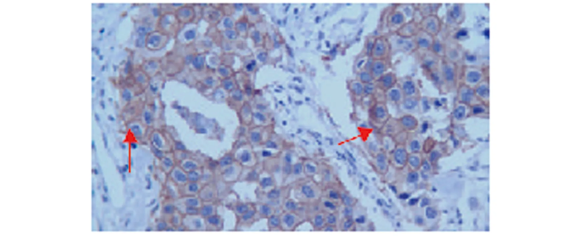
2.1乳腺癌和胃癌的Her2阳性表达情况对比经过观察,Her2的乳腺癌和胃癌标本中阳性表达率分别为40.0%和36.7%,对比差异无统计学意义(P>0.05)。见表1、图1~4。
2.2Her2表达与乳腺癌和肿瘤患者临床各病理特征间的关系检测和观察结果显示,Her2表达不受恶性肿瘤患者病理类型和年龄的影响,与TNM分期与淋巴结转移有明显相关性(P<0.05)。见表2与表3。
2.3相关性分析我们选择了恶性肿瘤的常规检测指标Survivin、Bcl-2进行了相关性分析,结果显示乳腺癌与胃癌中的HER2表达都与Survivin、Bcl-2表达存在明显正向相关性(P<0.05)。见表4。
表1乳腺癌和胃癌的Her2阳性表达情况对比(n)
Tab.1Her2 positive expression comparison in breast cancer and gastric cancer(n)

TissuesnPositiveexpressionPositiveexpressionrateBreastcancer602440.0%Gastriccancer602236.7%χ20.293P>0.05

图1 乳腺导管原位癌HER2表达强阳性于胞膜(×400)Fig.1 DCIS HER2 strong expression in membrane(×400)

图2 乳腺浸润性导管癌HER2表达强阳性于胞膜(×400)Fig.2 Invasive ductal carcinoma HER2 strong expression in membrane (× 400)

图3 胃鳞癌HER2表达强阳性于胞浆(×400)Fig.3 HER2 expression in squamous cell carcinoma of stomach strongly positive in cytoplasm (× 400)

图4 胃腺癌HER2表达强阳性于胞浆(×400)Fig.4 Gastric adenocarcinoma strong positive HER2 expression in cytoplasm (× 400)
表2乳腺癌HER2表达的临床病理特点分析(n)
Tab.2Clinical and pathological features of HER2 protein expression in breast cancer (n)

Pathologicalfeaturesn=60Positiveexpression(n=24)χ2PAge≥604518(40.0%)0.000>0.05<60156(40.0%)Pathologicaltype-invasiveductalcarcinoma5418(33.3%)0.293>0.05Invasivelobularcarcinoma21(50.0%)Invasivelobularcarcinomamixingduct85(62.5%)TNMinternationalstaging-Ⅰstage3312(36.4%)0.311>0.05Ⅱstage219(42.9%)Ⅲstage63(50.0%)Lymphnodemetastasis-Yes208(40.0%)0.000>0.05No4016(40.0%)
表3胃癌HER2表达的临床病理特点分析(n)
Tab.3Clinical and pathological features of HER2 protein expression in gastric cancer (n)

Pathologicalfeaturesn=60Positiveexpression(n=22)χ2PAge≥604416(36.4%)0.078>0.05<60166(37.5%)Pathologicaltype-invasiveductalcarcinoma104(40.0%)0.293>0.05Invasivelobularcarcinoma4216(38.1%)Invasivelobularcarcinomamixingduct82(25.0%)TNMinternationalstaging-Ⅰstage3412(35.3%)0.331>0.05Ⅱstage207(35.0%)Ⅲstage62(33.3%)Lymphnodemetastasis-Yes208(40.0%)0.236>0.05No4014(35.0%)
表4乳腺癌与胃癌中的HER2表达与传统指标的相关性分析
Tab.4Correlation analysis between HER2 expression and traditional indicators in breast cancer and gastric cancer

IndicatorsBreastcancerrPGastriccancerrPSurvivin0.522<0.050.500<0.05Bcl-20.467<0.050.387<0.05
3讨论
乳腺癌与胃癌都是最常见的恶性肿瘤之一,其中胃癌居消化系统恶性肿瘤首位,乳腺癌居妇科肿瘤的首位。我国是世界上乳腺癌与胃癌都高发的国家,目前乳腺癌与胃癌手术切除及放化疗治疗效果仍不尽人意,中晚期患者总体5年生存率仍然不高[12]。恶性肿瘤最根本的特征为转移与分期增加,该特征是将恶性肿瘤患者引向死亡的重要因素,是影响手术切除效果及术后预后效果的关键因素[13]。现代肿瘤学认为,恶性肿瘤的发生、发展不仅与肿瘤细胞增殖有关,还与信号传导通路的变化有关。表皮生长因子受体(EGFR)家族是癌症发生和发展相关的重要信号调节因子,EGFR是一种细胞表面受体,主要表达于上皮组织细胞组织。基础研究显示Her2的表达触发信号下游的蛋白磷酸化,进而调节血管生成和细胞增殖[14]。一些乳腺癌患者体内Her2蛋白表达显著上调,从而促发下游信号通路,引起下游蛋白磷酸化,从而加快肿瘤的发生和发展。比如恶性肿瘤中普遍存在凋亡信号途径的缺陷,提示凋亡的抑制可能促进了肿瘤的形成、进展和癌细胞转移的出现[15]。而常规的肿瘤检测指标Survivin、Bcl-2是细胞生长、分化和存活的重要调节因子,Survivin、Bcl-2均具有抑制细胞凋亡的作用,且目前对其抗凋亡作用机制的研究已较成熟[16]。
Her2编码一种跨膜酪氨酸激酶受体,该受体蛋白与EGFR家族成员及其配体之间相互作用,通过细胞间的信号传导,调节细胞生长、分化和增殖。不过Her2的过表达除了常规肺癌外,但乳腺癌与胃癌中的表达状况存在争议。相关研究表明,Her2的过度表达可加快肿瘤新生血管生成、抑制细胞凋亡、上调血管通透性因子(VPF)/血管内皮生长因子(VEGF)表达,提高肿瘤细胞迁移和侵袭能力,延长肿瘤细胞存活周期,破坏机体组织抗侵袭屏障等。可见,科学准确地分析Her2表达状态不仅能为乳腺癌与胃癌患者的预后提供依据,同时有助于乳腺癌和胃癌的有效诊治[17]。目前,HER2受体蛋白表达主要通过免疫组化法来检测,采用抗HER2的特异性抗体来分析肿瘤细胞表面HER2表达状况。本文Her2的乳腺癌和胃癌标本中阳性表达率分别为40.0%和36.7%,对比差异无统计学意义(P>0.05)。表明HER2的过表达存在于乳腺癌,也在胃癌中过度表达。同时当前也有研究表明30%以上的人类恶性肿瘤的上皮源性肿瘤都普遍过量表达HER2,可见,Her2基因的过表达可能同肿瘤的发生发展直接相关。
乳腺癌与胃癌患者在临床上都表现出组织学与TNM分期的相关差异,比如乳腺上皮细胞的可塑性产生了各种各样的细胞亚型,同时在为癌组织的侵袭处,Survivin、Bcl-2在细胞核高表达,而在中央癌巢区域Survivin、Bcl-2的核表达降低。这种表达模式使得癌组织中新克隆细胞的形态与那些原位癌巢中未发生转移的癌细胞有相似的组织学特性[18,19]。本文Her2表达与乳腺癌与胃癌患者的年龄、病理类型无关,与TNM分期与淋巴结转移有明显相关性(P<0.05)。TNM分歧越晚,Her2表达水平越高(P<0.05)。癌组织中Her2表达同肿瘤淋巴结转移正相关,具备统计学意义(P<0.05),可见,Her2同肿瘤细胞增殖或转移直接相关,表明乳腺癌与胃癌中Her2的高表达可增强肿瘤的细胞的迁移能力。有研究通过分析认为过表达Her2可触发多种转移机制,进而提高肿瘤细胞转移能力,过表达HER2可调节上皮细胞钙黏蛋白等黏附因子的表达,进而提高肿瘤细胞转移能力[20]。本文相关性研究显示乳腺癌与胃癌中的HER2表达都与Survivin、Bcl-2表达存在明显正向相关(P<0.05),提示其可能通过调控Survivin及Bcl-2的表达,进而参与乳腺癌与胃癌凋亡通路的调控,影响乳腺癌与胃癌的发生进展及转移等过程。
总之,乳腺癌和胃癌患者Her2都呈现高病理表达状况,与TNM分期与淋巴结转移有明显相关性,能通过调节部分凋亡抑制蛋白,参与肿瘤的发病与转移过程。
参考文献:
[1]Kontani K,Hashimoto SI,Murazawa C,etal.Factors responsible for long-term survival in metastatic breast cancer[J].World J Surg Oncol,2014,12(1):344.
[2]Qiu M,Zhou Y,Zhang X,etal.Lauren classification combined with HER2 status is a better prognostic factor in Chinese gastric cancer patients[J].BMC Cancer,2014,14(1):823.
[3]Stenehjem DD,Yoo M,Unni SK,etal.Assessment of HER2 testing patterns,HER2+ disease,and the utilization of HER2-directed therapy in early breast cancer[J].Breast Cancer (Dove Med Press),2014,29(6):169-177.
[4]Eryilmaz MK,Mutlu H,Salim DK,etal.The neutrophil to lymphocyte ratio has a high negative predictive value for pathologic complete response in locally advanced breast cancer patients receiving neoadjuvant chemotherapy[J].Asian Pac J Cancer Prev,2014,15(18):7737.
[5]Rakhshani N,Kalantari E,Bakhti H,etal.Valuation of HER-2/neu overexpression in gastric carcinoma using a tissue microarray[J].Asian Pac J Cancer Prev,2014,15(18):7597-7602.
[6]Hu X,Cao J,Hu W,etal.Multicenter phase II study of Apatinib in non-triple-negative metastatic breast cancer[J].BMC Cancer,2014,14(1):820.
[7]Clarkson J,Busby ER,Kirilov M,etal.Sexual differentiation of the brain requires perinatal kisspeptin-GnRH neuron signaling[J].J Neurosci,2014,34(46):15297-15305.
[8]Liu X,Wang D,Zheng L,etal.Is early oral feeding after gastric cancer surgery feasible? A systematic review and meta-analysis of randomized controlled trials[J].PLoS One,2014,9(11):112062.
[9]Cai H,Kong W,Zhou T,etal.Radiofrequency ablation versus reresection in treating recurrent hepatocellular carcinoma:a meta-analysis[J].Medicine (Baltimore),2014,93(22):e122.
[10]Chawki S,Ploussard G,Montlahuc C,etal.Bladder cancer in HIV-infected adults:an emerging concern?[J].J Int AIDS Soc,2014,17(4 Suppl 3):19647.
[11]Bilici A,Selcukbiricik F,Demir N,etal.Modified docetaxel and cisplatin in combination with capecitabine (DCX) as a first-line treatment in HER2- negative advanced gastric cancer[J].Asian Pac J Cancer Prev,2014,15(20):8661-8666.
[12]Wynn ML,Consul N,Merajver SD,etal.Inferring the effects of honokiol on the notch signaling pathway in SW480 colon cancer cells[J].Cancer Inform,2014,13(Suppl 5):1-12.
[13]Sarkis LM,Norlen O,Aniss A,etal.The Australian experience with the Bethesda classification system for thyroid fine needle aspiration biopsies[J].Pathology,2014,46(7):592-595.
[14]Aggarwal A,Liu ML,Krasnow SH.Breast cancer in male veteran population:an analysis from VA cancer registry[J].J Community Support Oncol,2014,12(8):293-297.
[15]Kowalczuk O,Kozlowski M,Niklinska W,etal.Increased MET gene copy number but not mRNA level predicts postoperative recurrence in patients with non-small cell lung cancer[J].Transl Oncol,2014,7(5):605-612.
[16]Wong H,Lau S,Cheung P,etal.Lobular breast cancers lack the inverse relationship between ER/PR status and cell growth rate characteristic of ductal cancers in two independent patient cohorts:implications for tumor biology and adjuvant therapy[J].BMC Cancer,2014,14(1):826.
[17]Jeong GA,Kim HC,Kim HK,etal.Perigastric lymph node metastasis from papillary thyroid carcinoma in a patient with early gastric cancer:the first case report[J].J Gastric Cancer,2014,14(3):215-219.
[18]Son HS,Shin YM,Park KK,etal.Correlation between HER2 overexpression and Cclinicopathological characteristics in gastric cancer patients who have undergone curative resection[J].J Gastric Cancer,2014,14(3):180-186.
[19]Fekih M,Petit T,Zarca D,etal.Use of guidelines and heterogeneity of decision making for adjuvant chemotherapy in hormone-receptor positive,HER2-negative,early breast cancer:results of a French national survey[J].Bull Cancer,2014,101(10):918-924.
[20]Tang Y,Liu X,Su B,etal.microRNA-22 acts as a metastasis suppressor by targeting metadherin in gastric cancer[J].Mol Med Rep,2015,11(1):454-460.
[收稿2015-09-20修回2015-12-25]
(编辑许四平)
Clinical significance of Her2 pathological expression in breast and gastric cancer patients
YAN Zhen-Yu,MAI Chun-Yang,GAO Peng,SU Bo,WANG Shi-Xuan.
Nanyang Central Hospital of He′nan Province,Nanyang 473009,China
[Abstract]Objective:To investigate the clinical significance of Her2 pathological expression in the breast and gastric cancer patients for providing a reference for cancer prevention.Methods: Selected surgical resection specimens of 60 diagnosed in advanced gastric cancer and 60 diagnosed with advanced breast cancer from March 2009 to May 2014 in our hospital,all specimens were given the Her2 pathological expression of immunohistochemical analysis and investigation the clinical and pathological data.Results: The Her2 positive expression rates in the breast cancer and gastric cancer specimens were 40.0% and 36.7%,respectively that compared to no significant difference (P> 0.05).The Her2 expression was not related to the cancer patient′s age,histological type,and there were significantly correlated to the TNM stage and lymph node metastasis (P<0.05).Spearman correlation analysis showed that Her2 expression in the breast cancer and gastric cancer were significant positive correlation to the Survivin,Bcl-2 expression (P<0.05).Conclusion: Breast cancer and gastric cancer patients have shown high pathological expression of Her2,and are related to the TNM stage and lymph node metastasis,it may be through inhibited the apoptosis regulating proteins involved in the pathogenesis and metastasis of tumors.
[Key words]Breast cancer;Stomach cancer;Her2;Pathological features
doi:10.3969/j.issn.1000-484X.2016.06.019
通讯作者②,E-mail:maicy0418@163.com。
作者简介:闫振宇(1971年-),男,主治医师,主要从事临床病理诊断研究,E-mail:yanzy0418@163.com。
中图分类号R737.9R735.2
文献标志码A
文章编号1000-484X(2016)06-0858-05
①本文为湖北省卫生厅科研基金重点项目(No:JX3A10)--乳腺癌淋巴管特异性分子标志物筛选及淋巴道转移早期诊断。
③华中科技大学同济医学院附属同济医院,武汉430030。

