
Changing the Way We “See” Scar:How Multimodality Imaging Fits in the Electrophysiology Laboratory
2016-05-22 02:35:38AnitaWokhluMD
Anita Wokhlu, MD
1North Florida/South Georgia VA Medical Center, Gainesville FL, USA
2Cardiology, Department of Medicine, University of Florida, Gainesville FL, USA
Abbreviations
CAD coronary artery disease
CT computed tomography
EAVM electroanatomic voltage mapping
EP electrophysiology
FDG fluorodeoxyglucose
LGE late gadolinium enhancement
MRI magnetic resonance imaging
PET positron emission tomography
TTE transthoracic echocardiography
VT ventricular tachycardia
SCD sudden cardiac death
SPECT single photon emission computed tomography
Introduction
With the widespread adaptation of substratebased ablation for ventricular tachycardia (VT),the electrophysiology (EP) laboratory has become increasingly integrative. In addition to fluoroscopic guidance, standard pacing maneuvers, and direct anatomic visualization with intracardiac ultrasonography, electroanatomic voltage mapping(EAVM) tools have enabled electrophysiologists to create three-dimensional renderings of myocardial scar in real time. These scar maps, performed in sinus or paced rhythms, can guide ablation, and therefore are highly valuable in the setting of VTs associated with hemodynamic instability, multiple morphologies, and inconsistent inducibility.Nonetheless, despite various ablative approaches to substrate modification, the overall success rates of these strategies for VT have been modest. This has led to interest in complementary methods of delineating scar.
Noninvasive methods of myocardial scar assessment, magnetic resonance imaging (MRI) in particular, have proven to be highly accurate. However,whether these promising modalities will demonstrate sufficient additive value to be incorporated into an already complex EP laboratory environment is uncertain. This review is designed to explain our current state of knowledge of myocardial scar assessment and its implications in the treatment of patients with ventricular arrhythmias. Various noninvasive modalities for myocardial scar assessment will be described, as will correlative information about how such scar relates to voltage mapping descriptions of substrate. A detailed discussion of integrating scar imaging into VT ablation will follow. Finally, the promising role of noninvasive imaging of scar to optimize risk strati fication for sudden cardiac death (SCD) will be addressed.
Multimodality Assessment of Myocardial Scar
From a pathology standpoint, “myocardial scar”refers to the fibrotic replacement of normal tissue that occurs after injury and associated necrosis. The presence of scar is not always apparent by imaging assessments of morphology. In the case of extensive infarct, a region of thinned myocardium or aneurysm may be observed by transthoracic echocardiography (TTE), computed tomography (CT), or MRI. In some cases, fatty deposition in the subendocardium can also occur. This lipomatous metaplasia may be observed by cardiac CT or MRI. Although it is typically observed in patients with a chronic infarct, it has also been described in idiopathic dilated cardiomyopathy [1]. Even wall motion assessment by TTE is not a consistent method to detect scar. In regions of dysfunction, it may be difficult to discern infarct from ischemia because of stunned or hibernating myocardium.
In the setting of coronary artery disease (CAD)the presence or absence of extensive scar can be determined by a variety of noninvasive tests that assess viability. Viability studies assess the potential of dysfunctional regions of the heart to produce functional recovery after revascularization. With dobutamine stress echocardiography, viable myocardium demonstrates contractile reserve in regions of dysfunction. Infarct, on the other hand, remains akinetic with progressively increasing dobutamine doses. The sensitivity of this technique to detect fully viable segments that will recovery function after revascularization is approximately 80%; however, the ability to detect partially viable segments with 25-50% scar is lower [2].
Another modality for scar detection is rest-redistribution single photon emission CT (SPECT) with thallium. Thallium-201, an analog of potassium, is actively taken up by myocytes with preserved membrane function. Although at rest perfusion in ischemic myocytes may be poor, they will demonstrate tracer uptake on delayed imaging so long as there are viable. Viable regions de fined by this modality have a high (>85%) chance of functional improvement with revascularization, whereas nonviable regions with persistent defects fail to demonstrate functional improvement in more than 75% of cases [3].
Positron emission tomography (PET) with [18F]fluorodeoxyglucose (FDG), a glucose analog, has a higher resolution than SPECT. Viable myocardium,with its relative affinity for glucose, demonstrates increased FDG uptake in regions of poor perfusion(i.e., a metabolic-perfusion mismatch). Nonviable or infarcted myocardium demonstrates poor FDG uptake, with matched perfusion and metabolic defects on PET. Compared with thallium scintigraphy, PET performs similarly in terms of predicting left ventricular functional improvement, although it likely has higher yield when there is signi ficant left ventricular dysfunction [4]. In addition, PET has been shown to detect fibrosis in nonischemic cardiomyopathy [5]. Although all three forms of viability assessment provide a macroscopic view of large territories that might not bene fit from revascularization, none of them directly visualize scar the way that MRI does.
Direct Visualization of Scar with MRI
Cardiac MRI is the most robust noninvasive modality for scar detection. The MRI technique of late gadolinium enhancement (LGE) makes use of gadolinium’s inability to cross intact cell membranes in normal tissue and its tendency to pool in regions of myocardial cell damage and fibrosis where the extracellular space has expanded. Because gadolinium alters the T1 relaxation properties of the surrounding tissue in a magnetic environment, regions of fibrosis demonstrate brightening or hyperenhancement, also known as LGE or delayed gadolinium enhancement, several minutes after contrast medium administration. In comparison, normal myocardium, because of its failure to retain contrast medium, appears blackened or nulled. In animal infarct models, regions of LGE closely match scar on pathology [6]. In patients with myocardial infarction, the difference in image intensity between abnormal and normal myocardium has been reported as more than six standard deviations with a high inplane resolution in the range of 1.5 mm [7].
MRI also allows a full-thickness assessment of fibrosis within ventricular myocardium. MRI provides information about the presence and size of LGE, its location, and its pattern of distribution (i.e.,CAD or non-CAD). In the presence of CAD, LGE is described as either subendocardial or transmural because of the wavefront physiology of ischemia.In nonischemic heart diseases, LGE classically spares the subendocardium and tends to be epicardial, mid-wall, or global, but may also be absent.For example, in idiopathic dilated cardiomyopathy,59% of patients demonstrate no enhancement and 28% demonstrate mid-wall striae, whereas 13%demonstrate patterns typical of CAD [8]. Approximately half of patients with nonischemic cardiomyopathies who present for VT ablation demonstrate scar on MRI [9]. If present, these regions are frequently home to critical substrate.
In some scenarios, LGE MRI can aid in identifying the cause of cardiomyopathy, which may have electrophysiological implications. The left ventricular hypertrophy in hypertrophic cardiomyopathy,for example, can appear concentric or symmetric in up to 42% of patients [10, 11], sometimes making this diagnosis difficult by TTE. The demonstration of a characteristic pattern of hyperenhancement at right ventricular septal insertion points or in regions of hypertrophy would support the diagnosis of hypertrophic cardiomyopathy. Classic findings have been described in other disease states as well. There is patchy intramural or epicardial involvement in cardiac sarcoidosis, myocarditis, Anderson-Fabry disease, and Chagas disease. More global or diffuse LGE may be seen in systemic conditions or in filtrative diseases such as amyloidosis, systemic sclerosis, and cardiac sarcoidosis. Figure 1 demonstrates examples of the various types of scar seen by MRI.
Scar Assessment with EAVM
Although MRI is the optimal noninvasive assessment method for scar, the invasive assessment of scar with EAVM serves a unique purpose in EP.Mapping is performed inreal timein the EP laboratory with the goal of identifying a critical region of arrhythmic substrate. To create an electroanatomic voltage map, a catheter makes point contact with a given region of tissue, and with use of reference points, a three-dimensional rendering of the touched surface typically with voltage information is created. If a region of critical tissue is identi fied,the same catheter can often be used to perform confirmatory EP maneuvers and direct ablation.
In EAVM, myocardium may be characterized as healthy/normal tissue, dense scar, or a border zone.These distinctions typically rely on the operator’s choice of voltage cutoffs and interpretation of the electrogram signals; therefore, they are not absolute. Some commonly used voltage cutoffs are derived from studies of VT entrainment, such as the study by Hsia et al. [12]. They reported that all entrance and isthmus sites corresponded to regions of abnormal bipolar voltage (<1.5 mV), with most of the sites being located in regions of electrically de fined dense scar (≤0.5 mV) rather than in its border zones (0.5-1.5 mV).
Although EAVM is good at de fining general regions of interest, it has its drawbacks. Achieving a sufficient point density of electrogram data can be tedious and time-consuming. It usually involves radiation exposure and occasionally can provoke arrhythmias. Most importantly, voltage mapping is subject to error. Normal tissue may yield a low-amplitude electrogram if contact is inadequate or where there is overlying fat, such as on the epicardium. Conversely,abnormal tissue may appear to produce a high-amplitude signal if its far- field qualities are not recognized,or the catheter slips between a normal and an abnormal structure. This can result, for example, if a catheter inadvertently touches a normal papillary muscle instead of an underlying region of inferior infarction.In addition, electrograms mapped from the endocardium do not represent the full myocardial thickness.Epicardial mapping is one approach to this problem;however, it has known risks and may still fail to identify mid-myocardial substrate.
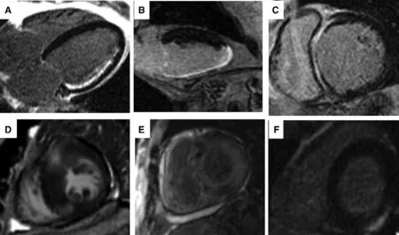
Figure 1 Various Patterns of Scar by Late Gadolinium Enhancement with Magnetic Resonance Imaging are Shown:(A) Subendocardial infarct consistent with coronary artery disease, (B) Transmural infarct consistent with coronary artery disease, (C) Mid-wall or mid-myocardial scar in a patient with dilated cardiomyopathy, (D) Right ventricular septal insertion scar pronounced in a region of hypertrophy consistent with hypertrophic obstructive cardiomyopathy, (E) Diffuse pattern with difficulty nulling in cardiac amyloidosis, and (F) epicardial late gadolinium enhancement in a patient with cardiac sarcoidosis.
Integrating MRI in the EP Laboratory
The great potential of integrating noninvasive scar data into EAVM would be to limit precise point mapping to only critical regions of substrate. Various methods to merge scar data into EAVM systems have been described [13-21]. It is possible to merge MRI and voltage datasets with vendor-integrated segmentation tools during the procedure. In one common approach, the endocardial volume on MRI can be registered as a shell. Then with the same dataset, the endocardial border of scar for each MRI short-axis image can be traced manually or in a semiautomated fashion with a signal intensity of two or three standard deviations above the threshold to de fine scar. A cutting tool can be used to extract the planimetered LGE regions to create a full-thickness slab of scar.This can be fused with an endocardial shell to create an endocardial map of scar. Then this LGE map can be fused with the electroanatomic voltage map through a landmark registration process [15]. Landmarks might include the left ventricular apex, left main coronary artery, aortic valve, or mitral annulus[18, 19]. Although LGE maps commonly display the presence or absence of endocardial scar, advanced maps based on signal intensity and transmurality, as shown in Figure 2, have also been described.
In some cases, the most challenging step in this integration process may be obtaining the preprocedure MRI image. In patients with conventional implantable cardiac de fibrillators, MRI is generally contraindicated. There is a concern for damaging the device, inadvertent harm to the patient, and lead-related artifacts. Relying on the growing body of experience of MRI being safely performed in patients with implanted devices, some groups have performed preablation MRI in patients with de fibrillators. In addition, it is likely that the emerging generation of MRI-conditional de fibrillator systems will potentially resolve the issue of MRI safety. In the meantime, integration of CT late contrast enhancement or PET-CT datasets may provide alternatives [20, 21].

Figure 2 An Example of Preprocedural Processing Magnetic Resonance Imaging for Integration with the Electroanatomic Mapping System.Endocardial and epicardial ventricular contours are identi fied with use of the short-axis contrast-enhanced magnetic resonance imaging slices (A), De fining intensity and transmurality of scar (yellow, gray zone; red, core infarction) (B). This can be used to generate color-coded endocardial (C and E) and epicardial (D and F) surface meshes. (C) and (D) show the percentage of transmurality and (E) and (F) show the same meshes coded for signal intensity. Higher signals suggest denser scar. Reproduced with permission from Winjmaalen et al. [16]. Copyright © 2011, Oxford University Press.
Correlating MRI and Voltage Data
Integration studies of MRI and voltage data have yielded insights into how voltage mapping findings relate to LGE abnormalities. Sites with low voltage correlate with the presence of LGE [9, 13, 14]. In one study of postinfarction VT ablation, Desjardins et al.[14] reported that sites with a bipolar voltage of 1.0 mV or less demonstrate 89% sensitivity and 84% specificity in detection of scar as de fined by LGE. In addition, a threshold of 1.3 mV seemed to yield the best correlation between infarct size assessment by EAVM and LGE. Furthermore, all identifiable isthmus sites were located in areas of LGE.More than 70% of those isthmus sites were located in the core zone of the infarct (where the MRI signal intensity is three or more standard deviations greater than the reference), whereas the remaining sites localized to the peripheral gray zone (where LGE signal intensity was less). Transmurality was associated with lower voltages in general but had a wide range of overlap with subendocardial infarcts.
Despite the relative correlation, considerable mismatch has been reported. Codreanu et al. [13]reported that with a 1.5-mV bipolar cutoff, there was a 20% mismatch in infarct size measurements by EAVM and LGE. specific regions of mismatch were in fluenced by the choice of transseptal or transaortic ablative approaches. Another study reported good correlation overall but with a tendency to underestimate infarct size by EAVM in inferior infarcts [16].
These findings suggest that EAVM by amplitude alone only roughly approximates scar. Abnormal electrogram characteristics besides low voltage,including prolonged duration, fragmentation or fractionation, sharp or “spiky” potentials, and isolated or late potentials, further correspond with the presence of LGE [13, 14, 22]. Desjardins et al. [14]reported that 89% of patients with fragmented electrograms and 95% of patients with isolated, late potentials had associated LGE.
In addition, regions of epicardial and intramural scar pose special problems from a voltage mapping perspective. These regions of interest may not be readily identi fied endocardially by standard voltage thresholds. One study reported that when epicardial regions of LGE were mapped from the endocardium, higher bipolar voltages (1.52±1.41 mV)were seen compared with the bipolar voltages for endocardial regions of LGE (0.94±1.07 mV) [14].Although epicardial mapping, when feasible, is bene ficial in these situations, intramural or midmyocardial scars represent blind spots that can go undetected by both epicardial and endocardial approaches. In a population of patients with nonischemic cardiomyopathies of various causes, Bogun et al.[9] found that endocardial voltage mapping failed to detect the presence of mid-myocardial scar that was seen by LGE. Furthermore, the presence of isolated mid-myocardial LGE was associated with a much higher rate of procedural failure.
MRI Insights into Infarct Architecture
In addition to scar location, scar heterogeneity is thought to be conducive to electrical dispersion and areas of slow conduction that are a substrate for arrhythmia. Within the core zone of an infarct,MRI identi fies heterogeneous areas that do not demonstrate maximal contrast enhancement. These gray zones correspond histologically to surviving muscle bundles within the scar tissue of an infarct,and it has been posited that they are more likely to contain critical conducting channels than regions of homogeneous scar [22-24]. In a study of heart failure patients by Lin et al. [25], the presence of these conductive channels, corridors of heterogeneous scar with relatively higher signal intensity within dense scar, was associated with a higher rate of ventricular fibrillation/VT attacks and mortality. In one integration study where LGE signal intensity maps were used to identify “channels” of heterogeneous tissue amid dense scar, these foci of heterogeneity were demonstrated to be critically related to clinical VT on the basis of their electrophysiological characteristics in 83% of patients [26].
Similarly, in a study of VT patients, critical isthmus sites, electrically de fined by excellent pace map profiles, evidence of concealed entrainment, or termination during ablation, tended to cluster in regions with near transmurality (more than 75% wall thickness)or at the transition between core and border zone regions, suggestive of heterogeneity. Sites ful filling both criteria contained all isthmus sites de fined by concealed entrainment, 77% of VT termination sites,and 56% identi fied by pace mapping [27].
MRI and Procedural Planning
Although real-time integration of MRI scar maps in the EP laboratory is the ideal application of this modality, MRI has demonstrated additional utility in the areas of procedural planning and the assessment of unsuccessful ablations. In a study of ablative strategies for ischemic VT, patients were divided on the basis of whether transmural or subendocardial LGE was seen on MRI. Acosta et al.[23] compared endocardial-only ablation in patients with subendocardial scar, endocardial-only ablation in patients with transmural scar, and an endocardial-epicardial approach in patients with transmural scar. Patients with transmural scar who did not have epicardial ablation had a signi ficantly higher rate of VT recurrence (41%) compared with the transmural scar group in which the endocardial-epicardial approach was used (12%). The ablative efficacy in the transmural scar group in which the endocardialepicardial approach was used was very similar to the efficacy in the subendocardial group that underwent endocardial ablation alone. The presence of epicardial or intramural scar, rather than transmurality, was the criterion used in another study of patients undergoing repeated VT ablation to plan an ablation strategy either with or without epicardial access. Njeim et al. [28] were able to maintain the strategy in 95% of patients. They concluded that MRI-guided decision making was preferable to an empiric epicardial approach in repeated ablations.
MRI can also be used to con firm effective and properly localized ablative lesion formation. Ilg et al. [29] assessed radiofrequency lesion formation in 35 patients before ablation and at more than 1 year after ablation of VT orpremature ventricular contractions, in nine of whom previous ablations had failed. With 83% reported ablation efficacy in the overall group, new scar corresponding to ablative lesions was seen in 71% of the total group. In this group, unsuccessful ablations were more common when papillary muscles were targeted, with ablation failure in one third of papillary VT cases.In these cases, MRI demonstrated LGE in the adjacent endocardium rather than on the papillary itself.This suggests poor tissue contact during attempts at papillary muscle ablation. In an additional third of unsuccessful ablations, where an initial endocardial approach failed, an epicardial site was the successful ablation target in a repeated procedure. The absence of scar, as was noted more often in right ventricular out flow tract sites, did not necessarily predict an unsuccessful ablation. Not surprisingly,larger regions of LGE indicated more complex ablative substrate.
MRI in Risk Strati fication
Beyond VT ablation, MRI has an even greater potential in the risk strati fication for SCD. The presence of LGE, its extent, and its heterogeneity have all demonstrated correlations with cardiac outcomes[30-33]. In a study of patients with a left ventricular ejection fraction greater than 35% and nonsustained VT, Dawson et al. [31] reported that the presence of scar was associated with more than a threefold increased risk of the combined end point of SCD,sustained VT, and de fibrillator firing. A meta-analysis in 2747 nonischemic subjects reported that the risk of major adverse cardiac events was threefold higher when scar was present, and in 1367 of these patients, the risk of arrhythmic events was fivefold higher [32]. Additional studies suggest that the extent of scar is predictive of adverse events [29].In combined cohorts of ischemic and nonischemic patients, it seems a critical volume of scar may portend risk, with values in the range of 5% or greater of left ventricular mass suggested as possible cutoffs [33].
Scar heterogeneity, in addition to its role in reentrant VT, likely portends greater risk of malignant arrhythmias. More extensive tissue heterogeneity has been found to correlate with increased inducibility by programmed EP study [33]. In addition,in a study of patients with ischemic cardiomyopathy who underwent MRI and subsequent de fibrillator implantation, the extent of the infarct gray zone was a stronger predictor of spontaneous ventricular arrhythmias than were total or core infarct sizes[34]. It is important to note, though, that in ischemic heart disease, the data are mixed as to whether the extent of total scar, core infarct, or peri-infarct is most predictive of adverse cardiovascular events.
The incremental risk associated with LGE in the setting of left ventricular systolic dysfunction is a critical issue in risk strati fication. Klem et al. [35]attempted to answer this question by analyzing their cohort of patients with a range of left ventricular ejection fractions. Their study showed that patients with a left ventricular ejection fraction greater than 30% and signi ficant scar (>5%) had higher risk of death or implantable cardiac de fibrillator discharge than those with minimal scar (hazard ratio 6.3, 95%con fidence interval 1.4-28), but that this risk was similar to that of the group with a left ventricular ejection fraction of 30% or less. Among patients with a left ventricular ejection fraction of 30% or less, those with scar demonstrated greater risk than those without scar (hazard ratio 3.9; 95% con fi-dence interval 1.2-13.1). Those with a left ventricular ejection fraction of 30% or less and minimal scarring had risk similar to that of patients with a left ventricular ejection fraction greater than 30%.These strategies need to be evaluated prospectively in larger cohorts but certainly suggest an important role for MRI in future SCD risk assessments.
Conclusion and Take-Home Messages
1. There is an emerging role for integrating noninvasive scar assessment, particularly MRI,in the treatment of patients with ventricular arrhythmia.
2. LGE with MRI allows the electrophysiologist to begin a procedure with an understanding of the topography and extent of myocardial scar.
3. With the use of MRI integration techniques, the precise nature of EAVM can be focused on critical regions of interest, which can save time and effort.
4. An epicardial or intramural location of scar can meaningfully change the procedural approach.5. Postprocedure MRI may help the next procedure determine whether ablative lesions were appropriately targeted.
6. The location, extent, and heterogeneity of LGE seem to add to our current rudimentary methods for SCD assessment.
Conflict of Interest
The author declares no Conflict of interest.
REFERENCES
1. Baroldi G, Silver MD, DeMaria R,Parodi O, Pellegrini A. Lipomatous metaplasia in left ventricular scar.Can J Cardiol 1997;13(1):65-71.
2. Baumgartner H, Porenta G, Lau YK, Wutte M, Klaar U, Mehrabi M, et al. Assessment of myocardial viability by dobutamine echocardiography, positron emission tomography and thallium-201 SPECT:correlation with histopathology in explanted hearts. J Am Coll Cardiol 1998;32(6):1701.
3. Iskandrian AS, Hakki A, Kane SA, Goel IP, Mundth ED, Hakki A, et al. Rest and redistribution thallium-201 myocardial scintigraphy to predict improvement in left ventricular function after coronary arterial bypass grafting. Am J Cardiol 1983;51:1312-6.
4. Marin-Neto JA, Dilsizian V,Arrighi JA, Perrone-Filardi P,Bacharach SL, Bonow RO. Thallium scintigraphy compared with 18F- fluorodeoxyglucose positron emission tomography for assessing myocardial viability in patients with moderate versus severe left ventricular dysfunction. Am J Cardiol 1998;82(9):1001.
5. Wang L, Yan C, Zhao S, Fang W. Comparison of99mTc-MIBI SPECT/18F-FDG PET imaging and cardiac magnetic resonance imaging in patients with idiopathic dilated cardiomyopathy: assessment of cardiac function and myocardial injury. Clin Nucl Med 2012;37(12):1163-9.
6. Kim RJ, Fieno DS, Parrish TB,Harris K, Chen EL, Simonetti O,et al. Relationship of MRI delayed contrast enhancement to irreversible injury, infarct age, and contractile function. Circulation 1999;100:1992-2002.
7. Kim RJ, Wu E, Rafael A, Chen E-L, Parker MA, Simonetti O, et al.The use of contrast-enhanced magnetic resonance imaging to identify reversible myocardial dysfunction.N Engl J Med 2000;343:1445-53.8. McCrohon JA, Moon JC, Prasad SK, McKenna WJ, Lorenz CH,Coats AJ, et al. Differentiation of heart failure related to dilated cardiomyopathy and coronary artery disease using gadolinium-enhanced cardiovascular magnetic resonance.Circulation 2003;108:54-9.
9. Bogun FM, Desjardins B, Good E,Gupta S, Crawford T, Oral H,et al. Delayed-enhanced magnetic resonance imaging in nonischemic cardiomyopathy: utility for identifying the ventricular arrhythmia substrate. J Am Coll Cardiol 2009;53:1138-45.
10. Cannavale A, Ordovás KG, Higgins CB. Magnetic resonance imaging of hypertrophic cardiomyopathy. J Thorac Imaging 2013;28(1):W12-8.
11. Wigle ED. Cardiomyopathy: the diagnosis of hypertrophic cardiomyopathy. Heart 2001;86:709-14.
12. Hsia HH, Lin D, Sauer WH, Callans DJ, Marchlinski FE. Anatomic characterization of endocardial substrate for hemodynamically stable reentrant ventricular tachycardia: identi fication of endocardial conducting channels. Heart Rhythm 2006;3:503-12.
13. Condreanu A, Odille F, Aliot E,Marie PY, Magnin-Poull I, Andronache M, et al. Electroanatomic characterization of post-infarct scars comparison with 3-dimensional myocardial scar reconstruction based on magnetic resonance imaging. J Am Coll Cardiol 2008;52(10):839-42.
14. Desjardins B, Crawford T, Good E, Oral H, Chugh A, Pelosi F, et al.Infarct architecture and characteristics on delayed enhanced magnetic resonance imaging and electroanatomic mapping inpatients with postinfarction ventricular arrhythmia.Heart Rhythm 2009;6(5):644-51.
15. Gupta S, Desjardin B, Baman T,Baman T, Ilg K, Good E, et al.Delayed-enhanced MR scar imaging and intraprocedural registration into an electroanatomic mapping system in post-infarction patients.JACC Cardiovascular Imaging 2012;5(2):207-10.
16. Wijnmaalen PA, van der Geest RJ, van Huls van Taxis CFB, Siebelink H-MJ, Kroft LJM, Bax JJ,et al. Head-to-head comparison of contrast-enhanced magnetic resonance imaging and electroanatomical voltage mapping to assess post-infarct scar characteristics in patients with ventricular tachycardias: real-time image integration and reversed registration. Eur Heart J 2011;32:104-14.
17. Arenal A, del Castillo S, Gonzalez-Torrecilla E, Atienza F, Ortiz M, Jimenez J, et al. Tachycardiarelated channel in scar tissue in patients with sustained monomorphic ventricular tachycardia: in fluences of voltage scar de finition.Circulation 2004;110:2568-74.
18. Tao Q, Milles J, Van Huls Van Taxis C, Lamb HJ, Reiber JH, Zeppenfeld K, et al. Toward magnetic resonance-guided electroanatomical voltage mapping for catheter ablation of scar-related ventricular tachycardia: a comparison of registration methods. J Cardiovasc Electrophysiol 2012;23(1):74-80.
19. Pandozi C, Dottori S, Lavalle S,Ficili S, Galeazzi M, Russo M,et al. Integration of MR images with electroanatomical maps: feasibility and utility in guiding left ventricular substrate mapping. J Interv Card Electr 2010;29(3):157-66.
20. Truong QA, Thai WE, Wai B,Cordaro K, Cheng T, Beaudoin J, et al. Myocardial scar imaging by standard single-energy and dual-energy late enhancement CT: comparison with pathology and electroanatomic map in an experimental chronic infarct porcine model. J Cardiovasc Comput Tomogr 2015;9(4):313-20.
21. Tian J, Smith MF, Chinnadurai P,Dilsizian V, Turgeman A, Abbo A,et al. Clinical application of PET/CT fusion imaging for three-dimensional myocardial scar and left ventricular anatomy during ventricular tachycardia ablation. J Cardiovasc Electrophysiol 2009;20(6):567-604.
22. Ashikaga H, Sasano T, Dong J,Muz Zviman M, Evers R, Hopenfeld B, et al. Magnetic resonancebased anatomical analysis of scarrelated ventricular tachycardia:implications for catheter ablation.Circ Res 2007;101(9):939-47.
23. Acosta J, Fernández-Armenta J,Penela D, Andreu D, Borras R, Vassanelli F, et al. Infarct transmurality as a criterion for first-line endo-epicardial substrate-guided ventricular tachycardia ablation in ischemic cardiomyopathy. Heart Rhythm.2016;13(1):85-95.
24. Perin EC, Silva GV, Sarmento-Leite R, Sousa AL, Howell M, Muthupillai R, et al. Assessing myocardial viability and infarct transmurality with left ventricular electromechanical mapping in patients with stable coronary artery disease: validation by delayed-enhancement magnetic resonance imaging. Circulation 2002;106:957-61.
25. Lin LY, Su MY, Chen JJ, Lai LP,Hwang JJ, Tseng CD, et al. Conductive channels identi fied with contrast-enhanced MR imaging predict ventricular tachycardia in systolic heart failure. JACC Cardiovasc Imaging 2013;6(11):1152-9.
26. Perez-David E, Arenal A, Rubio-Guivernau JL, del Castillo R, Atea L, Arbelo E, et al. Noninvasive identi fication of ventricular tachycardia-related conducting channels using contrast-enhanced magnetic resonance imaging in patients with chronic myocardial infarction: comparison of signal intensity scar mapping and endocardial voltage mapping. J Am Coll Cardiol 2011;57(2):184-94.
27. Piers SR, Tao Q, de Riva Silva M,Siebelink HM, Schalij MJ, van der Geest RJ, et al. CMR based identi fication of critical isthmus sites of ischemic and nonischemic VT. JACC Cardiovasc Imaging 2014;7(8):774-84.
28. Njeim M, Yokokawa M, Frank L,Crawford T, Good E, Morady F,et al. Value of cardiac magnetic resonance imaging in patients with failed ablation procedures for ventricular tachycardia. J Cardiovasc Electrophysiol 2015. doi:10.1111/jce.12848. [Epub ahead of print].
29. Ilg K, Baman T, Gupta SK, Swanson S, Good E, Chugh A, et al.Assessment of radiofrequency ablation lesions by CMR imaging after ablation of idiopathic ventricular arrhythmias. J Am Coll Cardiol Imaging 2010;2:278-85.
30. Scott PA, Rosengarten JA, Curzen NP, Morgan JM. Late gadolinium enhancement cardiac magnetic resonance imaging for the prediction of ventricular tachyarrhythmic events: a meta-analysis. Eur J Heart Fail 2013;15(9):1019-27.
31. Dawson DK, Hawlisch K, Prescott G, Roussin I, Di Pietro E, Deac M,et al. Prognostic role of CMR in patients presenting with ventricular arrhythmias. JACC Cardiovasc Imaging 201;6(3):335-44.
32. Kim EK, Chattranukulchai P, Klem I. Cardiac magnetic resonance scar imaging for sudden cardiac death risk strati fication in patients with non-ischemic cardiomyopathy.Korean J Radiol 2015;16(4):683-95.
33. Schmidt A, Azevedo CF, Cheng A,Gupta SN, Bluemke DA, Foo TK,et al. Infarct tissue heterogeneity by MRI identi fies enhanced cardiac arrhythmia susceptibility in patients with LV dysfunction. Circulation 2007;115(15):2006-14.
34. Roes SD, Borleffs CJ, van der Geest RJ, Westenberg JJ, Marsan NA, Kaandorp TA et al. Infarct tissue heterogeneity assessed with contrast-enhanced MRI predicts spontaneous ventricular arrhythmia in patients with ischemic cardiomyopathy and implantable cardioverter-de fibrillator. Circ Cardiovasc Imaging 2009;2(3):183-90.
35. Klem I, Weinsaft JW, Bahnson TD,Hegland D, Kim HW, Hayes B,et al. Assessment of myocardial scarring improves risk strati fi-cation in patients evaluated for cardiac de fibrillator implantation.J Am Coll Cardiol 2012;60(5):408-20.
 Cardiovascular Innovations and Applications2016年1期
- Cardiovascular Innovations and Applications的其它文章
- Implantable Cardiac De fibrillators: Who Needs Them and Who Does Not?
- The Subcutaneous Implantable Cardioverter-De fibrillator: A Practical Review and Real-World Use and Application
- Syncope and Early Repolarization: A Benign or Dangerous ECG Finding?
- Stroke Prevention in Atrial Fibrillation:Current Strategies and Recommendations
- Principles of Arrhythmia Management During Pregnancy
- Current Management of Ventricular Tachycardia:Approaches and Timing