IL-6对Raji和OCI-LY8细胞生长影响的分子机制**?
杨文秀,李品浩,陈 琴,裴媛媛
(贵州医科大学病理学教研室,贵州贵阳 550004)
IL-6对Raji和OCI-LY8细胞生长影响的分子机制**?
杨文秀,李品浩,陈琴,裴媛媛
(贵州医科大学病理学教研室,贵州贵阳550004)
[摘要]目的:观察白介素-6(IL-6)对伯基特淋巴瘤(BL) Raji细胞和弥漫大B细胞淋巴瘤(DLBCL) OCILY8细胞生长的影响,探讨信号转导子与转录活化-3(STAT3)及基质金属蛋白酶组织抑制因子-1(TIMP-1)分子在BL和DLBCL中发生发展的作用及其相互关系。方法:用不同浓度的IL-6(0、50、100、150及200 μg/L)分别培养Raji细胞和OCI-LY8细胞24 h、48 h及72 h(分别为对照组和不同浓度IL-6试验组),用MTT法检查2株细胞生长情况,用RT-qPCR检测培养48 h时2株细胞STAT3、TIMP-1的mRNA表达,用Western blot检测100 μg/L IL-6培养48 h时Raji细胞的STAT3、p-STAT3及TIMP-1蛋白的表达,流式细胞技术检测100 μg/L IL-6培养48 h 时2株细胞的细胞周期变化。结果:与相应的对照组比较,不同浓度IL-6试验组2株细胞在A490nm处OD值明显降低,呈现药物浓度依赖关系(P<0.01) ; 2株细胞内的STAT3,TIMP-1的mRNA表达明显升高(P<0.05),呈现出药物浓度依赖关系(P<0.05),2株细胞内TIMP-1与STAT3的mRNA表达呈正相关关系(r =0.982,P = 0.018) ; IL-6组Raji细胞中p-STAT3、STAT3和TIMP-1蛋白表达增高; IL-6组Raji和OCI-LY8细胞的G1期细胞都明显减少、S期细胞明显增多(P<0.05),G2-M期的Raji细胞无明显变化而OCI-LY8细胞则明显增多(P = 0.037)。结论: IL-6对Raji细胞和OCI-ly8细胞生长有明显作用,STAT3活化及其下游靶基因TIMP-1的上调表达可能是IL-6影响2株细胞生长的重要分子机制。
[关键词]淋巴瘤,B细胞;伯基特淋巴瘤;细胞培养;信号转导子与转录活化子-3;基质金属蛋白酶组织抑制因子-1;白细胞介素-6
网络出版时间: 2016-02-23网络出版地址: http: / /www.cnki.net/kcms/detail/52.5012.R.20160223.1909.022.html
信号转导子与转录活化子3(single transducer and transcription activater-3,STAT3)是STATS家族的重要成员,多项临床研究发现,肝癌、乳腺癌、肺癌、胃癌等肿瘤中STAT3活性均发生高频率的异常活化,且活化程度与患者的预后呈显著负相关[1-4]。STAT3是EGFR、IL-6/JAK、Src等多个致癌性酪氨酸激酶信号通道的汇聚焦点[5],阻断STAT3传导通路可以阻断多种肿瘤发生的机制。近年来,以STAT3为靶点的肿瘤治疗研究逐渐成为肿瘤基因治疗研究的热点。有文献报道,伯基特淋巴瘤(Burkitt lymphoma,BL)和弥漫大B细胞淋巴瘤(diffuse large B cell lymphoma,DLBCL)中存在STAT3分子的异常,但其活化对BL和DLBCL影响的分子机制尚未完全清楚[6-7]。本研究观察了STAT3激动剂白介素-6(interleukin-6,IL-6)对BLRaji细胞和DLBCL-OCI-LY8细胞生长的影响,并进一步探讨了STAT3和基质金属蛋白酶组织抑制因子-1 (tissue inhibitors of metalloproteinase-1,TIMP-1)的活化是否是药物作用的分子机制,为临床治疗BL和DLBCL寻找可能的基因靶点提供实验依据。
1 材料和方法
1.1实验材料
BL-Raji细胞株购自中国科学院上海细胞库,DLBCL-OCI-LY8细胞株由复旦大学肿瘤医院周晓燕教授惠赠。细胞培养基RPMI1640购自Hyclone公司,总RNA试剂盒购自上海碧云天公司,RTPCR试剂盒购自Invitrogen公司,抗STAT3(单克隆抗体)、抗TIMP-1和抗β-actin抗体购自Santa Cruz公司,抗p-STAT3抗体购自CST公司,IL-6购自Pepro TECH公司,PCR引物由上海生工合成。
1.2方法
1.2.1细胞培养和分组分别将Raji细胞和OCI-LY8细胞置于RPMI1640培养液(含10%胎牛血清和1%青霉素/链霉素)及混合培养基(含 10%胎牛血清、90% IMDM和1%双抗)中,在37℃、5% CO2饱和湿度的恒温培养箱中培养,每2~3 d换液1次,待细胞计数>1×10⁶/mL时,按1∶2~1∶3传代培养。收集细胞并调节浓度为2× 105/mL,2株细胞分别加入不同浓度的IL-6(0、50、100、150及200 μg/L),分别为对照组和不同IL-6浓度试验组,继续培养24、48及72 h。
1.2.2检测细胞活力用MTT法对细胞生长活力进行检测。分别于培养24、48及72 h后收集对照组和不同IL-6浓度试验组的Raji和OCI-ly8细胞,再于无血清培养基中培养24 h,同步化后于96孔板布板。每孔加入0.5% MTT 20 μL,继续培养4 h,加入三联溶剂(SDS 10 g +异丁醇5 mL + 10 mol/L HCl 0.1 mL,用双蒸水溶解配成100 mL溶液) 150 μL,置摇床上低速振荡10 min。镜下观察结晶物充分溶解后在酶联免疫检测仪490 nm处测量各孔的吸光值A。每组5个复孔,实验重复3次。
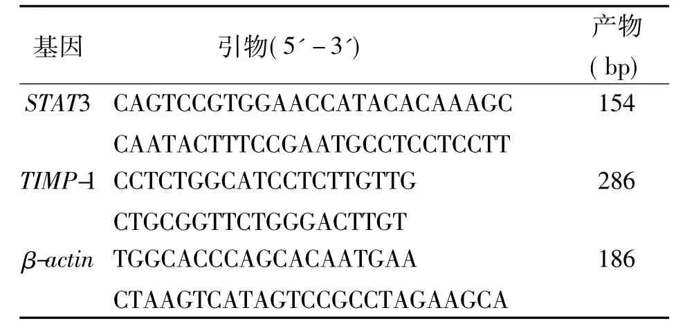
1.2.3检测STAT3和TIMP-1的mRNA按试剂盒说明提取细胞mRNA,检测mRNA 260/280光密度比值。RT-qPCR扩增检测STAT3和TIMP-1的mRNA相对表达量。PCR循环参数: 95℃10 min、90℃15 s、60℃1 min,40个循环。结果用2-ΔΔCT表示,以β-actin作为内参照。mRNA相对表达量2-ΔΔCt= 2-[实验组(Ct目的基因-Ct管家基因)-对照(Ct目的基因-Ct管家基因)]。PCR引物序列和产物见表1。

表1 RT-qPCR引物序列及相应产物Tab.1 The sequences of the primers for RT-qPCR and corresponding products
1.2.4 STAT3、p-STAT3和TIMP-1蛋白表达IL-6的最佳作用浓度取100 μg/L,收集该浓度下培养48 h足量的Raji细胞,用PBS洗涤3次后,加入一定量的细胞裂解液及蛋白酶抑制剂,冰上不时振荡充分裂解30 min,12 000 r/m离心5 min后取上清液。蛋白样品采用BCA法进行定量后,进行PAGE凝胶电泳,每孔上样30 mg,10%聚丙烯酰胺凝胶电泳后转至PVDF膜上,用5% BSA封闭p-STAT3、5%脱脂牛奶封闭其他目的蛋白后,分别加一抗孵育过夜(稀释度:抗STAT3、抗p-STAT3、抗TIMP-1均为1∶500,抗β-actin为1∶1 000),二抗稀释比例为1∶5 000,洗膜,加辣根过氧化酶偶联的二抗孵育1.5 h、再洗膜。加ECL发光液显色,暗室曝光,得到STAT3、p-STAT3和TIMP-1的条带胶片,胶片扫描后用Gelpro32软件分析。
1.2.5检测细胞周期分别将Raji细胞和OCILY8细胞以70 000 mL接种于6孔板中,加入最佳作用浓度(100 μg/L)的IL-6培养48 h后,收集Raji细胞和OCI-LY8细胞,用流式细胞仪检测细胞周期。
1.3统计学方法
使用SPSS 20.0统计软件包。STAT3,TIMP-1 的mRNA及蛋白表达水平采用两独立样本t检验,药物作用的多组间比较采用单因素方差分析,多组间均数的两两比较采用SNK q检验,多组均数与一个对照样本均数比较采用Dunnett t检验。各药物浓度组间两分子表达之间的关系及分子表与药物浓度的相关性关系采用Pearson相关性分析,以P <0.05为差异有统计学意义。
2 结果
2.1细胞活力
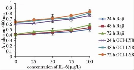
与相应的对照组比较,Raji细胞及OCI-LY8细胞490 nm处吸光度值在加入IL-6培养后显著增高(P<0.05)。Raji和OCI-LY8细胞吸光度值与IL-6呈现出药物浓度依赖关系,r和P依次为0.966、0.008及0.959、0.010。见图1。
2.2 STAT3 mRNA和TIMP-1mRNA
各组细胞mRNA的A260/A280光密度比值为1.8~2.0,符合后续试验要求。RT-qPCR结果显示,与相应的对照组比较,2株细胞的试验组STAT3,TIMP-1的mRNA表达明显升高,不同IL-6浓度试验组Raji细胞和OCI-LY8细胞之间STAT3 和TIMP-1的mRNA表达均有显著差异(P<0.05),且2种基因mRNA表达都呈现出药物浓度依赖关系(Raji细胞IL6与STAT3 r = 0.979,P = 0.021,IL-6与TIMP-1 r = 0.981,P = 0.019; OCILY8细胞IL6与STAT3 r = 0.996,P = 0.004,IL-6 与TIMP-1 r =0.973,P =0.027)。2株细胞TIMP-1与STAT3的mRNA表达呈正相关(r = 0.961、0.998,P =0.039、0.002)。见表2。
2.3 Raji细胞p-STAT3、STAT3及TIMP-1蛋白
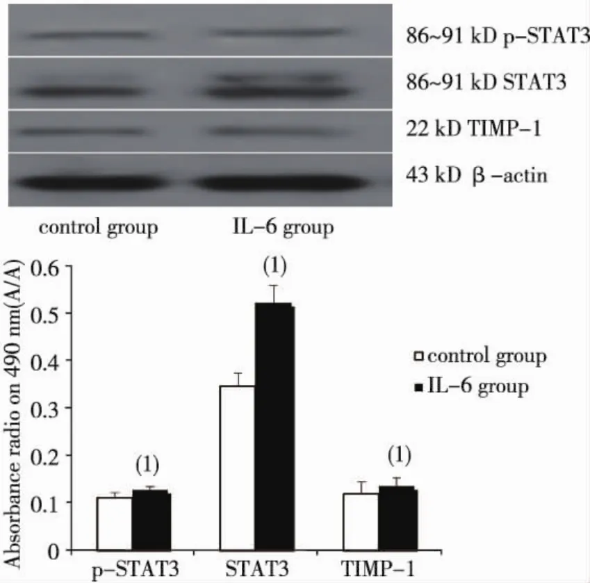
与对照组比较,100 μg/L IL-6培养48 h时的Raji细胞p-STAT3及STAT3蛋白升高,P分别为0.026、0.001,见图2。
2.4细胞周期
Raji细胞和OCI-LY8细胞G0/G1、S、G2-M各周期的细胞百分率差异有统计学意义(P =0.001、P =0.001、P =0.000)。与对照组相比,试验组Raji和OCI-LY8细胞的G1期细胞都明显减少(P = 0.031和P = 0.019),S期细胞明显增多(P = 0.038和P = 0.034),G2-M期的Raji细胞无明显变化(P = 0.63),G2-M期的OCI-LY8细胞则明显增多(P =0.037)。见表3。

表2 不同浓度IL-6培养Raji和OCI-LY8细胞48 h时STAT3 mRNA及TIMP-1 mRNA水平Tab.2 The mRNA expressions of STAT3 and TIMP-1 in the Raji and OCI-LY8 cells treated with IL-6 for 48 h
表3 100 μg/L IL-6培养Raji和OCI-LY8细胞48 h时的细胞周期变化(±s)Tab.3 Propotion of the cell cycle in OCI-LY8 and the Raji cells cultured with 100 μg/L IL-6

表3 100 μg/L IL-6培养Raji和OCI-LY8细胞48 h时的细胞周期变化(±s)Tab.3 Propotion of the cell cycle in OCI-LY8 and the Raji cells cultured with 100 μg/L IL-6
(1)与相应对照组比较,P<0.05
细胞 分组 细胞周期细胞(%) G1 S G2/M Raji Control 46.51±2.03 44.31±1.01 9.18±1 16.59±1.89 .90 IL-6 40.81±2.29(1)49.94±2.59(1)9.25±1.71 OCI-LY8 Control 55.74±2.55 30.42±2.27 13.88±0.40 IL-6 49.02±1.91(1)34.39±2.09(1)

图1 IL-6对Raji、OCI-LY8细胞生长的影响Fig.1 The effects of IL-6 at different concentrations for different times on the growth of the two kind of cells

图2 100 μg/L IL-6培养48 h时Raji细胞中STAT3、p-STAT3、TIMP-1蛋白表达Fig.2 The protein levels of STAT3,p-STAT3 and TIMP-1 in the Raji cells with 100 μg/L IL-6
3 讨论
有研究发现,JAK/STAT3信号转导通路持续激活可导致细胞异常增殖或恶性转化,该通路在侵袭性B细胞淋巴瘤侵袭及转移过程中发挥重要作用[8-10]。用该通路的抑制剂可抑制STAT3在侵袭性恶性淋巴瘤细胞系中的活性。TIMP-1是基质金属蛋白酶组织抑制因子(tissue inhibitors of metalloproteinases,TIMPs)家族中的一员。TIMPS是一个多基因家族编码蛋白基质金属蛋白酶(matrix metalloproteinase,MMPs)的内源性特异性抑制因子。关于TIMP-1的研究,一方面认为它具有抗有丝分裂的活性,能促进内皮细胞增生,能与基质胶原酶、基质分解素及明胶酶(主要是明胶酶B)形成可逆的复合物,通过抑制MMPs的活化及其对ECM的降解、抑制ECM中相关凋亡蛋白的释放,发挥调节细胞凋亡及促进肿瘤细胞生长的功能[11]。还有研究认为,TIMP-1作为一种转录抑制剂抑制MMPs基因的转录;另一方面TIMP-1位于JAK/STAT信号通路的下游,其表达受JAK/STAT信号通路调控。后者可通过上调TIMP-1的表达而抑制大鼠肾小球系膜细胞凋亡[12]。近年来,在人红白血病细胞研究中又发现TIMP-1和JAK/STAT存在双向调控作用[13]。在肝的纤维化研究中发现敲除STAT3基因之后,CCl4导致的肝纤维化中TIMP-1的高表达消失[14]。同时有研究发现,TIMP-1可以由淋巴瘤细胞自分泌或旁分泌产生,通过B细胞生长分化因子IL-10以及原癌基因bcl-xL来抑制细胞程序性死亡,从而延长正常扁桃体B细胞、Burkitt淋巴瘤细胞的寿命[15]。还有研究发现TIMP-1与非霍奇金淋巴瘤的临床分级相关。
IL-6是IL-6/JAK信号通路的激活剂[16-17],激活JAK后可促进STAT3的表达及磷酸化,以磷酸化二聚体的形式进入细胞核,与下游凋亡及细胞周期调控基因、基质金属蛋白酶等靶基因的启动子结合,调节肿瘤细胞的增殖凋亡和迁移等过程。本研究在培养的Raji和OCI-LY8细胞中分别加入不同浓度的IL-6后,发现细胞生长活力增强,呈现出药物浓度依赖关系。2株细胞G1期均体现出减少的趋势,S期细胞明显增多,处于G2-M的Raji细胞无明显改变,而OCI-ly8细胞明显增多。这表明JAK/STAT3信号通路对Raji和OCI-LY8细胞的生长可能有明显的影响。进一步探讨IL-6影响2株细胞生长的相关分子机制,本研究检测了细胞内STAT3和TIMP-1的mRNA及蛋白表达情况。Raji细胞和OCI-LY8细胞内STAT3基因的mRNA表达水平随着IL-6浓度的增加而增高,TIMP-1和STAT3的mRNA表达呈正相关关系。在IL-6作用48 h后Raji细胞内p-STAT3,STAT3和TIMP-1蛋白表达明显升高,三者变化趋势一致。提示IL-6作用下,TIMP-1蛋白的表达可能与STAT3的活化有关。
综上,IL6能显著促进淋巴瘤Raji及OCI-LY8细胞的生长,并促进细胞周期的运行。其作用可能与细胞内STAT3分子活化有关,细胞内TIMP-1的上调表达也可能是IL-6活化STAT3的结果。2种细胞内STAT3的活化及其引起的TIMP-1的表达上调可能是IL-6影响淋巴瘤细胞生长的重要机制,这可能是Burkitt和DLBCL淋巴瘤治疗的新思路。
4 参考文献
[1]Morikawa T,Baba Y,Yamauchi M,et al.STAT3 expression,molecular features,inflammation patterns,and prognosis in a database of 724 colorectal cancers[J].Clin Cancer Res,2011(6) : 1452-1462.
[2]Grivennikov S,Karin E,Terzic J,et al.IL-6 and STAT3 are required for survival of intestinal epithelial cells and development of colitis-associated cancer[J].Cancer Cell,2009(2) : 103-113.
[3]Berishaj M,Gao SP,Ahmed S,et al.STAT3 is tyrosinephosphorylated through the interleukin-6/glycoprotein 130/Janus kinase pathway in breast cancer[J].Breast Cancer Res,2007(3) : R32.
[4]Kim DY,Cha ST,Ahn DH,et al.STAT3 expression in gastric cancer indicates a poor prognosis[J].J Gastroenterol Hepatol,2009(4) : 646-651.
[5]侯嘉杰,孙倍成.STAT3:慢性炎症介导肿瘤发生和进展的关键节点[J].生物化学与生物物理进展,2014 (1) : 69-78.
[6]Soldini D,Montagna C,Schüffler P,et al.A new diagnostic algorithm for Burkitt and diffuse large B-cell lymphomas based on the expression of CSE1L and STAT3 and on MYC rearrangement predicts outcome[J].Ann Oncol,2013(1) : 193-201.
[7]Mizowaki T,Sasayama T,Tanaka K,et al.STAT3 activation is associated with cerebrospinal fluid interleukin-10 (IL-10) in primary central nervous system diffuse large B cell lymphoma[J].Journal of neuro-oncology,2015(2) : 165-74.
[8]Al Zaid Siddiquee K,Turkson J.STAT3 as a target for inducing apoptosis in solid and hematological tumors[J].Cell Res,2008(2) : 254-67.
[9]Siveen KS,Sikka S,Surana R,et al.Targeting the STAT3 signaling pathway in cancer: role of synthetic and natural inhibitors[J].Biochim Biophys Acta,2014(2) : 136-154.
[10]Lam LT,Wright G,Davis RE,et al.Cooperative signaling through the signal transducer and activator of transcription 3 and nuclear factor-κB pathways in subtypes of diffuse large B-cell lymphoma[J].Blood,2008 (7) : 3701-3713.
[11]Udayakumar TS,Stratton MS,Nagle RB,et al.Fibroblast growth factor-1 induced promatrilysin expression through the activation of extracellular-regulate kinases and STAT3[J].Neoplasia,2002(4) : 60-67.
[12]温文斌,林洪丽,吴泰华,等.JAK/STAT通路在金属蛋白酶1组织抑制剂抑制肾小球系膜细胞凋亡中的作用[J].中华肾脏病杂志,2008(1) : 24-29.
[13]Lambert E,Boudot C,Kadri Z,et al.Tissue inhibitor of metalloproteinases-1 signalling pathway leading to erythroid cell survival[J].Biochem J,2003 (3) : 767-774.
[14]Wang H,Lafdil F,Wang L,et al.Tissue inhibitor of metalloproteinase 1 (TIMP-1) deficiency exacerbates carbon tetrachloride-induced liver injury and fibrosis in mice: involvement of hepatocyte STAT3 in TIMP-1 production[J].Cell&bioscience,2011(1) : 14.
[15]樊嵘,金冶宁.TIMP-1在乳腺癌中的作用机制和临床意义[J].现代肿瘤医学,2009(1) : 154-158.
[16]Garbers C,Aparicio-Siegmund S,Rose-John S.The IL-6/gp130/STAT3 signaling axis: recent advances towards specific inhibition[J].Curr Opin Immunol,2015 (34C) : 75-82.
[17]Munoz J,Dhillon N,Janku F,et al.STAT3 inhibitors: finding a home in lymphoma and leukemia[J].Oncologist,2014(5) : 536-544.
(2015-12-20收稿,2015-12-31修回)
中文编辑:戚璐;英文编辑:赵毅
Molecular Mechanism of IL-6 Effect on the Growth of Two Lymphoma Cells
YANG Wenxiu,LI Pinhao,CHEN Qin,PEI Yuanyuan
(Department of Pathology,Guizhou Medical University,Guiyang 550004,Guizhou,China)
[Abstract]Objective: To observe the effects of IL-6 (interleukin-6) on the growth of Burkitt lymphoma (BL) cell line Raji and Diffuse large B cell lymphoma (DLBCL) cell line OCI-LY8,and to investigate their effect on cellular growth and the relationship between changes of STAT3 (single transducer and transcription activater-3) and TIMP-1 (tissue inhibitors of metalloproteinase-1) expression.
[Key words]lymphoma,B cell; Burkitt lymphoma; cell culture; single transducer and transcription activator-3; tissue inhibitors of metalloproteinase-1; interleukin-6
*[基金项目]国家自然科学基金(No.81160299) ;贵州省优秀人才省长基金(No.2011.125)
[中图分类号]R559; R34
[文献标识码]A
[文章编号]1000-2707(2016) 02-0125-05
Methods: Raji and OCI-LY8 cells were cultured by varied concentration of IL-6(0 g/L,50 g/L,100 g/L,150 g/L and 200 g/L) for 24 h,48 h and 72 h.Viability of 2 strains of cells was measured by MTT.The mRNA expressions of STAT3 and TIMP-1 of 2 strains of cells were detected by RT-PCR,and p-STAT3,STAT3 and TIMP-1 protein expression of Raji cell cultivated by 100 g/L IL-6 for 48 h were detected by Western blotting.Cell cycle was examined by flow cytometry.Results: Comparing with control group,OD value at 490 nm decreased the two strain of cells treated by varied concentration of IL-6.It was presented concentration-dependence relationship(P<0.01) ; mRNA expressions of STAT3 and TIMP-1 in both cells exhibited positive correlation (r = 0.982,P = 0.018) ; protein expression of p-STAT3,STAT3 and TIMP-1 of Raji cells in IL-6 groups increased.Cells were distinctly reduced at phase G1 and increased at phase S in two types of cell of IL-6(P<0.05).The Raji cell atphase G2-M showed no significant change while OCI-LY8 cells increased significantly(P = 0.037).Conclusions: IL-6 has an obvious effect on the growth of Raji and OCI-LY8 cell.The activation of STAT3 and up-regulated expression of TIMP-1 might be important molecular mechanism for promoting the viability of Burkitt and DLBCL cell.

