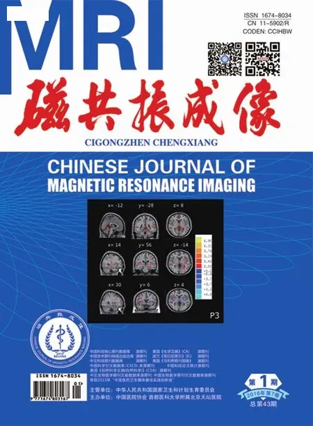
基于体素形态学测量技术对高原地区正常成人脑结构的研究
李超伟,鲍海华,孔德民,李伟霞,员强
基于体素形态学测量技术对高原地区正常成人脑结构的研究
李超伟,鲍海华*,孔德民,李伟霞,员强
[摘要]目的 基于体素形态学测量(VBM)技术,分析久居高原地区(>3000 m) 正常成人脑结构体积的变化。材料与方法 选取两组正常成人参与本次研究,其中包括高原组[男8例,女8例,平均年龄(21.81 ± 2.07)岁]和与之年龄、受教育年限相匹配的平原组[男7例、女13例,平均年龄(21.85 ± 1.90)岁],对每个被试行全脑扫描,获取3D-T1结构图像,利用VBM方法对全脑灰、白质图像进行统计学分析。结果 与平原组比较,高原组正常成人左侧后扣带回、颞上回灰质体积增加;右侧岛叶灰质体积减低;白质体积增加区域为左侧丘脑、右侧额上回、左侧豆状核、左侧枕叶。结论 利用VBM技术对MRI结构图像分析,能够客观显示高原地区相对平原地区正常成人脑部特定区域体积的变化,从而全面的评价高原长期低氧对脑结构的影响。
[关键词]脑;人体测量术;高原;磁共振成像
国家自然科学基金(编号:81060117);青海省科技厅国际合作项目(编号:2012-H-807)
作者单位:
青海大学附属医院影像中心,西宁810001
鲍海华,E-mail: baohelen2@sina.com
接受日期:2015-11-11
李超伟, 鲍海华, 孔德民, 等.基于体素形态学测量技术对高原地区正常成人脑结构的研究.磁共振成像, 2016, 7(1): 1–5.
Study of brain structure in nomal plateau area adult with voxel-based morphometry
LI Chao-wei, BAO Hai-hua*, KONG De-min, Li Wei-xia, YUAN Qiang
The Affiliated Hospital of Qinghai University, Medical Imaging Center, Xining 810001, China
*Correspondence to: Bao HH, E-mail: baohelen2@sina.com
Received 17 Oct 2015, Accepted 11 Nov 2015
ACKNOWLEDGMENTS National Natural Science Foundation of China(No.81060117).International cooperation project of science and Technology Department of Qinghai Province (No.2012-H-807).
Abstract Objective: The aim of the study was to investigate the brain structure volumes alterations in born and raised high altitude (HA) (>3000 m) normal adult by using voxel-based morphometry method (VBM).Materials and Methods: Two groups of adults participated in the study, including an HA group [8 males and 8 females, mean age=(21.81±2.07) years] and an age- and education-matched sea level (SL) group [7 males and 13 females, mean age=(21.85±1.90) years].3D-T1 structural images of all subjects who were underwent the whole brain scan were acquired.Then we used the VBM method to compare the whole brain GM and WM images differences between HA group and SL group.Results: HA acclimatization (vs.SL) showed increased gray matter volume in the left posterior cingulate, the left superior temporal gyrus,decreased GM volumes was found in the right insular lobe in highland group and we also found increased WM volumes in left thalamus, the right superior frontal gyrus, the lentiform nucleus, the left occipital lobe.Conclusions: The VBM method was applied to the analysis of the magnetic resonance structural images and it could objectively display the volume changes of specific brain areas in HA group and could get us a comprehensive evaluation of the impact of altitude hypoxia on brain structure.
Key words Brain; Anthropometry; Altitude; Magnetic resonance imaging
青藏高原号称“世界屋脊”,是我国面积最大,海拔最高,居住人口最多的高原地区。缺氧是高原环境影响人体的最关键的因素之一。近年来,有关高原低氧对脑部影响的文献报道比较多,例如对慢性高原病患者脑部的病理生理及神经影像学表现进展[1]做一综述;研究发现,慢性高原病患者灰质体积增加的脑区为右侧舌回、后扣带回、双侧海马及左侧颞下回[2];平原移居至青藏高原地区并适应生活2年的正常成人的不同脑区灰、白质结构发生变化[3];也有学者发现,长期生活在海拔2600~4200 m地区的正常成人下达平原地区1年的脑灰白质结构发生改变[4]。但是久居高原从未下达平原地区的正常成人的脑结构的变化尚未报道。
基于体素形态学测量(voxel-based morphometry, VBM)技术是一种全自动化、客观进行全脑形态分析的技术,能够定量和全面的评估大脑结构差异[5];此技术已经广泛地应用于研究多种疾病导致的脑形态学改变[6]。笔者应用优化的VBM技术,对高原及平原地区正常成人进行全脑结构的形态学测量,分析全脑结构变化。
1 材料与方法
1.1临床资料
1.1.1研究对象
搜集两组正常成人参与本次研究,并均为汉族人;高原组共计16例,来自两代以上均出生并长期居住在高原地区[居住海拔高度平均为(3628.88±295.72) m]的正常成人,其中男8 例,女8例,年龄18~24岁,平均(21.81±2.07)岁;受教育时间平均(11.69±0.48)年。平原组共计20例,其中男7例,女13例,平均年龄(21.85±1.90)岁,居住海拔高度平均为(391.72±373.24) m;受教育时间平均(13.50±3.56)年。两组的平均年龄和受教育时间差异无统计学意义(P值分别为0.96和0.59)。所有受检者在检查时均就读于青海大学医学院,到青海后1周内进行MRI检查,检查前均了解了检查内容和意义并签署知情同意书,并由青海省伦理委员会批准。
1.1.2纳入标准和排除标准
纳入标准:(1)常规头颅MRI扫描无其他脑实质病变;(2)患者无MRI检查的相关禁忌证;(3)临床各项检查确认无精神异常等;(4)均为右利手。排除标准:(1)有慢性高原病;(2)被确诊的脑神经失调;(3)过去有脑部损伤致意识丧失。
1.2MRI检查方法
应用3.0 T超导MR成像系统(PHILIPS),标准头颅8通道相控阵线圈完成所有扫描序列;研究对象首先进行常规头部MR成像获得T1WI和T2WI;同时采用超快速场回波(TFE)序列获取3D-T1结构像,TR 7.5 ms,TE 3.7 ms,层厚2 mm,层间隔–1 mm,矩阵256 × 256,激发角度7°,扫描全脑将连续获得176层矢状面图像;所有的扫描均由同一名资深影像科医师操作完成。
1.3数据处理及统计分析
将所有被试的原始数据导入个人电脑工作站,采用统计参数图(SPM8)的嵌套软件VBM8 toolbox进行数据处理;计算和处理图像矩阵在Matlab平台上运行;数据处理包括:(1)对头位偏差大的图像进行位置校正;(2)每个被试3D-T1图像分割成灰质、白质、脑脊液后,将所有被试者的MR图像都配准至模板图像,进行空间标准化处理;(3)采用半高全宽为8 mm的三维高斯核进行图像平滑,并对处理后的结果进行统计分析。采用两样本t检验进行比较(检验参数t>3.7459,P<0.05;校正),选取相邻像素(voxels)大于389个以上的组块(cluster)才视为有差异的脑区,将检验结果叠加到T1结构图的模板上,分析研究对象全脑结构体积的变化。

表1 高原组与平原组脑区相比灰质增加的统计分析结果Tab.1 The statistic analysis results of increased gray matter voxels in HA group contrasted with SL group
2 结果
VBM分析显示高原地区正常成年人与平原组相比:(1)高原组较平原组的左侧后扣带回、左侧颞上回灰质体积增加(表1,图1);(2)高原组较平原组的右侧岛叶灰质体积减低(表2,图1);(3)高原组较平原组的左侧丘脑、右侧额上回、左侧豆状核、左侧枕叶白质体积增加(表3,图2)。

图1 高原组与平原组相比灰质体积有差异的脑区的统计参数图。红色代表HA正常成人脑区相对于SL正常成人脑区灰质体积增加的区域,包括:左侧后扣带回;左侧颞上回;蓝色代表HA正常成人脑区相对于SL正常成人脑区灰质体积减少的区域:右侧岛叶 (统计阈值设定为P<0.05,cluster size>389,校正) 图2 高原组与平原组相比白质体积有差异脑区的统计参数图。红色代表HA正常成人脑区相对于SL正常成人脑区白质体积增加的区域,包括:左侧丘脑、右侧额上回、左侧豆状核、左侧枕叶 (统计阈值设置为P<0.05,cluster size>389,校正)Fig.1 The statistical parametric map showed difference between HA group and SL group in gray matter volume.Compared with SL group, red display the increased regions of GM volume in HA group, include left posterior cingulate, the left superior temporal gyrus and blue display the reduced regions in the right insular lobe.The statistical threshold is set to P<0.05, cluster size>389, corrected.Fig.2 The statistical parametric map showed difference between HA group and SL group in white matter volume.Compared with SL group, red display the increased regions of WM volume in HA group, include in left thalamus, the right superior frontal gyrus, the lentiform nucleus, the left occipita lobe.The statistical threshold is set to P<0.05, cluster size>389, corrected.

表 2 高原组与平原组脑区相比灰质减低的统计分析结果Tab.2 The statistic analysis results of decreased gray matter voxels in HA group contrasted with SL group

表 3 高原组与平原组脑区相比白质减低的统计分析结果Tab.3 The statistic analysis results of increased white matter voxels in HA group contrasted with SL group
3 讨论
一个世纪以来,从临床方面对脑缺氧后的生理学、组织解剖学、神经化学等进行了大量的宏观和微观研究;但从影像学的角度上报道较少。
研究表明高海拔环境(低温,低压,紫外线,寒冷,脱水)下的土著居民和移民在呼吸道和心血管方面产生了适应性的改变,这直接与氧气的运输有关[7-10]。大脑是人体的控制中心,通过其传入反馈,心血管和呼吸道系统的适应性改变作用于大脑,也可能引起相应脑结构的改变[11]。另一方面,中枢神经系统是高度氧化的,它不可避免的遭受含氧量低的压力。血红蛋白浓度以及动脉血氧饱和度的变化使大脑血流的氧输送发生改变,最后导致脑结构的积累性改变[12-14]。许多研究者对高原地区居民的研究主要注重于脑葡萄糖代谢率[13]和脑自身调节[15-16]。但到目前为止,高原地区的居民脑部结构的适应性改变仍然不清楚。
目前研究表明,高海拔适应与大脑结构的改变有关,包括某些区域皮层灰质体积和白质结构的改变;根据Zatorre等[17]的研究发现,正常成人灰质增加可能与以下几个方面有关:神经细胞的增加、胶质细胞再生、突触发生及血管生成。然而,灰质的减少可能与缺氧新陈代谢副产物和低氧环境下谷氨酸能神经细胞释放的谷氨酸盐增多有关[18]。本研究结果示,高海拔地区正常成人较平原组左侧后扣带回、颞上回灰质体积增加;前脑岛叶灰质体积减少;说明高原正常成人的此脑区对缺氧极为敏感。
后扣带回是默认网络中心节点之一,是情节处理和工作记忆的重要构成部分;颞上回是视听觉中枢的一部分。登山运动员[18-22]、生活在适度海拔高度的世居者[23]以及高海拔移民的后代[24-25]中有短期记忆,视觉结构,程序学习,工作记忆的减弱并且反应时间的增加情况,该区域灰质增加也可能解释上述机制,到目前为止为什么发生及怎样发生还不完全清楚。
岛叶皮层已被证明与心血管疾病控制有关[26-28],前脑岛在呼吸困难中起重要作用[29],呼吸困难常常发生在对高海拔低氧的适应过程中。最近,Paulus等[30]提出一种假设:前脑岛在高海拔环境中处理自身平衡稳定是必需的;有氧代谢能力与右侧前脑岛灰质有很强的相关性[31],高海拔地区居民在生长发育期间需氧容量降低[32-33],因而岛叶灰质体积减少。所以,笔者推测居民在适应于高海拔会有前脑岛部灰质的减少。
白质由神经纤维构成,位于大脑皮质与基底核之间。弥散张量成像是研究脑白质的方法之一,但本文章采用基于VBM方法测量脑白质体积的研究。本研究结果显示,白质体积增加区域为左侧丘脑、右侧额上回、左侧豆状核、左侧枕叶。丘脑与脑内许多结构有着丰富的纤维连接,并且是脑的中继站,起着过滤器的作用,通过排除多余或者无关的刺激来传递重要的或相关的信息。前额叶的主要功能是记忆、判断、分析、思考、操作,人类完成高级认知任务主要与前额叶有关。左侧豆状核与右侧运动和神经传导有关。枕叶为视觉皮质中枢,以上白质的改变可能与情感的调节、认知功能、地处高原环境、基因和生活环境等有关。
我们初步探讨了长期低氧对高原地区正常成人脑结构的影响,证明了高海拔适应性与脑部结构改变有关;但本研究样本量较小,没有设计认知功能测试,本研究中对结果的解释主要依靠其他学者的研究结果。今后, 笔者将扩大样本量,行脑认知功能测验等,为高原地区正常成人脑结构改变提供更多证据;增进我们对高原长期低氧改变脑结构的全面认识。
参考文献[References]
[1]Yang CX, Bao HH.Pathophysiology and neuroimaging development of brain alterations in chronic mountain sickness.Chin J Magn Reson Imaging, 2015, 6(2): 151-154.
杨丛珊, 鲍海华.慢性高原病脑部改变的病理生理及神经影像学进展.磁共振成像, 2015, 6(2): 151-154.
[2]Liu CX, Bao HH, Li WX, et al.Voxel-based morphometry MRI study of gray Matter’s alteration in patients with chronic mountain sickness.Chin J Magn Reson Imaging, 2014, 5(3): 211-215.
刘彩霞, 鲍海华, 李伟霞, 等.慢性高原病患者脑灰质变化的VBM-MRI研究.磁共振成像, 2014, 5(3): 211-215.
[3]Zhang J, Zhang H, Li J, et al.Adaptive modulation o f adult brain gray and white matter to high altitude: structural MRI studies.PLoS ONE, 2013, 8(7): e68621.
[4]Zhang J, Yan X, Shi J, et al.Structural modifications of the brain in acclimatization to high-altitude.PLoS One, 2010, 5(7): e11499.
[5]Ashburner J, Friston KJ.Voxel-based morphometry-the methods.Neuroimage, 2000, 11(6 Pt 1): 805-821.
[6]Zhang J, Zhang CZ, Zhang YT.Advanced clinical application of voxel based morphometery.Int J Med Radiol, 2010, 33(4): 314-316.
张敬, 张成周, 张云亭.基于体素的形态学测量技术临床应用进展.国际医学放射学杂志, 2010, 33(4): 314-316.
[7]Zhuang J, Droma T, Sun S, et al.Hypoxic ventilatory responsiveness in Tibetan compared with Han residents of 3658 m.J Appl Physiol (1985), 1993, 74(1): 303-311.
[8]Curran LS, Zhuang J, Sun SF, et al.Ventilation and hypoxicventilatory responsiveness in Chinese-Tibetan residents at 3658 m.J Appl Physiol (1985), 1997, 83(6): 2098-2104.
[9]Beall CM.Two routes to functional adaptation: tibetan and Andean high-altitude natives.Proc Natl Acad Sci USA, 2007, 104(Suppl 1): 8655-8660.
[10]Penaloza D, Arias-Stella J.The heart and pulmonary circulation at high altitudes: healthy highlanders and chronic mountain sickness.Circulation, 2007, 115(9): 1132-1146.
[11]Zhang J, Yan X, Shi J, et al.Structural modifications of the brain in acclimatization to high-altitude.PLoS One, 2010, 5(7): e11499.
[12]Iwasaki K, Zhang R, Zuckerman JH, et al.Impaired dynamic cerebral autoregulation at extreme high altitude even after acclimatization.J Cereb Blood Folw Metab, 2011, 31(1): 283-292.
[13]Yan X, Zhang J, Gong Q, et al.Cerebrovascular reactivity among native-raised high altitude residents: an fMRI study.BMC Neurosci, 2011, 12: 94.
[14]Hochachka PW, Clark CM, Brown WD, et al.The brain at high altitude: hypometabolism as a defense against chronic hypoxia? J Cereb Blood Flow Metab, 1994, 14(4): 671-679.
[15]Jansen GF, Krins A, Basnyat B, et al.The role of the altitude level on cerebral autoregulation in man resident at high altitude.J Appl Physiol, 2007, 103(2): 518-523.
[16]Claydon VE, Gulli G, Slessarev M, et al.Cerebrovascular responses to hypoxia and hypocapnia in Ethiopian high altitude dwellers.Stroke, 2008, 39(2): 336-342.
[17]Zatorre RJ, Fields RD, Johansen-Berg H.Plasticity in gray and white: neuroimaging changes in brain structure during learning.Nat Neurosci, 2012, 15(4): 528- 536.
[18]Virues-Ortega J, Buela-Casal G, Garrido E, et al.Neuropsychological functioning associated with high-altitude exposure.Neuropsychol Rev, 2004, 14(4):197-224.
[19]Wilson MH, Newman S, Imray CH.The cerebral effects of ascent to high altitudes.Lancet Neurol, 2009, 8(2): 175-191.
[20]Hornbein TF, Townes BD, Schoene RB, et al.The cost to the central nervous system of climbing to extremely high altitude.N Engl J Med, 1989, 321(25): 1714-1719.
[21]Nelson TO, Dunlosky J, White DM, et al.Cognition and metacognition at extreme altitudes on Mount Everest.J Exp Psychol Gen, 1990, 119(4): 367-374.
[22]Regard M, Oelz O, Brugger P, et al.Persistent cognitive impairment in climbers after repeated exposure to extreme altitude.Neurology, 1989, 39(2 Pt 1): 210-213.
[23]Zhang J, Liu H, Yan X, et al.Minimal effects on human memory following long-term living at moderate altitude.High Alt Med Biol, 2011, 12(1): 37-43.
[24]Yan X, Zhang J, Gong Q, et al.Adaptive influence of long term high altitude residence on spatial working memory: an fMRI study.Brain Cogn, 2011, 77(1): 53-59.
[25]Yan X, Zhang J, Gong Q, et al.(2011b) Prolonged high-altitude residence impacts verbal working memory: an fMRI study.Exp Brain Res, 2011, 208(3): 437-445.
[26]Verberne AJ, Owens NC.Cortical modulation of the cardiovascular system.Prog Neurobiol, 1998, 54(2): 149-168.
[27]Green AL, Paterson DJ.Identification of neurocircuitry controllingcardiovascular function in humans using functional neurosurgery: implications for exercise control.Exp Physiol, 2008, 93(9): 1022-1028.
[28]Wager TD, Waugh CE, Lindquist M, et al.Brain mediators of cardiovascular responses to social threat: part I: Reciprocal dorsal and ventral sub-regions of the medial prefrontal cortex and heart-rate reactivity.Neuroimage, 2009, 47(3): 821-835.
[29]Davenport PW, Vovk A.Cortical and subcortical central neural pathways in respiratory sensations.Respir Physiol Neurobiol, 2009, 167(1): 72-86.
[30]Paulus MP, Potterat EG, Taylor MK, et al.A neuroscience approach to optimizing brain resources for human performance in extreme environments.Neurosci Biobehav Rev, 2009, 33(7): 1080-1088.
[31]Peters J, Dauvermann M, Mette C, et al.Voxel-based morphometry reveals an association between aerobic capacity and grey matter density in the right anterior insula.Neuroscience, 2009, 163(4): 1102-1108.
[32]Frisancho AR, Martinez C, Velasquez T, et al.Influence of developmental adaptation on aerobic capacity at high altitude.J Appl Physiol, 1973, 34(2): 176-180.
[33]Marconi C, Marzorati M, Grassi B, et al.Second generation Tibetan lowlanders acclimatize to high altitude more quickly than Caucasians.J Physiol, 2004, 556(Pt 2): 661-671.
DOI:10.12015/issn.1674-8034.2016.01.001
文献标识码:A
中图分类号:R445.2;R322.81
收稿日期:2015-10-17
通讯作者:
基金项目:

