3D打印技术在儿童狭颅症矫治中的应用研究
刘天甲 顾 硕 吴水华 范双石 陈朝晖
3D打印技术在儿童狭颅症矫治中的应用研究
刘天甲 顾 硕 吴水华 范双石 陈朝晖
目的探讨应用3D打印技术辅助儿童狭颅症矫治的效果。方法回顾性分析2015年9月至2016年8月,应用3D打印技术辅助手术的15例狭颅症患儿。术前对患儿行头颅CT扫描进行三维颅骨重建,应用3D打印技术对畸形进行模拟整复,并打印1∶1的颅骨模型。术前应用模型与家属沟通并制定手术计划,术后行三维头颅CT观察矫治效果。结果15例患儿均成功按预定设计完成手术,手术时长1.5~3.5 h。术后患儿头颅外观及三维头颅CT均显示手术效果良好。术后随访2~12个月,无明显并发症,患儿语言及智力发育进步明显。结论将3D打印技术应用于儿童狭颅症的矫治,可节省手术时间,减小手术创伤,保证了足够的颅腔容积,术后头颅外形良好,明显促进脑部发育,有利于推动个性化治疗。
3D打印狭颅症手术儿童
狭颅症(Craniostenosis)又称颅缝早闭、颅缝骨化症等,是指颅骨骨缝的过早融合或缺失,属于先天性颅骨发育障碍性疾病。因早闭的颅缝不同而呈现不同临床表现,最常见的是矢状缝早闭引起的舟状头畸形,和因单侧冠状缝早闭引起的斜头畸形。此外,还存在短头畸形、三角头畸形、尖头畸形等。畸形采用手术矫正,目的是为了扩展颅腔、为脑正常发育提供空间[1-2],同时改善外观。畸形颅骨精确复位,并减少手术创伤,是继续努力的目标。我们从2015年9月至2016年8月,采用3D打印技术,进行个体化设计,以指引狭颅症手术,取得满意效果。
1 资料与方法
1.1 临床资料
本组共15例,男11例,女4例;年龄19个月至7岁,平均33个月。矢状缝早闭9例,单侧冠状缝早闭3例,额缝早闭2例,单侧人字缝早闭1例。
1.2 术前准备
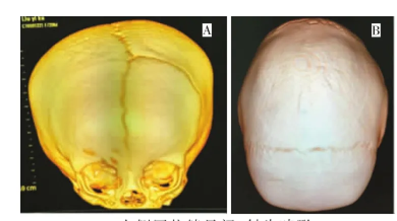
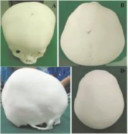
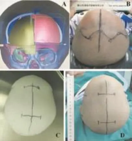
术前均行三维CT,Mimics软件处理数据,1∶1制作3D打印模型,设计切口和截骨方案(图1~3)。
1.3 手术方法
按照术前设计的手术切口和截骨方案,进行骨瓣的截取;根据预制的目标骨板,如额眶桥带,对已截取骨瓣进行塑形,随后将塑型骨瓣放置于目标位置连接固定。术后随访均行头颅CT扫描。
2 结果
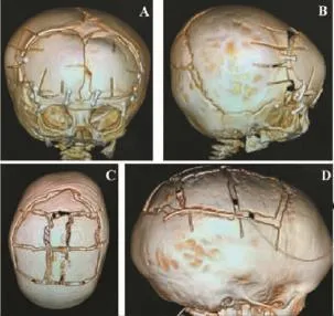
15例患儿均成功按预定设计完成手术,手术时长1.5~3.5 h。术后头颅CT三维颅骨成像(图4)显示,骨瓣成形位置准确,与术前设计相符,术后患儿头颅外形效果满意。本组患儿术后随访2~12个月,未发现明显并发症,患儿语言及智力发育进步明显(图5)。

图1 狭颅症患儿术前三维CT扫描颅骨重建Fig.1Three-dimensional computed tomographic evaluation of Craniostenosis in Children before operation

图2 3D打印模型Fig.2Three-dimensional printed model

图3 手术方案设计Fig.3Surgical plan design

图4 术后三维CT扫描Fig.4Three-dimensional computed tomographic evaluation

图5 典型病例:狭颅症(右侧斜头畸形)患儿Fig.5 Typical case:anterior plagiocephaly
3 讨论
3D打印技术起源于上世纪80年代,以数字化模型为基础,运用黏合材料,经逐层打印的方式来构造实体[3],属于增材制造。3D打印技术已被应用于器官模型的制造、个性化组织工程支架材料和假体植入物的制造、细胞或组织打印等方面[4-7]。骨科领域中,3D打印关节、假肢以及个体定制钢板已经开始应用于临床;耳鼻喉科、口腔科中,人工外耳道、种植牙等方面也有了初步的应用[8];在颌面外科中,被用于指导外科导板的设计制作、应用,个体化植入物也已出现。
颅缝早闭是婴幼儿时期起病的颅骨发育畸形,发生率约为1/2 500[9],可表现为颅内高压、发育缓慢、智力低下、精神活动异常、癫痫发作等症状。其好发部位依次为矢状缝(40%~55%)、冠状缝(20%~25%)、额缝(5%~15%)以及人字缝(1%~5%)[10]。矢状缝、冠状缝早闭较常见,矢状缝早闭导致的舟状头畸形,表现为颅部穹窿前后拉长,左右狭窄(即双顶径线过短);单侧冠状缝早闭引起斜头畸形的特征性表现是前额外观不对称,患侧前额、顶骨扁平,健侧额骨代偿性向前隆起,又被称为“小丑样畸形”,同样可以引起面部畸形,表现为鼻根部朝向患侧,患侧颧弓前移,下颌朝向健侧[11]。头颅畸形的诊断除颅部外观外,主要通过影像学检查,特别是CT扫描三维重建,可清晰显示过早闭合的骨缝以及头颅畸形。在治疗方面,手术是唯一的治疗途径,对于不同形式的颅缝早闭有不同的手术方式,但基本的要求都是合理地进行颅骨切开,重建早闭颅缝,重塑颅骨外形,从而保证合理的颅腔容积[11]。我们将头颅CT扫描的数据转换成3D打印数据,进行电脑模拟整复,进而利用3D打印技术在斜头、舟状头、三角头等畸形矫正术前制备出1∶1的颅骨模型,有效指导了手术过程,减少了手术时间,减轻了手术损伤及术中出血,患者外形和功能修复效果良好。
3D打印具有以下优点:①打印精度较高,可较完美地还原畸形部位,提供较为真实的手术模拟环境,对手术设计,选择最佳手术方案,减少手术风险具有积极意义[12];②模型制备快捷方便[13];③模型可用于手术操作演示[14];④便于术前医患沟通,有助于患方理解手术方案[15];⑤在个性化治疗方面具有显著优势[16]。
通过临床应用,我们认为3D打印技术在狭颅症矫治术中具有重要的辅助作用,尤其是在斜头及其他复杂畸形的矫治中具有非常好的手术引导作用。在我们的实际应用中,我们体会到3D打印技术在狭颅症矫治术中,可明显缩短手术时间,减少了手术创伤,优化了手术效果。当然,目前3D打印模型仍存在一定的误差,医学影像学数据生成的文件可直接应用于3D打印,但打印应用软件集成度及功能与3D打印设备,尚无法无缝对接[17]。相信随着关联学科的发展,3D打印技术在医学领域中将会获得广为广泛的应用,对于进一步开展个性化治疗、改善手术效果,具有极为重要的作用。
[1]Renier D,Lajeunie E,Arnaud E,et al.Management of craniosynostoses[J].Childs Nerv Syst,2000,16(10-11):645-658.
[2]Weinzweig J,Baker SB,Whitaker LA,et al.Delayed cranial vault reconstruction for sagittal synostosis in older children:an algorithm for tailoring the reconstructive approach to the craniofacial deformity[J].Plast Reconstr Surg,2002,110(2):397-408.
[3]Leong KF,Cheah CM,Chua CK.Solid freeform fabrication of three-dimensional scaffolds for engineering replacement tissues and organs[J].Biomaterials,2003,24(13):2363-2378.
[4]王成焘.医学的3D打印革命[J].中国经济和信息化,2013(13): 48-49.
[5]Derby B.Printing and prototyping of tissues and scaffolds[J]. Science,2012,338(6109):921-926.
[6]Minns RJ,Bibb R,Banks R,et al.The use of a reconstructed three-dimensional solid model from CT to aid the surgical management of a total knee arthroplasty:a case study[J].Med Eng Phys,2003,25(6):523-526.
[7]Mahaisavariya B,Sitthiseripratip K,Oris P,et al.Rapid prototyping model for surgical planning of corrective osteotomy for cubitus varus:Report of two cases[J].Injury Extra,2006,37(5):176-180.
[8]陈雪.3D打印技术在医学中的发展应用[J]广东科技,2014(15): 60-63.
[9]Knoll B,Persing JA.Craniosynostosis[M],2nd ed.St Louis: Quality Medical Publishing,2008:541-563.
[10]Cohen MM.Epidemiology of craniosynostosis[M],2nd ed.New York:Oxford University Press,2000:112-118.
[11]侯智,杨辉.狭颅症手术治疗新进展[J].实用医院临床杂志,2013, 10(5):22-25.
[12]张云峰,杨栋.3D打印骨科模型临床应用的初步探索[J].中国临床研究,2014,27(10):1260-1261.
[13]张海荣,鱼泳.3D打印技术在医学领域的应用[J].医疗卫生装备, 2015,36(3):118-120.
[14]McMenamin PG,Quayle MR,Mehenry CR,et al.The production of anatomical teaching resources using three-dimensional(3D) printing technology[J].Anat Sci Educ,2014,7(6):479-486.
[15]Olszewski R.Three-dimensional rapid prototyping models in craniomaxillofacial surgery:systematic review and new clinical applications[J].P Belg Roy Acad Med,2013,11(2):43-77.
[16]邓滨,欧阳汉斌,黄文华.3D打印在医学领域的应用进展[J].中国医学物理学杂志,2016,33(4):389-392.
[17]Vayrea B,Vignata F,Villeneuvea F.Designing for additive manufacturing[J].Procedia Cirp,2012,3(1):632-637.
The Effects of Three-dimensional Printing Technique in the Treatment of Craniostenosis in Children
LIU Tianjia1, GU Shuo1,WU Shuihua2,FAN Shuangshi2,CHEN Zhaohui2.1 Department of Neurosurgery,Shanghai Children's Medical Center,Shanghai Jiaotong University School of Medicine,Shanghai 200127,China;2 Department of Neurosurgery,Hunan Children's Hospital,Changsha 410007,China.Corresponding author:GU Shuo(E-mail:gushuo@shsmu.edu.cn).
ObjectiveTo explore the effects of 3D printing technique in the treatment of Craniostenosis in Children. MethodsThe clinical data of 15 cases underwent the operation with the guidance of 3d printing between September 2015 and August 2016 were analyzed retrospectively.Children underwent Computed Tomography(CT)and 3D reconstruction of the shell before operations,the imaging data was transferred into 3d printing data through professional software,the deformity was reconstructed on the computer and the 1∶1 skull model was printed by 3d printing technology,then the model was used to communicate with patients and make operative plans.The operations were finished as predetermined design,and the cranium CT scan was performed to observe the clinical efficacy.ResultsFifteen cases underwent operations successfully as predetermined design,operative time ranged from 1.5 to 3.5 hours.According to postoperative cranial CT imaging and realistic appearances,the results were satisfactory.All the children were followed up for 2 to 12 months,no obvious complications were observed,and the progress of language and intellectual development was obvious.ConclusionMaking use of 3d printing technology in the treatment of craniostenosis of children could save the operation time,reduce the surgical trauma,ensure the enough intro-cranial volume and postoperative appearances,promote the brain development obviously, and drive the trend of personalized therapy.
Three-dimensional printing;Craniostenosis;Surgery;Child
R622
A
1673-0364(2016)06-0346-03
2016年8月9日;
2016年9月20日)
10.3969/j.issn.1673-0364.2016.06.004
200127上海市上海交通大学医学院附属上海儿童医学中心神经外科(刘天甲,顾硕);410007湖南省长沙市湖南省儿童医院神经外科(吴水华,范双石,陈朝晖)。
顾硕(E-mail:gushuo@shsmu.edu.cn)。

