兔脊髓缺血再灌注损伤后能量代谢与NF-κ Bp65表达变化*
章建平方 华张竞超章放香
兔脊髓缺血再灌注损伤后能量代谢与NF-κ Bp65表达变化*
章建平①方 华①张竞超①章放香①
目的:探讨脊髓缺血再灌注损伤(SCIRI)中能量代谢与NF-κBp65的变化规律。方法:40只新西兰大白兔采用肾下腹主动脉阻断45 min,建立脊髓缺血再灌注损伤模型。分别于缺血前、缺血45 min及再灌注30、60、120 min时取脊髓测定腺苷酸(ADP、AMP、ATP)、活性氧(ROS)、核转录因子-κBp65(NF-κBp65)、抑制蛋白-κBα(I-κBα)和细胞间黏附分子-1(ICAM-1)蛋白表达变化。结果:与缺血前比较,缺血45 min及再灌注30、60、120 min时脊髓腺苷酸及I-κBα均降低(P<0.01),ROS、NF-κBp65和ICAM-1均升高(P<0.01)。结论:SCIRI中ICAM-1和NF-κBp65表达上调加重脊髓能量代谢障碍。
脊髓缺血再灌注损伤; 能量代谢; NF-κBp65
脊髓缺血再灌注损伤(spinal cord ischemiareperfusion injury, SCIRI)中脊髓神经细胞能量代谢异常将影响术后脊髓功能恢复[1-3]。研究证实,炎性反应是引起SCIRI的主要机制之一[4-5]。本研究通过观察SCIRI中脊髓腺苷酸代谢的变化来探讨抑制蛋白-κBα(inhibitor-kappa Bα,I-κBα)、核转录因子-κBp65 (nuclear factor-kappa Bp65,NF-κBp65)和细胞间粘附分子(intercellular adhesion molecule, ICAM-1)调控炎性信号转导在脊髓能量代谢障碍中的作用机制。
1 材料与方法
1.1 主要试剂 鼠抗兔ICAM-1、I-κBα及NF-κBp65多克隆抗体(均为Sigma公司);单磷酸腺苷(adenosine monophosphate,AMP)、二磷酸腺苷(adenosine diphosphate,ADP)和三磷酸腺苷(adenosine triphosphate,ATP)标准品(美国Sigma公司);链霉亲和素-生物素复合物(strept avidin-biotin complex,SABC)试剂盒及活性氧(reactive oxygen species,ROS)试剂盒(均为中国上海研吉生物科技有限公司)。
1.2 动物模型的建立 40只健康新西兰大白兔 (贵阳医学院实验动物中心提供),雌雄不限,体重2~2.5 kg。采用左肾动脉下方腹主动脉夹闭法建立SCIRI模型。阻断45 min后松钳开放腹主动脉,恢复脊髓血液灌注,以造成腰段脊髓再灌注损伤。术中以生理盐水浸润的温纱垫覆盖腹腔脏器,直肠温度维持于36~37 ℃。
1.3 评价方法 分别于缺血前、缺血45 min及再灌注30、60、120 min时间点取脊髓L3~L4节段脊髓组织,其中一部分脊髓组织加入4 ℃生理盐水超声匀浆及3500 r/min离心5 min后取上清液置于-80 ℃冰箱冻存,备测脊髓组织ROS活力变化。另一部分脊髓组织固定于10%福尔马林中,作为脊髓组织免疫组织化学检测。按照试剂盒说明书催化L-Arg氧反应法测定ROS活力以及SABC免疫组织化学法测定ICAM-1表达、I-κBα和NF-κBp65的表达及核移位,使用Olympus BX51图像采集分析系统等距随机抽样摄片并定量分析各时间点ICAM-1、I-κBα和NF-κBp65细胞核阳性表达率和胞核平均灰度值,计算阳性细胞平均百分率,作为此片的阳性细胞计数。
1.4 统计学处理 采用SPSS 16.0软件统计处理,计量资料以(±s)表示,组间比较采用单因素方差分析和q检验,以P<0.05为差异有统计学意义。
2 结果
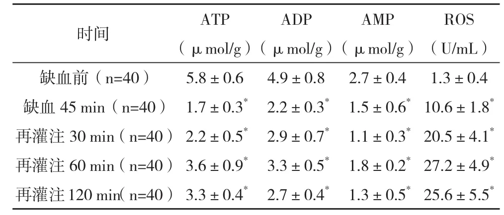
2.1 脊髓组织ATP、ADP和AMP含量的比较 缺血45 min及再灌注30、60、120 min时ATP、ADP和AMP均明显降低,ROS活力显著升高,与缺血前比较差异均有统计学意义(P<0.01)。见表1。
表1 ATP、ADP和AMP含量的变化(±s)

表1 ATP、ADP和AMP含量的变化(±s)
*与缺血前比较,P<0.01
ROS(U/mL)缺血前(n=40)5.8±0.64.9±0.82.7±0.41.3±0.4缺血45 min(n=40) 1.7±0.3*2.2±0.3*1.5±0.6*10.6±1.8*再灌注30 min(n=40) 2.2±0.5*2.9±0.7*1.1±0.3*20.5±4.1*再灌注60 min(n=40) 3.6±0.9*3.3±0.5*1.8±0.2*27.2±4.9*再灌注120 min(n=40) 3.3±0.4*2.7±0.4*1.3±0.5*25.6±5.5*时间ATP(μmol/g)ADP(μmol/g)AMP(μmol/g)
2.2 脊髓组织NF-κBp65、I-κBα和ICAM-1表达的比较 缺血45 min及再灌注30、60、120 min时间时I-κBα表达均明显下降,NF-κBp65及ICAM-1表达显著上升,与缺血前比较差异均有统计学意义(P<0.01),见表2。
表2 脊髓NF-κ Bp65、I-κ Bα和ICAM-1表达变化(±s)

表2 脊髓NF-κ Bp65、I-κ Bα和ICAM-1表达变化(±s)
*与缺血前比较,P<0.01
时间NF-κBp65I-κBαICAM-1平均灰度值阳性细胞百分率 (%)平均灰度值 阳性细胞百分率(%)平均灰度值阳性面积百分率(%)缺血前(n=40)44.3±10.18.5±1.9188.6±13.285.3±10.10.0±0.00.0±0.0缺血45 min(n=40)175.8±18.6*68.3±7.2*79.4±9.4*34.5±6.7*126.2±19.3*65.4±8.7*再灌注30 min(n=40)190.4±21.1*69.6±8.4*61.6±9.5*26.2±5.9*146.6±22.7*70.1±10.4*再灌注60 min(n=40)197.3±18.9*84.5±7.6*32.1±8.5*13.7±4.6*180.3±20.4*74.3±9.4*再灌注120 min(n=40)183.5±16.1*77.4±9.3*31.8±6.6*11.4±5.3*183.9±22.1*76.2±9.6*
3 讨论
SCIRI期间脊髓神经功能的维持依赖于充足的ATP合成与代谢,脊髓神经细胞能量代谢障碍不仅参与脊髓神经功能不全的发生, 而且是术后截瘫发生率上升的主要因素[6-8]。本研究发现,缺血后脊髓组织ROS活力显著升高,与此同时,缺血脊髓组织ATP、ADP和AMP水平下降,提示脊髓缺血后神经细胞线粒体损伤引起ATP、ADP和AMP合成与代谢异常,而再灌注后虽然脊髓组织的血供得到恢复,但SCIRI期间大量释放的ROS炎性介质进一步损害脊髓神经细胞能量转移与利用,说明脊髓缺血可引起脊髓神经细胞线粒体合成ATP、ADP和AMP能力受限,而再灌注时ROS炎性介质可加重损害脊髓神经细胞能量代谢,造成脊髓神经细胞能量代谢障碍。
本研究观察到脊髓缺血后脊髓组织I-κBα蛋白表达逐渐降低,而NF-κBp65核移位和ICAM-1蛋白表达逐渐升高且出现脊髓神经细胞能量代谢障碍,说明SCIRI期间脊髓组织NF-κBp65/I-κBα炎性信号转导系统启动,通过上调脊髓组织ICAM-1表达引起脊髓组织炎性反应,进而损害脊髓神经细胞能量转移与利用。研究证实ROS是白细胞活化过程中脂质过氧化反应的炎性介质之一[9-13],ROS可诱导I-κBα蛋白水解磷酸化启动NF-κBp65/I-κBα炎性信号转导系统[14]。本研究中,SCIRI脊髓神经细胞能量代谢障碍中ROS活力升高的同时ICAM-1及NF-κBp65蛋白表达也明显升高,说明NF-κBp65/I-κBα炎性信号启动可能是引起脊髓神经细胞能量代谢障碍的发生机制之一。
[1] Lowe M T,Kim E H,Faull R L,et al.Dissociated expression of mitochondrial and cytosolic creatine kinases in the human brain: a new perspective on the role of creatine in brain energy metabolism[J].J Cereb Blood Flow Metab,2013,33(8):1295-306.
[2] Kurtoglu T,Basoglu H,Ozkisacik E A,et al.Effects of cilostazol on oxidative stress, systemic cytokine release, and spinal cord injury in a rat model of transient aortic occlusion[J].Ann Vasc Surg, 2014 ,28(2):479-88.
[3] Zhou Y F,Li L,Feng F,et al.Osthole attenuates spinal cord ischemia-reperfusion injury through mitochondrial biogenesisindependent inhibition of mitochondrial dysfunction in rats[J].J Surg Res,2013 ,185(2):805-814.
[4] Breckwoldt M O,Pfister F M,Bradley P M,et al.Multiparametric optical analysis of mitochondrial redox signals during neuronal physiology and pathology in vivo[J].Nat Med,2014,20(5):555-560.
[5] Phillips J P,Cibert-Goton V,Langford R M,et al.Perfusion assessment in rat spinal cord tissue using photoplethysmography and laser Doppler flux measurements[J].J Biomed Opt,2013,18(3):037 005.
[6] Koriyama Y,Nakayama Y,Matsugo S,et al.Anti-inflammatory effects of lipoic acid through inhibition of GSK-3β in lipopolysaccharide-induced BV-2 microglial cells[J].Neurosci Res,2013 ,77(1-2):87-96.
[7] Liang C L,Lu K,Liliang P C,et al.Ischemic preconditioning ameliorates spinal cord ischemia-reperfusion injury by triggering autoregulation[J].J Vasc Surg, 2012,55(4):1116-1123.
[8] Kubota K,Saiwai H,Kumamaru H,et al.Neurological recovery is impaired by concurrent but not by asymptomatic pre-existing spinal cord compression after traumatic spinal cord injury[J].Spine, 2012, 37(17):1448-1455.
[9] Ye B,Kroboth S L,Pu J L,et al.Molecular identification and functional characterization of a mitochondrial sulfonylurea receptor 2 splice variant generated by intraexonic splicing[J].Circ Res, 2009,105(11):1083-1093.
[10] Cui Y,Zhang H,Ji M,et al.Hydrogen-rich saline attenuates neuronal ischemia-reperfusion injury by protecting mitochondrial function in rats[J].J Surg Res,2014, S0022-4804(14):00 529.
[11] Koriyama Y,Nakayama Y,Matsugo S,et al.Anti-inflammatory effects of lipoic acid through inhibition of GSK-3β in lipopolysaccharide-induced BV-2 microglial cells[J].Neurosci Res,2013 ,77(1-2):87-96.
[12]王义生,张振,张鑫,等. 乙醇对兔骨髓间充质干细胞6种神经递质mRNA表达的影响[J].郑州大学学报:医学版, 2011,46(3):357-360.
[13] Xu Y Q,Jin S J,Liu N L,et al.Aloperine attenuated neuropathic pain induced by chronic constriction injury via anti-oxidation activity and suppression of the nuclear factor kappa B pathway[J].Biochem Biophys Res Commun, 2014 ,451(4):568-573.
[14]张鹏,宋来君,杨波,等. 人脐血间质干细胞移植对脑创伤大鼠行为学和学习、记忆评分的影响[J].郑州大学学报:医学版,2006,41(2):233-235.
Changes of Energy Metabolism and the Expression of NF-κ Bp65 after Spinal Cord Ischemia-Reperfusion Injury in Rabbits
ZHANG J ian-ping,FANG Hua,ZHANG J ing-chao,et al.//Medical Innovation of China,2015,12(09):024-026
Objective:To assess the changes of energy metabolism and NF-κBp65 during spinal cord ischemia and reperfusion injury(SCIRI). Method:The SCIRI model was made by clamping the infrarenal aortic for 45 minutes in 40 rabbits.Reactive oxygen species(ROS),the expression of nuclear factor-kappa Bp65(NF-κBp65),inhibitor-Kappa Bα(I-κBα), intercellular adhesion molecule(ICAM-1) and the content of adenine nucleotide(ADP,AMP and ATP) in spinal cord tissue were determined before ischemia,45 minutes after ischemia,30,60 minutes and 120 minutes after reperfusion. Result: The spinal cord content of adenine nucleotide and I-κBα decreased while ROS,the expressions of NF-κBp65and ICAM-1 increased at 45 minutes after ischemia,30,60 minutes and 120 minutes after reperfusion as compared to the value before ischemia (P<0.01). Conclusion:The upregulation of the expression of ICAM-1 and NF-κBp65 aggravates the energy metabolism disorder of spinal cord during SCIRI.
Spinal cord ischemia-reperfusion injury; Energy metabolism; NF-κBp65
10.3969/j.issn.1674-4985.2015.09.008
2014-09-29) (本文编辑:蔡元元)
贵州省卫生厅基金资助项目(gzwkj2010-1-006);贵州省科技厅基金资助项目(黔科合SY字[2011]008号);贵州省科技厅基金资助项目(黔科SY字[2012]3090号);贵州省科技厅基金资助项目(黔科SY字[2012]001号)
①贵州省人民医院 贵州 贵阳 550002
方华
First-author’s address:Guizhou Provincial People’s Hospital, Guiyang 550002,China

