跟骨定量超声与股骨近端特定区域骨密度相关性研究
2015-10-14 01:56:55李明吕厚辰张里程尹鹏滨胡坚兴唐佩福张立海
中国骨与关节杂志 2015年12期
李明 吕厚辰 张里程 尹鹏滨 胡坚兴 唐佩福 张立海
跟骨定量超声与股骨近端特定区域骨密度相关性研究
李明 吕厚辰 张里程 尹鹏滨 胡坚兴 唐佩福 张立海
目的 探讨跟骨定量超声 ( QUS ) 参数与股骨近端特定区域骨密度的相关性。方法 连续纳入34 例患股骨颈骨折行髋关节置换的老年女性,行跟骨定量超声、髋部双能 X 线检测 ( DXA ),探讨上述检测结果相关程度。结果 根据 DXA 检测患者髋部骨密度 ( BMD ) T 值显示,骨质疏松患者 13 例 ( T ≤ -2.5 ),跟骨骨密度 ( Est. BMD ) 与股骨转子间 BMD 呈现正相关 ( r=0.355,P<0.05 );跟骨超声振幅衰减平均值 ( BUA ) 与股骨转子间 BMD 呈现正相关 ( r=0.450,P<0.01 )。结论 跟骨定量超声检测参数与股骨近端特定区域股骨转子间 BMD 存在相关性,对骨质疏松特定区域的骨量的评价及骨折风险预测有一定参考价值。
跟骨;股骨;骨密度;超声检查;骨折
随着人口老龄化的加剧,骨质疏松性骨折已成为老年人健康的巨大威胁,60 岁以上的高龄人群中,约 1 / 2 的女性和 1 / 3 的男性将至少发生一次骨质疏松性脆性骨折[1],骨质疏松性骨折具有较高的致残率及死亡率[2-3],以髋部骨折为例,1 年内死亡率男性为 27.6%~40.5%,女性为 15.8%~23.3%,约 40% 患者需长期护理[4]。因此骨质疏松早期的诊断和治疗对降低骨折风险至关重要。目前评价骨质疏松常用的方法包括双能 X 线吸收仪( DXA ) 骨密度测定、定量计算机断层扫描 ( QCT ) 骨密度测定以及定量超声 ( QUS ) 骨密度测定[5-8]。自1984 年 Langton 等[9]首次报道采用定量超声检测骨质疏松患者以来,定量超声因其无辐射、操作简便、低成本等显著优势已得到了广泛应用并逐渐推广,在评价骨质疏松中具有重要的价值。相关研究证实,定量超声相关参数如宽带超声衰减 ( BUA ),超声传导速度 ( SOS ) 与髋部、腰椎骨折风险相关[10]。然而就当前多发的髋部骨折而言,特定区域的骨量变化可更好地预测骨折风险,跟骨定量超声参数虽能够很好地反应跟骨松质骨骨量性质,但其与髋部股骨近端特定区域 ( 股骨颈、转子间、Ward's 区 ) 骨量变化的相关性却研究甚少。故本研究通过对 34 例骨质疏松性股骨颈骨折行关节置换的患者行 DXA 及跟骨定量超声检测,探讨跟骨定量超声参数与股骨近端特定区域骨密度相关性,进而证实跟骨定量超声对髋部特定区域骨折风险预测能力。
资料与方法
一、一般资料
连续纳入 2014 年 1 月至 6 月于中国人民解放军总医院骨科行关节置换的 34 例,56~93 岁的骨质疏松性股骨颈骨折的绝经女性为研究对象,平均年龄 ( 75.0±10.8 ) 岁,患肿瘤、关节感染、糖尿病和其它可能影响骨代谢的疾病患者均被排除。
二、方法
定量超声测定 BMD:所用设备为 Sahara 型跟骨定量超声仪 ( Sahara Clinical Bone Sonometer,HOLOGIC,Bedford,MA,USA ),测试前使用体模校正调试仪器。检查时将受检者基本信息输入仪器,取端坐位 ( 无法取坐位患者行站立位 ),用超声凝胶涂抹足跟部,按检测要求放置并固定于仪器凹槽中进行检测,记录测定结果:超声传播速度( SOS,m / s )、超声振幅衰减平均值 ( BUA,dB / MH2)、estimated BMD ( Est. BMD, g / cm2),定量超声硬度指数 ( QUI / stiffness ),QUI / stiffness 计算公式为QUI=0.41×( BUA+SOS )-571,无计量单位[11]。上述检测均由同一检查者完成,Est. BMD、SOS、BUA的变异系数分别为 3%、0.22% 和 3.7%。
DXA 测定 BMD:所用设备为 Discovery 双能X 线吸收仪 ( DXA,HOLOGIC Discovery-A,Apex software version 13.3,Bedford,MA,USA )。受检者均为术后 1~3 天内进行检测,检测时将受检者基本信息输入仪器,受检者取仰卧位于检测台上,测定研究对象的健侧股骨近端 ( 包括全髋部、股骨颈、转子间及三角区 ) BMD 值。上述检测均由同一检查者完成,髋部骨密度的变异系数为 0.8%。
三、统计学分析
采用 SPSS 22.0 软件进行统计学分析。定量数据用±s 表示,相关性用简单相关及偏相关分析,相关系数 r 表示参数间的相关性。P<0.05 为差异有统计学意义。
结 果
一、基本资料
研究对象均为股骨颈骨折行髋关节置换术的老年绝经女性。年龄 56~93 岁,平均年龄 ( 75.0± 10.8 ) 岁,平均绝经年龄 ( 50.1±2.6 ) 岁,其中年龄>60 岁患者占 91.2%,骨质疏松患者 13 例 ( T ≤-2.5 ) ( 表 1 )。
二、检测指标
跟骨超声检测参数示:患者 Est. BMD 为 ( 0.249± 0.175 ) g / cm2,SOS 为 ( 1080.8±18.2 ) m / s,BUA 为( 45.9±8.3 ) dB / MH2,Stiffness 为 47.8±8.4。DXA 检查患者健侧髋部示:总 BMD 为 ( 0.685±0.114 ) g / cm2,股骨颈 BMD 为 ( 0.568±0.130 ) g / cm2,转子间 BMD( 0.536±0.104 ) g / cm2,Ward's 区 BMD 为 ( 0.427± 0.175 ) g / cm2( 表 2 )。
表 1 研究对象基本资料 ( n = 34,x- ± s )Tab.1 Baseline characteristics of patients ( n=34,±s )
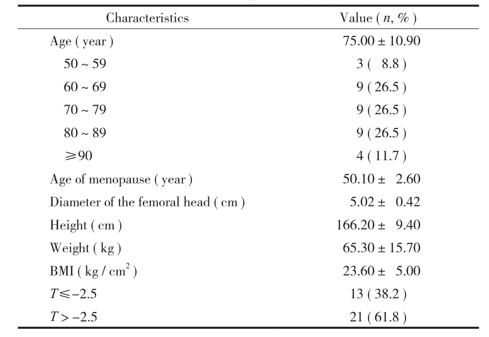
表 1 研究对象基本资料 ( n = 34,x- ± s )Tab.1 Baseline characteristics of patients ( n=34,±s )
CharacteristicsValue ( n, % )Age ( year ) 75.00±10.90 50~59 3 ( 8.8 )60~69 9 ( 26.5 )70~79 9 ( 26.5 )80~89 9 ( 26.5 )≥90 4 ( 11.7 )Age of menopause ( year ) 50.10± 2.60 Diameter of the femoral head ( cm ) 5.02± 0.42 Height ( cm )166.20± 9.40 Weight ( kg ) 65.30±15.70 BMI ( kg / cm2) 23.60± 5.00 T≤-2.513 ( 38.2 )T > -2.521 ( 61.8 )
表 2 研究对象检测指标 ( n = 34,x- ± s )Tab.2 Detection index of patients ( n=34,±s )
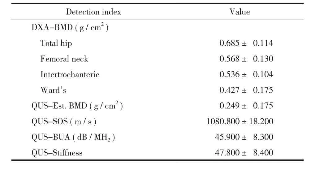
表 2 研究对象检测指标 ( n = 34,x- ± s )Tab.2 Detection index of patients ( n=34,±s )
Detection indexValue DXA-BMD ( g / cm2)Total hip 0.685± 0.114 Femoral neck 0.568± 0.130 Intertrochanteric 0.536± 0.104 Ward's 0.427± 0.175 QUS-Est. BMD ( g / cm2) 0.249± 0.175 QUS-SOS ( m / s )1080.800±18.200 QUS-BUA ( dB / MH2) 45.900± 8.300 QUS-Stiffness 47.800± 8.400
三、跟骨超声参数与股骨近端骨密度相关性分析
跟骨超声参数与股骨近端特定区域骨密度( total、femoral neck、Intertrochanteric、Ward's ) 相关性分析,控制了患者股骨头直径后行偏相关分析,结果显示跟骨 Est. BMD、BUA 与股骨转子间 BMD 呈现正相关 ( r=0.355,P<0.05;r=0.450,P<0.01 )( 表 3,图 1a,b )。
四、跟骨超声参数与股骨近端骨密度分组相关性分析
根据患者髋部总 BMD ( Total hip ) T 值分层,分为骨质疏松 ( T ≤ -2.5,n=13 ) 和骨量降低或正常 ( T>-2.5,n=21 ) 两组,探讨跟骨超声参数与股骨近端特定区域骨密度 ( total、femoral neck、Intertrochanteric、Ward's ) 相关性,控制患者股骨头直径后行偏相关分析,结果显示两组数据无明显相关关系。但当 T ≤ -2.5 时,Est. BMD、BUA 与股骨转子间 BMD 相关性为 ( r=0.339,P=0.144;r= 0.429,P=0.059 )。
表 3 跟骨定量超声参数与股骨近端特定区域骨密度相关性 ( n = 34,x- ± s )Tab.3 Correlations between calcaneus quantitative ultrasound parameters and specifc areas of proximal femoral bone mineral density ( n=34,±s )
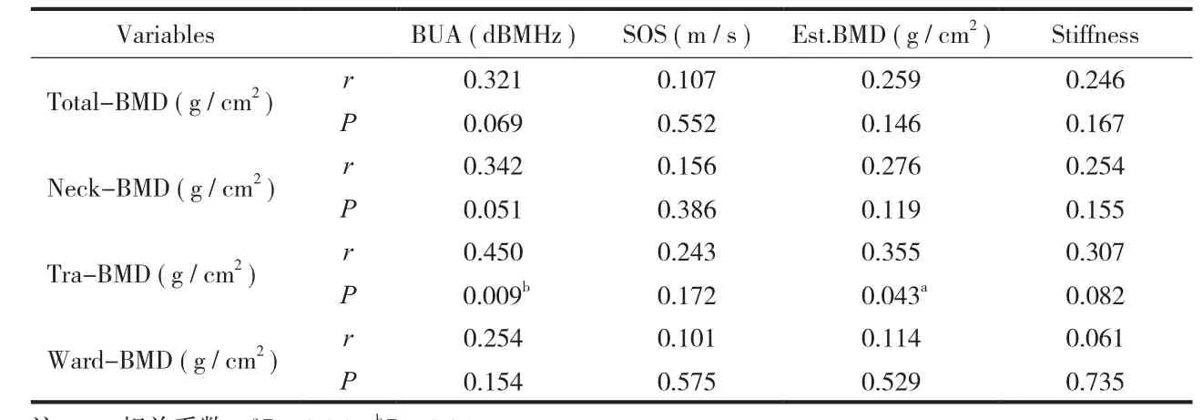
表 3 跟骨定量超声参数与股骨近端特定区域骨密度相关性 ( n = 34,x- ± s )Tab.3 Correlations between calcaneus quantitative ultrasound parameters and specifc areas of proximal femoral bone mineral density ( n=34,±s )
注:r:相关系数,aP < 0.05,bP < 0.01Notice: R: Related coefficient,aP<0.05,bP<0.01
VariablesBUA ( dBMHz )SOS ( m / s )Est.BMD ( g / cm2)Stiffness Total-BMD ( g / cm2)r P 0.246 0.167 Neck-BMD ( g / cm2)r P 0.321 0.069 0.107 0.552 0.259 0.146 0.254 0.155 Tra-BMD ( g / cm2)r P 0.342 0.051 0.156 0.386 0.276 0.119 0.061 0.735 0.307 0.082 Ward-BMD ( g / cm2)r P 0.450 0.009b0.243 0.172 0.355 0.043a0.254 0.154 0.101 0.575 0.114 0.529
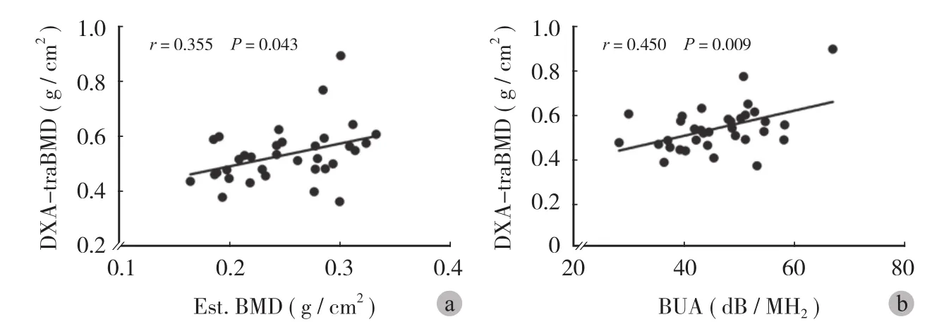
图 1 线性回归分析 a:跟骨 Est. BMD 与股骨转子间 BMD 相关关系;b:跟骨 BUA与股骨转子间 BMD 相关关系Fig.1 Linear regression analysis a: Correlations between QUS-BMD and intertrochanteric BMD; b: Correlations between QUS-BUA and intertrochanteric BMD
讨 论
骨质疏松症是全身性的骨骼疾病,其特征为骨量减少,骨小梁变细、断裂、数量减少,皮质骨多孔、变薄为特征,导致骨强度下降、骨脆性增加及骨折的危险性增加的全身性骨病[12]。骨强度主要由骨密度和骨质量两方面因素所决定。目前 DXA 作为临床上诊断骨质疏松的金标准,通过测定骨密度及 T 值反应骨骼中的矿物质含量从而诊断骨质疏松[8],但对骨骼强度及微观结构等因素却缺乏有效评价。跟骨定量超声不仅可反映骨密度,还可反映骨弹性、强度和显微结构,对骨质疏松的诊断具有一定意义[13]。
当前已有研究表明,股骨头的骨密度值明显高于转子间及 Word's 区域[14]。并且上述区域内骨代谢及骨丢失率也不尽相同[15]。所以笔者认为股骨近端不同特定区域骨质变化对股骨整体生物力学性质及骨折风险有着重要的意义。而当前跟骨超声研究多为不同人群骨折风险预测及预后判断[16],探讨定量超声较 DXA 之间不同方面的比较,探索临床进一步推广使用[17-18]。仅有少数研究探讨了骨骼不同部位 BMD 与跟骨超声参数之间的关系[19]。而定量超声参数与髋部特定区域 ( 股骨颈、转子间、Word's 区 ) 之间的关系却尚未有明确研究。
故本研究通过对 34 例绝经后老年女性行跟骨定量超声及健侧髋部 DXA 检查,结果显示跟骨Est. BMD 与股骨转子间 BMD 有相关 ( r=0.355,P<0.05 ),但其与髋部其它部位 BMD 无明显相关关系,考虑原因为跟骨、转子间区皆为高含量松质骨结构,其骨改建周期短,对骨质变化敏感,可能存在一定程度的相关性。而股骨颈及股骨内部较之皮质骨含量较多,Ward's 区多为空虚结构。因此,跟骨与上述区域 BMD 相关性不明显。此外,通过分析患者跟骨定量超声检测参数,发现 BUA 与股骨转子间 BMD 呈现正相关。上述结果初步解释了跟骨超声参数与股骨近端骨量之间的联系,也提示其可能与髋部骨折风险之间的相关性[17,20-21]。
进一步通过 T 值分层后,在 T>-2.5 组,股骨转子间 BMD 与跟骨 Est. BMD 及 BUA 存在有近似相关关系 ( r=0.339,P=0.144;r=0.429,P= 0.059 );而对于 T<-2.5 组 ( N=13 ),并未发现跟骨超声参数与股骨近端特定区域存在统计学差异的相关关系。本研究结果显示:经 DXA 评价后,对于T>-2.5 的老年股骨颈骨折患者,其跟骨超声的相关参数与股骨粗隆间处的密度存在相关关系。对于通过 DXA 评价可以诊断为骨质疏松的患者 T<-2.5,本研究由于样本量不足,未发现具有统计学意义的相关关系,下一步应进一步扩大样本量,评价两者之间的联系。
本研究存在不足:( 1 ) 本次实验样本均选自股骨颈骨折老年女性患者,故研究结论适用于股骨颈骨折人群,若将结论扩大到更大的范围的骨质疏松人群或者正常人群,其外部真实性需要进一步研究证实。( 2 ) 本研究仅评价了定量超声检测与股骨近端密度之间的联系,其结构学、力学参数未涉及,可在后续研究中补充完善。( 3 ) 校正因素为骨骼大小 ( 股骨头直径 )。考虑到骨骼大小对测量的影响,及其它影响因素 ( 如骨代谢等 ),可能对跟骨超声参数对髋部特定区域骨质变化也存在一定的影响,须进一步研究。
本研究结果提示在股骨颈骨折人群中,同为负重区松质骨结构,跟骨定量超声检测参数与股骨转子间区域存在相关性。因此,笔者推测跟骨超声参数在一定程度上可以反映股骨转子间骨质变化,对骨质疏松患者早期特定区域的诊断及预测转子间骨质疏松性骨折相关风险具有一定的参考价值。
[1] Nguyen ND, Ahlborg HG, Center JR, et al. Residual lifetime risk of fractures in women and men. J Bone Mineral Res, 2007,22(6):781-788.
[2] Bliuc D, Nguyen ND, Milch VE, et al. Mortality risk associated with low-trauma osteoporotic fracture and subsequent fracture in men and women. Jama, 2009, 301(5):513-521.
[3] Center JR, Bliuc D, Nguyen TV, et al. Risk of subsequent fracture after low-trauma fracture in men and women. Jama,2007, 297(4):387-394.
[4] Morin S, Lix LM, Azimaee M, et al. Mortality rates after incident non-traumatic fractures in older men and women. Osteoporos Int, 2011, 22(9):2439-2448.
[5] Adams JE. Quantitative computed tomography. Euro J Radiol,2009, 71(3):415-424.
[6] Hans D, Dargent-Molina P, Schott AM, et al. Ultrasonographic heel measurements to predict hip fracture in elderly women: the EPIDOS prospective study. Lancet, 1996, 348(9026):511-514.
[7] Kanis JA. Assessment of fracture risk and its application to screening for postmenopausal osteoporosis: synopsis of a WHO report. WHO Study Group. Osteoporos Int, 1994, 4(6):368-381.[8] Punda M, Grazio S. Bone densitometry--the gold standard for diagnosis of osteoporosis. Reumatizam, 2014, 61(2):70-74.
[9] Langton CM, Palmer SB, Porter RW. The measurement of broadband ultrasonic attenuation in cancellous bone. Eng Med,1984, 13(2):89-91.
[10] Chan MY, Nguyen ND, Center JR, et al. Absolute fracture-risk prediction by a combination of calcaneal quantitative ultrasound and bone mineral density. Calcifed Tissue International, 2012,90(2):128-136.
[11] Krieg MA, Cornuz J, Ruffieux C, et al. Comparison of three bone ultrasounds for the discrimination of subjects with and without osteoporotic fractures among 7562 elderly women. J Bone Miner Res, 2003, 18:1261-1266.
[12] Cummings SR, Melton LJ. Epidemiology and outcomes of osteoporotic fractures. Lancet, 2002, 359(9319):1761-1767.
[13] Cortet B, Boutry N, Dubois P, et al. Does quantitative ultrasound of bone reflect more bone mineral density than bone microarchitecture? Calcified Tissue International, 2004,74(1):60-67.
[14] Schott AM, Cormier C, Hans D, et al. How hip and whole-body bone mineral density predict hip fracture in elderly women: the EPIDOS Prospective Study. Osteoporos Int, 1998, 8:247-254.
[15] Emaus N, Wilsgaard T, Ahmed LA. Impacts of body mass index, physical activity, and smoking on femoral bone loss: the Tromso study. J Bone Miner Res, 2014, 29:2080-2089.
[16] Khaw KT, Reeve J, Luben R, et al. Prediction of total and hip fracture risk in men and women by quantitative ultrasound of the calcaneus: EPIC-Norfolk prospective population study. Lancet, 2004, 363:197-202.
[17] Hadji P, Imani P, Wuster C, et al. Comparison of dual-energy X-ray absorptiometry with six quantitative ultrasonometry devices in women with hip fractures. Climacteric, 2015, 18(3): 411-418.
[18] Kwok T, Khoo CC, Leung J, et al. Predictive values of calcaneal quantitative ultrasound and dual energy X ray absorptiometry for non-vertebral fracture in older men: results from the MrOS study (Hong Kong). Osteoporos Int, 2012,23:1001-1006.
[19] Jorgensen HL, Warming L, Bjarnason NH, et al. How does quantitative ultrasound compare to dual X-ray absorptiometry at various skeletal sites in relation to the WHO diagnosis categories? Clin Physiol, 2001, 21:51-59.
[20] Trimpou P, Bosaeus I, Bengtsson BA, et al. High correlation between quantitative ultrasound and DXA during 7 years of follow-up. Euro J Radiol, 2010, 73(2):360-364.
[21] Albanese CV, De Terlizzi F, Passariello R. Quantitative ultrasound of the phalanges and DXA of the lumbar spine and proximal femur in evaluating the risk of osteoporotic vertebral fracture in postmenopausal women. La Radiologia Medica,2011, 116(1):92-101.
( 本文编辑:王永刚 )
Correlations between quantitative ultrasound of calcaneus and specific areas of proximal femoral bone mineral density
LI Ming, LU Hou-chen, ZHANG Li-cheng, YIN Peng-bin, HU Jian-xing, TANG Pei-fu, ZHANG Li-hai. Department of Orthopaedics, Chinese PLA General Hospital, Beijing, 100853, PRC
ZHANG Li-hai, Email: zhanglihai74@qq.com
Objective To discuss correlations between quantitative ultrasound parameters of the calcaneus and specifc areas of proximal femoral bone mineral density. Methods Thirty-four elderly female with femoral neck fractures, who underwent total hip replacement surgery, were selected. The bone mass of the proximal femur was tested by dualenergy X-ray absorptiometry ( DXA ) and ultrasonic properties of the calcaneus by quantitative ultrasound( QUS ). Relevant results above were discussed. Results According to T value of bone mineral density ( BMD )testing, 13 patients were of osteoporosis ( T≤-2.5 ). BMD of the calcaneus and intertrochanter was positively correlated ( r=0.355, P<0.05 ). BUA was positively correlated with BMD of the intertrochanter ( r=0.450, P<0.01 ). Conclusions Parameters of BUA are correlated with specifc areas of proximal femoral BMD ( intertrochanteric )with reference value for the diagnosis of osteoporosis and risk prediction of the specifc areas of proximal femoral fracture.
Calcaneus; Femur; Bone density; Ultrasonography; Fractures, bone
10.3969/j.issn.2095-252X.2015.12.013
R681, R445.1
2014-2017 国家自然科学基金面上项目 ( 31370947 )
100853 北京,解放军总医院骨科 ( 李明、吕厚辰、张里程、尹鹏滨、唐佩福、张立海 ),影像科 ( 胡坚兴 )
张立海,Email: zhanglihai74@qq.com
2015-07-16 )
猜你喜欢
中国伤残医学(2022年14期)2022-12-23 06:39:10
中老年保健(2022年3期)2022-08-24 03:00:58
中老年保健(2021年10期)2021-08-24 06:42:46
中老年保健(2021年8期)2021-08-24 06:23:40
中老年保健(2021年9期)2021-08-24 03:51:10
保健医苑(2021年7期)2021-08-13 08:48:02
中国生殖健康(2019年2期)2019-08-23 08:12:02
灾害医学与救援(电子版)(2016年2期)2016-03-11 20:18:04
中国医学装备(2015年10期)2015-12-29 12:00:32
湖北科技学院学报(医学版)(2014年2期)2014-02-28 19:42:39