f/t PSA、PSAD、(f/t)/PSAD在PSA 4~20 μg/L区间对前列腺癌的诊断预测价值
马明辉 黄庆波 高宇 赵超飞 刘侃 史涛坪 马鑫 张旭
1中国人民解放军总医院泌尿外科 100853 北京
论 著
f/t PSA、PSAD、(f/t)/PSAD在PSA 4~20 μg/L区间对前列腺癌的诊断预测价值
马明辉1黄庆波1高宇1赵超飞1刘侃1史涛坪1马鑫1张旭1
1中国人民解放军总医院泌尿外科 100853 北京
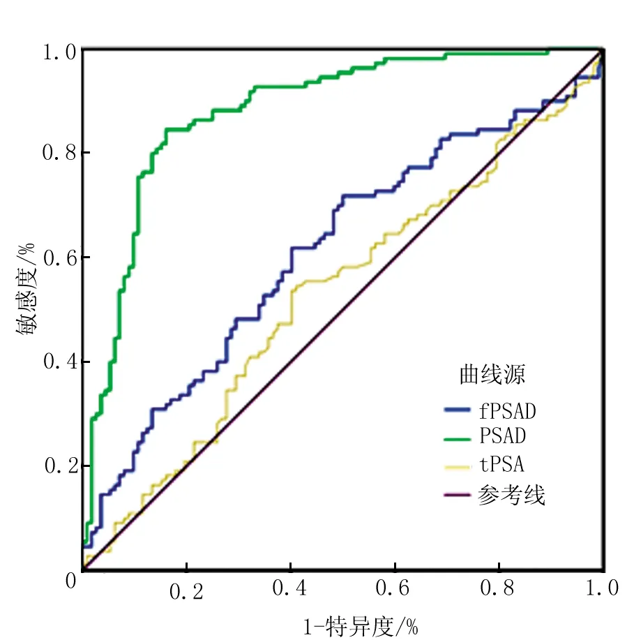
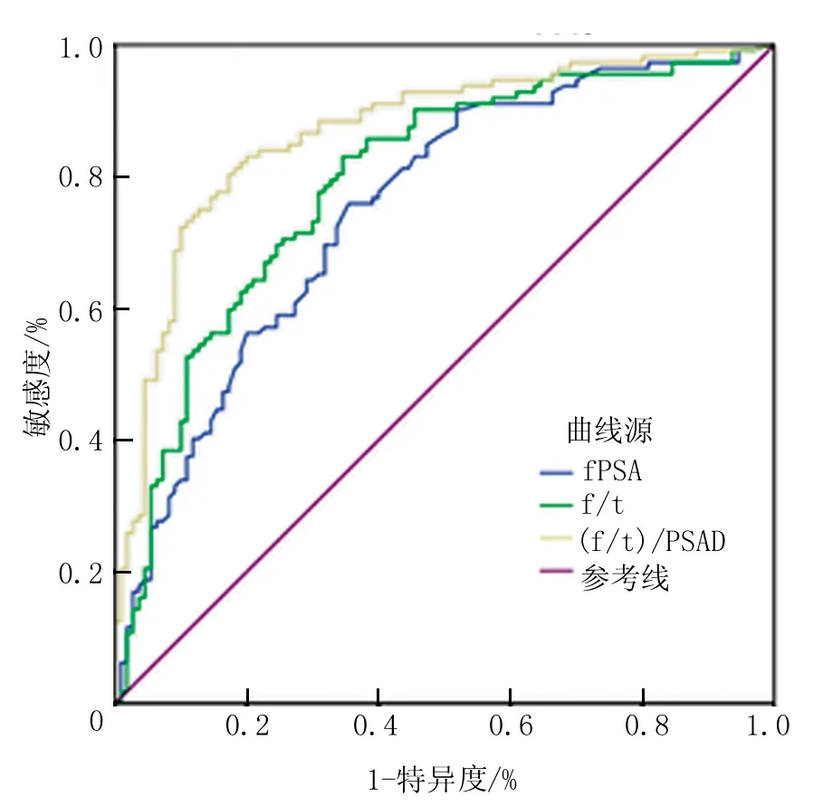
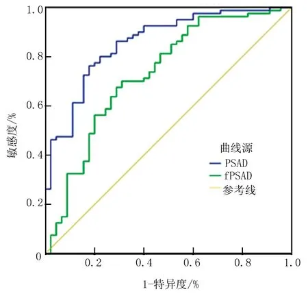
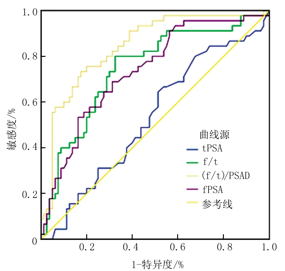
目的:通过对fPSA、f/t PSA、PSAD、fPSAD、(f/t)/PSAD指标的分析,评价对PSA灰区(4~20 μg/L)前列腺癌的诊断预测价值。方法:回顾性分析根据筛选条件纳入病例347例,按tPSA水平分为4 μg/L 前列腺特异性抗原;前列腺特异性抗原密度;前列腺癌;诊断 前列腺癌发病率在全球男性恶性肿瘤中位居第二[1]。我国前列腺癌的发生率近年来呈现上升趋势,在男性恶性肿瘤中发病率排名第六,病死率第九[2,3]。前列腺特异性抗原(PSA,或称总PSA、tPSA)作为单一检测指标,与直肠指诊、经直肠前列腺超声等比较,具有更高的前列腺癌阳性诊断预测率。目前国内外普遍认同的观点是PSA>4 μg/L为异常,而PSA 4 μg/L 1.1 临床资料 回顾性分析2012年1月~2014年12月期间因前列腺相关疾病、高度怀疑前列腺癌风险、在中国人民解放军总医院院泌尿外科住院患者的临床资料。筛选tPSA为4~20 μg/L,病理诊断明确,tPSA、fPSA、前列腺B超、病理诊断等临床资料完整,且无前列腺增生外科手术史、无急慢性前列腺炎、无尿潴留等疾病者,最终纳入病例347例。 1.2 研究分层 按tPSA水平将病例分为4 μg/L 1.3 检测指标 血清PSA检测:在行直肠指诊、前列腺按摩、导尿、经直肠超声、膀胱镜、前列腺穿刺等操作前,抽取静脉血,按说明书采用免疫荧光法(PSA检测试剂盒,意大利Sorin公司)、电化学发光法(PSA定量测定试剂盒,德国Roche诊断公司)测定。 前列腺测量及穿刺方法:由我院超声科具有高级职称医师负责,选用Acuson Sequoia512(Siemens Medical Solutions, USA)超声诊断仪,端式变频探头,频率为5~10 MHz,配备专用穿刺架,美国BARD全自动可调试活检枪,及18G、长20 cm的Trucut活检针。运用超声诊断仪测量前列腺前后径、上下径及左右径。超声引导下行系统6针穿刺,必要时可疑病灶加穿1~2针。穿刺后标本放入10%甲醛中固定,送病理检查。 f/t、前列腺体积(V)、前列腺特异性抗原密度(PSAD)、fPSAD的计算:f/t=fPSA/tPSA;V(ml)=前后径(cm)×左右径(cm)×上下径(cm)×0.52;PSAD=tPSA/V;fPSAD=fPSA/V;(f/t)/PSAD=(fPSA/tPSA)/PSAD。 1.4 统计学方法 2.1 前列腺癌阳性率 入选患者中,tPSA为4 μg/L 表1 不同区间前列腺癌阳性率分布 例 2.2 年龄与BMI指数 患者年龄、BMI指数的分布如表2所示,tPSA值在4 μg/L 2.3 tPSA、fPSA、f/t、PSAD、fPSAD、(f/t)/PSAD 对腺癌、增生两组患者的tPSA、fPSA、f/t、PSAD、fPSAD、(f/t)/PSAD水平进行统计学分析,tPSA在4 μg/L 2.4 ROC曲线面积的比较 绘制ROC曲线图(图1~4),得出不同tPSA区间各个检测指标的曲线下面积AUC值(表4)。当tPSA在4 μg/L 2.5 评估检测指标的敏感度、特异度及约登指数 分析ROC曲线坐标,计算约登指数。最大约登指数对应的坐标,即为最佳截点值,此时敏感度和特异度均最高,结果见表5。PSAD、(f/t)/PSAD值的敏感度、特异度均高;f/t值有较高的敏感度,但特异度一般;fPSA、fPSAD在tPSA 4 μg/L 表2 前列腺癌与前列腺增生患者年龄、BMI指数比较 表3 tPSA、fPSA、f/t、PSAD、fPSAD、(f/t)/PSAD比较 图1 tPSA、PSAD、fPSAD的ROC曲线图(tPSA 4 μg/L 图2 fPSA、f/t、(f/t)/PSAD的ROC曲线图(tPSA 4 μg/L 图3 PSAD、fPSAD的ROC曲线图(tPSA 10 μg/L 图4 tPSA、fPSA、f/t、(f/t)/PSAD的ROC曲线图(tPSA 10 μg/L PSA并非是前列腺癌的特异性抗原,它是由前列腺组织(包括正常组织、增生组织和癌组织)分泌的一种蛋白酶,具有前列腺上皮细胞的特异度。正常情况下,PSA几乎全部分泌到精液内。而前列腺癌时,由于癌组织对正常腺管上皮层、基底层、基底膜的破坏,使PSA渗漏到血液中,因此,作为一种血液标志物,血清PSA的检测对于前列腺癌的诊断具有实用价值。但是由于各种前列腺组织均可分泌PSA,如前列腺疾病、对前列腺操作刺激、年龄、种族等因素可以也导致PSA升高,因此PSA用于诊断早期前列腺癌缺乏特异度和敏感度[7~9]。尤其是在4~20 μg/L灰区内,前列腺癌与前列腺增生的PSA值容易发生重叠,单纯应用PSA无法实现前列腺癌的准确诊断预测。文献及指南中多推荐PSA相关变数如fPSA、f/t、PSAD、PSA速率(PSA velocity,PSAV)等互相参考[4,5]。 PSA在血清中主要以两种形式存在,其中绝大部分与α1抗糜蛋白酶、丝氨酸蛋白酶抑制剂、α2巨球蛋白等结合,少量以游离形式存在,即fPSA[10]。前列腺癌细胞中α1抗糜蛋白酶含量较前列腺增生细胞高,容易与PSA形成稳定结合,导致游离fPSA含量相对较少,因此fPSA、f/t与前列腺癌发生呈负相关,常用于提高PSA处于灰区时前列腺癌的检出率的一种方法[11]。国内推荐f/t>0.16为正常参考值[4,12]。PSA水平赖于前列腺细胞的数量,即与前列腺体积相关。良性细胞的平均PSA含量比癌细胞稳定,而单位体积的癌组织PSA值是良性组织的10倍[13,14]。因此,PSAD有助于区分前列腺增生与前列腺癌造成的PSA升高。目前推荐PSAD正常值为>0.15[15]。 表4 不同tPSA区间各个检测指标的曲线下面积值 表5 tPSA、fPSA、f/t、PSAD、fPSAD、(f/t)/PSAD的最佳截点 在我们的研究中,单纯以tPSA作为检测指标,在tPSA 10 μg/L 本研究还引入了两个检测指标:(f/t)/PSAD 及fPSAD。国内外文献显示(f/t)/PSAD的有关研究不多。Venezian等[16]首先报道了(f/t)/PSAD,发现其比单独使用f/t、PSAD预测前列腺癌效果更好。我国学者韩刚[17]、燕东亮等[18]等也通过研究证实在tPSA 4 μg/L 因此,在PSA灰区内,fPSA、f/t、PSAD、fPSAD、(f/t)/PSAD等指标的运用对于诊断预测前列腺癌具有重要价值,可以提高敏感度、特异度,降低漏诊、误诊概率,减少更多不必要的穿刺活检。其中f/t、PSAD、(f/t)/PSAD的诊断效能更好,值得进一步开展研究。 该研究病例来源仅局限于本单位泌尿外科的患者资料,未能实现多中心研究,且为回顾性研究,导致统计结果不能全面反映实际情况。后续可开展前瞻性、多中心的研究,开发PSA灰区内的其他检测指标,确立更科学的截值,提高灰区中前列腺癌的诊断水平。 [1]Center MM, Jemal A, Lortet-Tieulent J, et al. International variation in prostate cancer incidence and mortality rates. Eur Urol, 2012, 61(6):1079-1092. [2]韩苏军,张思维,陈万青,等.中国前列腺癌发病现状和流行趋势分析.临床肿瘤学杂志,2013,18(4):330-334. [3]赫捷,陈万青.2012中国肿瘤登记年报.北京:军事医学科学出版社,2012. [4]那彦群,叶章群,孙颖浩,等.中国泌尿外科疾病诊断治疗指南(2014版).北京:人民卫生出版社, 2014:61-89. [5]N. Mottet, J. Bellmunt, Erik Briers, et al. Guidelines on Prostate Cancer. European Association of Urology, 2015. [6]陈汉民,蔡联明,刘联斌,等.血清PSA联合PSAD检测对前列腺癌的诊断价值.现代肿瘤医学, 2014, 22(7):1640-1643. [7]Harvey P, Basuita A, Endersby D, et al. A systematic review of the diagnostic accuracy of prostate specific antigen. BMC Urology, 2009,9:14. [8]SP Balk, GJKo YJ Bubley. Biology of prostate-specific antigen. J Clin Oncol, 2003, 21(2):383-391. [9]Mistry K, Cable G. Meta-analysis of prostate-specific antigen and digital rectal examination as screening tests for prostate carcinoma, J Am Board FamPract. 2003, 16(2):95-101. [10]Christensson A, Laurell CB, Lilja H. Enzymatic activity of prostate specific antigen and its reactions with extracellular serine proteinase inhibitors. Eur J Biochem, 1990, 194(3):755-763. [11]邢金春.前列腺癌诊断治疗学.北京:人民卫生出版社, 2011:60-69. [12]李鸣, 那彦群.不同水平前列腺特异抗原的前列腺癌诊断率.中华医学杂志, 2008, 88(1):16-18. [13]Joshi N, Bissada NF, Bodner D, et al. Association between periodontal disease and prostate-specific antigen levels in chronic prostatitis patients. J Periodontol, 2010, 81(6):864-869. [14]Hans L, Cronin AM, Anders D, et al. Prediction of significant prostate cancer diagnosed 20 to 30 years later with a single measure of prostate-specific antigen at or before age 50. Cancer, 2011, 117(6):1210-1219. [15] Zheng XY, Zhang P, Xi LP, et al. Prostate-specific antigen velocity (PSAV) and PSA V per initial volume (PSAVD) for early detection of prostate cancer in Chinese men. Asian Pac J Cancer Prev, 2012, 13(11):5529-5533. [16]Veneziano S, Pavlica P, Compagnone G, et al. Usefulness of the (F/T)/PSA density ratio to detect prostate cancer. Urol Int, 2005, 74(1):13-18. [17]韩刚,高江平,曹希亮,等.游离前列腺特异抗原百分比/前列腺特异抗原密度在前列腺癌诊断中的应用.中华外科杂志,2006, 44(6):379-381. [18]燕东亮,胡恩平,张海涛,等.新参数(F/T)/PSAD对PSA在4~10 μg/L区间前列腺癌诊断价值. 医学研究杂志,2007,36(9):63-65. [19]赵旭旻.总PSA、游离PSA及复合PSA密度可增加触诊阴性前列腺癌患者前列腺外浸润疾病的检出率.中华医学信息导报,2004, 19(2):5-5. Predicted value of free-to-total prostate specific antigen (f/t PSA), prostate specific antigen density (PSAD) and (f/t)/PSAD in the diagnosis of prostate cancer with PSA between 4 and 20 μg/L MaMinghui1HuangQingbo1GaoYu1ZhaoChaofei1LiuKan1ShiTaoping1MaXin1ZhangXu1 (1Department of Urology, General Hospital of PLA, Beijing 100853, China) Zhang Xu, xzhang@foxmail.com Objective: To evaluate predicted value of prostate specific antigen (PSA) and its related indexes in the diagnosis of prostate cancer (PCa). Methods: A total of 347 cases with tPSA range of 4-20 μg/L, were divided into two groups according to the tPSA levels. The various indexes, including fPSA, f/t PSA, PSAD, fPSAD, (f/t)/PSAD were analyzed. Results: In 222 patients with tPSA range of 4-10 μg/L, the positive rate of prostatic adenocarcinoma was 49.6%, and in the remaining 125 patients with tPSA range of 10-20 μg/L, that was 64.0% (P<0.05). There was no significant difference between PCa group and benign prostatic hyperplasia (BPH) group in age, BMI, tPSA, f/tPSA, and fPSAD. fPSA and (f/t)/PSAD were significantly lower in PCa group. ROC analysis indicated that PSAD and (f/t)/PSAD had the highest AUC value. For the cutoff values of 0.13 for PSAD, the sensitivity was 84.5% and specificity was 83.9%, and the AUC was 0.685. For the cutoff values of 1.26 for (f/t)/ PSAD, the sensitivity was 80.4% and specificity was 82.7%, and the (AUC) was 0.631. Conclusions: f/t PSA, PSAD and (f/t)/PSAD might be more effective in the differential diagnosis between PCa and BPH with tPSA range of 4-20 μg/L. prostate specific antigen; prostate specific antigen density; prostatic neoplasms; diagnosis 国家高技术研究发展计划(863计划) (2014AA020607) 张旭,xzhang@foxmail.com 2015-09-17 R737.2 A 2095-5146(2015)06-366-061 资料与方法
2 结果


3 讨论


