
野苋菜提取物抗肿瘤作用及诱导人肝癌HepG2细胞凋亡的分子机制
2015-02-26 06:55刘金娟曹成亮蒋继宏
中国药理学通报 2015年11期
刘金娟,曹成亮,丁 盼,蒋继宏
(江苏师范大学江苏省药用植物生物技术重点实验室,江苏徐州 221116)
野苋菜提取物抗肿瘤作用及诱导人肝癌HepG2细胞凋亡的分子机制
刘金娟,曹成亮,丁 盼,蒋继宏
(江苏师范大学江苏省药用植物生物技术重点实验室,江苏徐州 221116)
中国图书分类号:R284.1;R329.25;R735.702.2;R979.1
摘要:目的 研究野苋菜提取物的抗肿瘤作用及诱导人肝癌HepG2细胞凋亡的可能机制。方法 采用Alamar blue法检测野苋菜提取物对肿瘤细胞的抑制作用;光学显微镜和Ho-echst 33258荧光染色观察HepG2细胞的形态变化;流式细胞术检测该提取物对HepG2细胞凋亡的影响;Western blot 和caspase-3活性检测试剂盒分析野苋菜提取物诱导HepG2细胞凋亡的作用机制;caspase-9、caspase-3抑制剂(Z-LEHD-FMK和Ac-DEVD-CHO)验证相关的调控信号转导通路。结果 野苋菜提取物对多种肿瘤细胞均有抑制作用,其中HepG2细胞最敏感;经提取物处理过的HepG2细胞出现明显的凋亡特征,流式细胞术进一步证实野苋菜提取物能够诱导HepG2细胞的凋亡;野苋菜提取物处理48h后的HepG2细胞中Bcl-2、survivin表达下调,Bax、PARP、Apaf-1、caspase-9表达量增加,并且caspase-3、caspase-9酶活性明显提高;caspase-9和caspase-3的抑制剂能够逆转野苋菜提取物对HepG2细胞的抑制活性。结论 野苋菜提取物具有抗肿瘤作用,并且能够激活caspase-9内源性凋亡信号通路,发挥其促进HepG2细胞凋亡的作用。
关键词:野苋菜提取物;抗肿瘤;细胞凋亡;caspase-9;特异性抑制剂;分子机制
网络出版时间:2015-10-16 9:52 网络出版地址:http://www.cnki.net/kcms/detail/34.1086.R.20151016.0952.032.html
野苋菜学名刺苋(Amaranthus spinosus L.),又称银杏苋,为苋科苋属的一年生草本植物,全世界均有分布,是一种十分优良的药食两用作物[1],具有开发价值,受到广大学者的密切关注。野苋菜富含棕榈酸、亚油酸、亚麻酸、多种必需氨基酸等,是非常受欢迎的时尚野菜。《本草纲目》中有记载苋菜可入药治疗痢疾、肠炎、痔疮等疾病[2]。近年来有研究表明,野苋菜在调节血脂、调低心血管病发病风险等方面也有重要作用[3],但其是否具有预防肿瘤的作用,国内外研究较少。本文通过体外培养的肿瘤细胞为模型,探讨野苋菜的抗肿瘤作用及其可能的分子机制,为肿瘤的防治提供食疗依据,也为野苋菜资源的综合利用提供理论依据。
1 材料与方法
1.1材料
1.1.1供试细胞株 人肝癌HepG2细胞株、人胃癌SGC-7901细胞株、人乳腺癌MDA-MB-231细胞株、人肺癌NCI-H460细胞株购于上海中科院细胞库,由本实验室冻存。
1.1.2研究对象 野苋菜采自郊外田间,并由专业人员鉴定。
1.1.3主要试剂与仪器 Alamar Blue(美国Sigma公司);caspase-9、Bax、Bcl-2、survivin、Apaf-1和GAPDH抗体(美国Bioworld公司);Z-LEHD-FMK (BioVision公司);HRP标记羊抗兔IgG、PARP抗体、caspase-3活性检测试剂盒、Ac-DEVD-CHO、细胞裂解液(碧云天生物技术研究所);化学发光检测试剂盒(Thermo公司);其余试剂均为国产分析纯。
IBE2000显微镜(COIC公司);CO2细胞培养箱(Thermo公司);SpectroMax M2荧光检测仪(Molecu-lar Device公司);流式细胞仪(BD公司);荧光显微镜(德国Leica公司);电泳仪及转膜仪(美国Bio-Rad公司);化学发光凝胶成像系统(Protein Simple公司)。
1.2方法
1.2.1野苋菜提取物的制备 野苋菜提取物的制备参照文献[4],将野苋菜切碎,然后采用家用榨汁机将其榨成汁液,用低温离心机4℃、5 000 r· min-1离心10 min,取上清,无菌滤膜抽滤后保藏于-20℃冰箱备用。
1.2.2细胞培养与体外抗肿瘤活性实验 采用Alamar blue法检测各浓度提取物体外抗肿瘤活性,具体实验方法参照文献[5]。
1.2.3 Hoechst 33258染色法观察细胞形态 取状态良好的HepG2细胞,胰酶消化后接种于6孔培养板,待贴壁后,加入含野苋菜提取物培养液,48 h后倒置相差显微镜观察并拍照;收集细胞,加入Ho-echst 33258染液(终浓度5 mg·L-1),染色10 min,取10 μL悬液滴于载玻片上,荧光显微镜下观察拍照。实验重复3次。
1.2.4流式细胞仪检测细胞凋亡 参照Annexin V-FITC/PI细胞凋亡检测试剂盒说明书。收集经过野苋菜提取物处理48 h后的细胞,PBS重悬;2 000 r·min-1离心5 min,弃上清;加入195 μL的FITC-Annexin V结合液,轻轻将细胞吹成悬液;加入5 μL 的PI,轻轻混匀;室温避光孵育10 min,1 000 r· min-1离心5 min,弃上清,加入190 μL的FITC-An-nexin V结合液轻轻重悬细胞;最后向细胞悬液中加入10 μL的PI染色液并充分混匀,冰浴中避光放置,流式细胞仪检测。
1.2.5Western blot检测凋亡相关蛋白质表达 收集野苋菜提取物处理48 h后的细胞,提取蛋白,12%SDS-PAGE分离,转膜,5%脱脂奶粉封闭,一抗(1∶500)封闭过夜,二抗(1∶2 000)37℃孵育2 h,化学发光凝胶成像系统检测。实验重复3次。
1.2.6caspase-3和caspase-9酶活性的检测caspase-3和caspase-9酶活性的测定按照caspase-3 和caspase-9活性检测试剂盒说明书进行。
1.2.7caspase-3和caspase-9抑制剂对野苋菜提取物抗肿瘤活性的影响 将HepG2细胞接种于96孔培养板中,细胞贴壁后,加入10 μmol·L-1caspase-9和caspase-3抑制剂(Z-LEHD-FMK和Ac-DEVD-CHO),孵育2 h后,加入野苋菜提取物继续培养48 h,Alarmar blue法检测细胞活性。
1.2.8统计学分析 采用SPSS 19.0统计分析软件进行单因素方差分析,数据以±s表示。
2 结果
2.1野苋菜提取物对各肿瘤细胞增殖的抑制作用不同浓度(1、2.5、5、10、20、40、80 mL·L-1)的野苋菜提取物处理HepG2、SGC-7901、NCI-H460、MDA-MB-231细胞48 h后,IC50值分别为32.70、41.28、51.34、48.27 mL·L-1(Tab 1)。由于HepG2细胞最敏感,因此选择HepG2细胞作为研究对象,进一步探讨野苋菜提取物抑制肿瘤细胞增殖的可能分子机制。
2.2野苋菜提取物对HepG2细胞形态的影响 对照组细胞生长贴壁较牢固,细胞间紧密相连,细胞边界清晰,细胞膜完整,细胞核被Hoechst 33258染成均匀的蓝色;而苋菜提取物处理组(IC50=32.7 mL ·L-1),细胞体积变小,形态逐渐变圆,细胞间接触松散,形态不规则,Hoechst 33258染色呈亮蓝色,表现出凋亡的明显特征(Fig 1)。

Tab 1 IC50values of cancer cells induced by Amaranthus spinosus L.extract at different concentrations with alarmar blue assay

Fig 1 Morphology changes of HepG2 cells induced by Amaranthus spinosus L.extract(20×)
2.3野苋菜提取物对HepG2细胞凋亡的影响 用野苋菜提取物(32.7 mL·L-1)处理HepG2细胞48 h后,细胞凋亡率(32.61%±0.43%)明显高于对照组(2.57%±0.10%),且差异有显著性(P<0.05),提示野苋菜提取物能够促进HepG2细胞的凋亡(Fig 2)。
2.4野苋菜提取物对HepG2细胞内凋亡相关因子表达的影响 野苋菜提取物处理HepG2细胞48 h后,Western blot检测发现Bax、Apaf-1、PARP、caspase-9、caspase-3蛋白水平表达明显上调,而抑凋亡因子Bcl-2和survivin表达下调(Fig 3)。
2.5野苋菜提取物对HepG2细胞内caspase-9、caspase-3酶活性的影响 野苋菜提取物处理HepG2细胞48 h后,caspase-9、caspase-3的活性均高于对照组,差异有显著性(P<0.01),见Fig 4。
2.6caspase-9和caspase-3抑制剂对野苋菜提取物促凋亡的影响 caspase-9和caspase-3抑制剂Z-LEHD-FMK和Ac-DEVD-CHO能够明显提高处理组中HepG2细胞的活性(P<0.05),说明caspase-9和caspase-3抑制剂可明显逆转野苋菜抑制肿瘤细胞增殖的作用,进一步验证了野苋菜提取物是通过caspase-9介导的内源性凋亡信号通路发挥抗肿瘤作用。见Fig 5。

Fig 2 Apoptotic rate of HepG2 cells treated with Amaranthus spinosus L.extract

Fig 3 Effect of Amaranthus spinosus L.extract on expression of apoptotic protein in HepG2 cells

Fig 4 Effect of Amaranthus spinosus L.extract on caspase-3 and caspase-9 activity in HepG2 cells
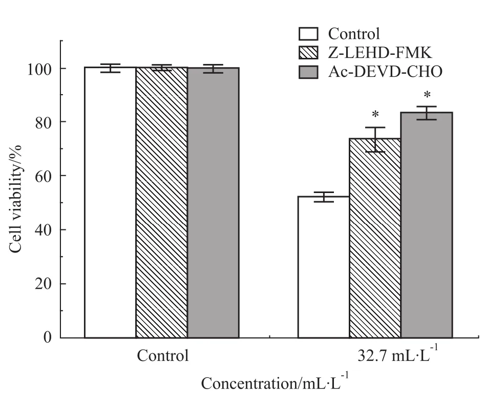
Fig 5 Effect of Z-LEHD-FMK and Ac-DEVD-CHO on the inhibited proliferation of Amaranthus spinosus L.extract
3 讨论
随着消费观念的变化,人们开始追求食品的内在营养、保健食疗,要求无污染、食用安全、方便等。苋菜是一种十分优良的菜、粮、药兼用作物。本文研究结果显示,苋菜提取物可明显抑制各种肿瘤细胞的增殖,其中对HepG2最敏感。有研究表明[6],肿瘤的发生主要是由于细胞的无限增殖和细胞凋亡受
到抑制引起的。本实验Hoechst 33258和流式细胞术检测结果表明,苋菜提取物可诱导HepG2细胞的凋亡。肿瘤细胞的凋亡过程是由多种凋亡相关蛋白参与的。Bcl-2家族蛋白是线粒体膜上调控细胞凋亡的一类蛋白[7],该类蛋白的结合状态将调节线粒体膜电位的变化,改变细胞色素C等相关蛋白的释放,结合Apaf-1等蛋白,进而激活caspase-9凋亡信号通路,激活caspase-3,剪切PARP,导致细胞凋亡[8]。Bcl-2和Bax是Bcl-2家族的两个重要成员,二者可通过形成同源或异源二聚体来调节细胞凋亡[9]。survivin是凋亡抑制蛋白家族的成员之一,具有抑制细胞凋亡的作用[10]。本实验结果表明,苋菜提取物上调抑癌蛋白Bax、Apaf-1,下调促癌蛋白Bcl-2、survivin的表达,同时提高caspase-9、caspase-3的活性,进而诱导HepG2细胞的凋亡。caspase-9和caspase-3特异性抑制剂处理细胞后,抑制了野苋菜提取物引起的细胞凋亡,进一步验证了野苋菜提取物是通过调控caspase-9介导的线粒体凋亡信号转导途径调控细胞凋亡的。
目前,对于苋菜所含成分及药理作用和其制剂的临床应用等方面的研究还不够完善,今后应对苋菜的有效成分,特别是单体成分进行深入研究,进一步使苋菜发挥出更高的药用价值和经济价值。
参考文献:
[1] Hilou A,Nacoulma O G,Guiguemde T R.In vivo antimalarial ac-tivities of extracts from Amaranthus spinosus L.and Boerhaavia erecta L.in mice[J].J Ethnopharmacol,2006,103(2):236-40.
[2] 玄永浩,金银哲,刘 旭,金英善.苋菜药理作用研究进展[J].长江蔬菜,2010,22(2):1-4.
[2] Xuan Y H,Jin Y Z,Liu X,Jin Y S.Research advances in phar- macology of Amaranthus spinosus L.[J].J Changjiang Vegeta-bles,2010,22(2):1-4.
[3] 岳大海,朱毓卉,蔺新英,等.野苋菜对高脂血症大鼠血脂水平的影响[J].中国老年学杂志,2012,32(6):1198-200.
[3] Yue D H,Zhu Y H,Lin X Y,et al.The effect of wild amaranth on serum lipid level in hyperlipidemia rats[J].Chin J Geront,2012,32(6):1198-200.
[4] Ray R B,Raychoudhuri A,Steele R,Nerurkar P.Bitter melon (Momordica charantia)extract inhibits breast cancer cell prolifera-tion by modulating cell cycle regulatory genes and promotes apopto-sis[J].Cancer Res,2010,70(5):1925-31.
[5] Chen Y Q,Liu J J,Yuan B,et al.Methylated actinomycin D,a novel actinomycin D analog induces apoptosis in HepG2 cells through Fas-and mitochondria-mediated pathways[J].Mol Car-cinog,2013,52(12):983-96.
[6] Ouyang L,Shi Z,Zhao S,et al.Programmed cell death pathways in cancer:a review of apoptosis,autophagy and programmed nec-rosis[J].Cell Prolif,2012,45(6):487-98.
[7] 张贝贝,刘文洪,李俊峰,叶志青.铁皮石斛多糖对高糖诱导的血管内皮细胞Bax、Bcl-2表达的影响[J].中国药理学通报,2015,31(1):64-70.
[7] Zhang B B,Liu W H,Li J F,Ye Z Q.Effects of polysaccharides from Dendrobium officinale on expression of Bax and Bcl-2 in vas-cular endothelial cells induced by high sugar[J].Chin Pharmacol Bull,2015,31(1):64-70.
[8] Weyhenmeyer B,Murphy A C,Prehn J H,Murphy B M.Targe-ting the anti-apoptotic Bcl-2 family members for the treatment of cancer[J].Exp Oncol,2012,34(3):192-9.
[9] Hao C,Gao L,Zhang Y,et al.Acetylated chitosan oligosaccha-rides act as antagonists against glutamate-induced PC12 cell ceath via Bcl-2/Bax signal pathway[J].Mar Drugs,2015,13(3):1267-89.
[10]Dang S C,Feng S,Wang P J,et al.Overexpression of Survivin mutant Thr34Ala induces apoptosis and inhibits gastric cancer growth[J].Neoplasma,2015,62(1):81-7.
Anticancer activity and mechanism of apoptosis induced by Amaranthus spinosus L.extract in HepG2 cells
LIU Jin-juan,CAO Cheng-liang,DING Pan,JIANG Ji-hong
(Key Laboratory of Biotechnology for Medicinal Plants of Jiangsu Province,Jiangsu Normal University,Xuzhou Jiangsu 221116,China)
Abstract:Aim To investigate the anticancer activity and the mechanism of the apoptosis induced by Ama-ranthus spinosus L.extract(ASE)in human hepatic carcinoma cell line HepG2.Methods Alamar blue assay was used for detecting the influence of ASE on the proliferation of the cancer cells.The morphological changes of cells were observed under inverted micro- scope and Hoechst 33258 stainning.The apoptosis of HepG2 cells was detected by flow cytometry.Western blot and caspase-3 activity kit were used to detect the protein expression in HepG2 cells.The specific inhibi-tor of caspase-9 and caspase-3(Z-LEHD-FMK and Ac-DEVD-CHO)was used to validate the signal transduc-tion pathyway.Results The results indicated that the
cell proliferation was inhibited by ASE,especicially the HepG2 cells.The HepG2 cells showed obvious apop-totic characteristics.Flow cytometry analysis further validated the apoptosis of HepG2 cells.The expression of Bcl-2 and survivin was downreagulated in HepG2 cells treated with ASE,and Bax,caspase-9,caspase-3,Apaf-1 and PARP were upregualted.Besides,the caspase-3 activity was also increased.Z-LEHD-FMK and Ac-DEVD-CHO significantly increased the cell vi-abilty of HepG2 cells induced by ASE.Conclusion These results confirm that ASE induces the apoptosis of HepG2 through mitochondria-mediated pathway.
Key words:Amaranthus spinosus L.extract;anti-cancer;apoptosis;caspase-9;specific inhibitor;mo-lecular mechanism
作者简介:刘金娟(1982-),女,博士,讲师,研究方向:抗肿瘤药物药理学,E-mail:jjlbest@163.com;蒋继宏(1962-),男,博士,教授,研究方向:生物技术,通讯作者,E-mail:jhjiang@jsnu.edu.cn
基金项目:国家自然科学基金资助项目(No 31370646);江苏师范大学自然科学基金项目(No 14XLA02,13XLA02);江苏省药用植物生物技术重点实验室开放课题(No KLBMP1404)
收稿日期:2015-07-18,修回日期:2015-08-18
文献标志码:A
文章编号:1001-1978(2015)11-1558-05
doi:10.3969/j.issn.1001-1978.2015.11.016
猜你喜欢
中老年保健(2022年2期)2022-08-24
课外语文·中(2022年1期)2022-02-16
中老年保健(2021年5期)2021-12-02
天津医科大学学报(2021年4期)2021-08-21
老友(2021年6期)2021-07-01
现代临床医学(2021年2期)2021-03-29
军民两用技术与产品(2021年10期)2021-03-16
百科知识(2021年2期)2021-02-24
天然产物研究与开发(2018年7期)2018-08-21
中成药(2017年10期)2017-11-16
