组织多普勒及应变率成像技术对二尖瓣狭窄患者右心室收缩功能的评价
商志娟,丛 涛,孙颖慧,王 珂,张树龙
(大连医科大学附属第一医院心内科,辽宁大连 116011)
组织多普勒及应变率成像技术对二尖瓣狭窄患者右心室收缩功能的评价
商志娟,丛 涛,孙颖慧,王 珂,张树龙
(大连医科大学附属第一医院心内科,辽宁大连 116011)
目的 应用组织多普勒(TDI)及应变率成像(SRI)技术评价无右心衰症状的二尖瓣狭窄(MS)患者的右心室(RV)整体及局部心肌收缩功能。方法 单纯性MS患者40例,健康对照组20例。测量左心房前后径(LAD)、肺动脉收缩压(PASP),描记二尖瓣口面积(MVA),M型超声测量三尖瓣环的收缩期位移(TAPSE),计算右室面积变化率(RVFAC)。应用TDI技术测量三尖瓣环收缩期速度(Tric-s),等容收缩期速度(IVV),等容收缩期加速度(IVA)。SRI测量右心室游离壁(RVFW)及室间隔的收缩期应变(S)及应变率(SR),RVFW及室间隔的S及SR的均值作为RV整体长轴的S和SR。并将S和SR与PASP进行相关性分析。结果 与对照组相比,MS组的LAD及PASP明显增高(P<0.01)。MS组的 TAPSE及 Tri-s、IVV、IVA明显低于对照组(P<0.01或P<0.05)。RV长轴整体S及SR,RVFW基底段及中段的S及SR,室间隔基底段S明显低于对照组(P<0.05或P<0.01),室间隔中段S及室间隔各段SR在两组间无差别。RV长轴整体S、SR均与PASP呈负相关,相关系数分别为r=-0.71,r=-0.59(P均<0.05)。结论 应用TDI及SRI技术可以早期发现MS患者RV整体及局部功能的受损情况。
组织多普勒;应变;应变率;二尖瓣狭窄;右心室功能;超声心动图
右心室(right ventricle,RV)功能状况直接影响二尖瓣狭窄(mitral stenosis,MS)患者的症状和生存率[1],在没出现体循环淤血之前,亚临床的RV功能不全容易被忽视[2]。RV形态不规则,解剖结构复杂,收缩功能的定量评价较困难[3],MRI及核素显像能够用来评价RV功能,但这些方法费时,而且价格昂贵,不能广泛应用于临床[4-5]。超声二维应变也可用来评价右室功能,但MS患者多数是房颤心律,因此,超声二维应变在这部分人群中应用受到限制。组织多普勒(tissue doppler imaging,TDI)及其衍生的应变率成像技术(strain rate imaging,SRI)可用来评价 RV 收缩功能[6-7],该技术成熟,方法简单,可行性好,而且不受患者心律的影响,可以广泛应用。本文的目的是应用TDI及其衍生的SRI研究无右心衰症状和体征的MS患者的右心室形态及功能的变化,从而了解MS对右心室整体及局部的收缩功能的影响。
1 资料与方法
1.1 研究对象
1.1.1 MS组:入选2012年1月—2013年3月大连医科大学附属第一医院住院及门诊的单纯性MS患者40例,女性27例,男性13例。轻度狭窄10例、中度狭窄16例、重度14例,年龄42~69岁,平均(57.0 ±8.9)岁,窦性心律11 例,房颤29 例,心功能分级Ⅰ~Ⅱ级,排除标准:具有右心衰的临床表现,糖尿病,高血压,冠心病,除二尖瓣狭窄外的中度或中度以上程度的瓣膜反流或狭窄,肺疾病,左心室射血分数<50%。
1.1.2 健康对照组:同期体检健康人20例。女性13例,男性7例。年龄41~71岁,平均(54.2±8.6)岁,临床、化验、辅助检查排除心肺疾患。
两组间年龄、性别比例、心率及体表面积的差异无显著性意义(P>0.05)。
1.2 仪器与方法
1.2.1 仪器:GE Vivid7超声心动图仪,S4探头,探头频率2.5 ~4.0 MHz,配有定量分析软件。
1.2.2 方法:受检者均左侧卧位、连接同步心电图,在呼气末采集3~5个心动周期图像。测量左心室舒张末内径(left ventricular end-diastolic diameter,LVEDD)、左心房前后径(left atrial end-systolic diameter,LAD)、心尖四腔切面右心室中部横径(right ventricular diameter,RVD)、描记二尖瓣口面积(mitral valve area,MVA),跨二尖瓣口的平均压差(mean pressure gradient,MPG)、通过三尖瓣反流法压差法估测肺动脉收缩压(pulmonary artery systolic pressure,PASP),用双平面 Simpson法测量左心室射血分数(left ventricular ejection fraction,LVEF)。获得心尖四腔心切面,描记右室舒张末和收缩末期的面积,并计算面积变化率(right ventricular fractional area change,RVFAC),将M型超声的取样线通过右心室游离壁与三尖瓣环交界处,测量三尖瓣环收缩期位移(tricuspid annular plane systolic excursion,TAPSE)。进入TVI模式,将脉冲TDI的取样容积置于三尖瓣环与右室游离壁交界处,可获得三尖瓣环收缩期速度(tricuspid annulus S-wave velocity,Tric-s)、等容收缩期速度(peak isovolumic contraction velocity,IVV)及IVV与加速时间的比值即等容收缩期加速度(isovolumic myocardial acceleration,IVA)。采集心尖四腔动态图像,帧频>120 f/s,清晰显示右心室游离壁(right ventricular free wall,RVFW)及室间隔,选择strain及strain rate程序,测量RVFW及室间隔的基底段及中间段的收缩期应变(strain,S)及应变率(strain rate,SR),取 RVFW及室间隔的S和SR的平均值作为RV整体长轴的S和SR值。上述测量值在窦性心律时取3个心动周期的平均值,房颤心律时取5个心动周期的平均值。
1.3 统计学方法
应用SPSS11.5分析软件,计量资料以±s表示,两组间比较用独立样本的t检验;变量间的相关性分析应用Pearson直线相关分析,分类变量用百分数来表示,两组间比较用χ2检验,P<0.05为差异有统计学意义。
2 结果
2.1 MS与对照组常规超声心动图比较
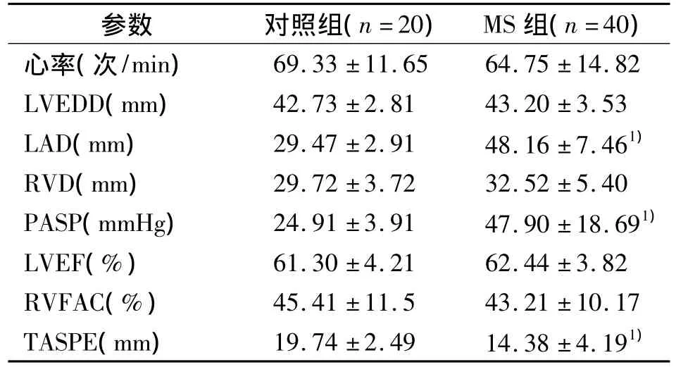
MS组的 MPG 为(9.40 ±5.80)mmHg,MVA 为(1.07 ±0.31)cm2。MS组 LVEDD、LVEF 及 RVFAC与对照组比较差异无显著性意义(P>0.05),MS组LAD、PASP、TASPE与对照组比较差异有显著性意义(P<0.01),MS组的RVD与对照组比较差异无显著性意义(P=0.05)。见表1。

表1 MS组与对照组常规超声参数Tab 1 Conventional echocardiographic parameters in MS group and control group
2.2 MS组与对照组的TDI参数的比较
与对照组相比,MS组的Tri-s、IVV及IVA明显低于对照组(P<0.05)。见表2。
表2 MS组与对照组TDI参数的比较Tab 2 TDI parameters in MS group and control group(±s)

表2 MS组与对照组TDI参数的比较Tab 2 TDI parameters in MS group and control group(±s)
1)与对照组比较,P均<0.05
参数 对照组(n=20) MS组(n=40)Tri- s(cm/s) 14.52 ±2.12 12.52 ±2.431)IVV(cm/s) 12.32 ±2.13 10.34 ±2.331)IVA(m/s2) 4.41 ±1.02 3.85 ±1.111)
2.3 MS组与对照组的SRI参数的比较
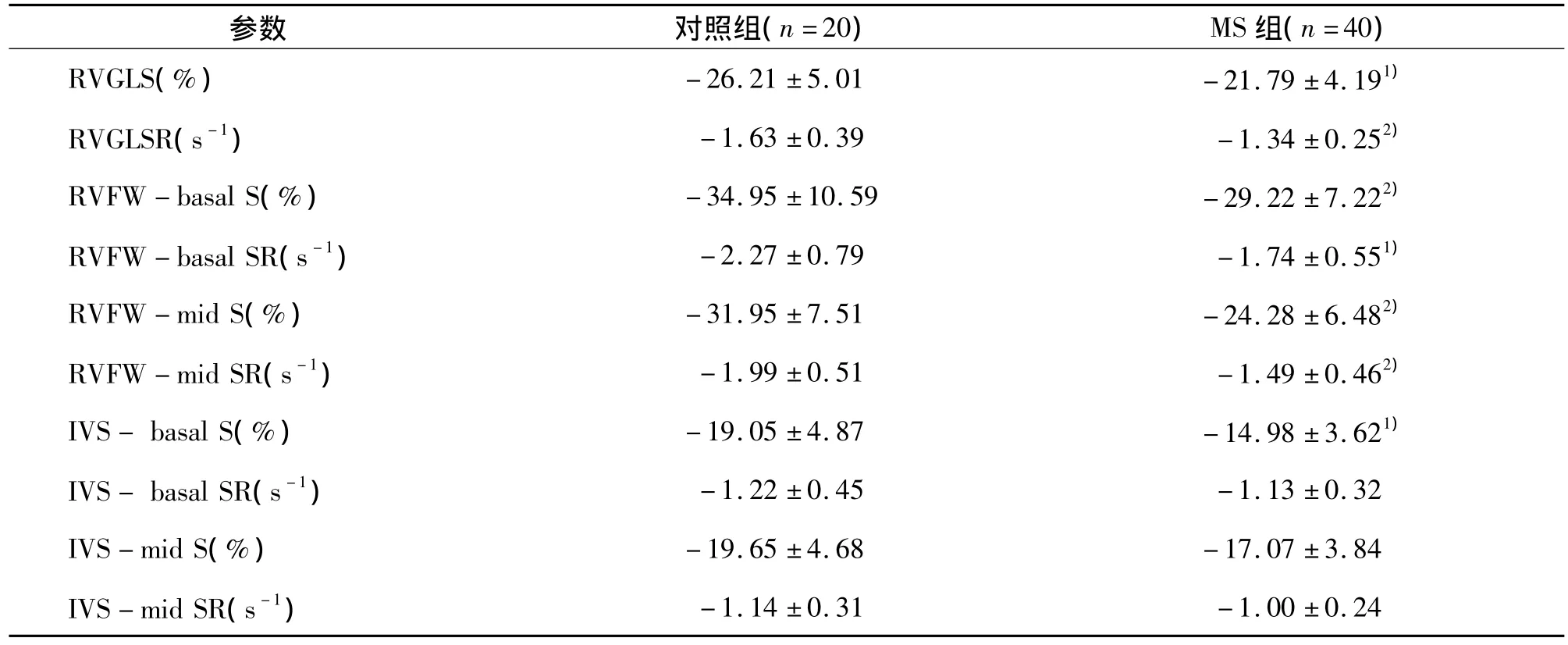
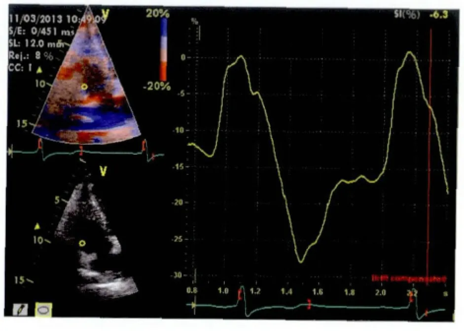
如表3所示,MS组RV长轴整体S及SR显著低于对照组(P<0.01或 P<0.05),节段性分析结果是,RVFW基底段及中段S及SR,室间隔基底段S明显降低(P<0.01或 P<0.05),室间隔中段 S、室间隔基底段及中段SR呈降低趋势,但差异无显著性意义。相关性分析表明RV长轴整体S、SR与PASP呈负相关,r= -0.71和 r= -0.59(P均 <0.05),与 LAD 呈负相关,r= -0.51(P <0.05),与MVA均无相关性。MS患者及正常对照组的应变曲线见图1和图2。
表3 MS组与对照组SRI参数的比较Tab 3 SRI parameters in MS group and control group (±s)

表3 MS组与对照组SRI参数的比较Tab 3 SRI parameters in MS group and control group (±s)
1)与对照组比较,P<0.05;2)与对照组比较,P<0.01;RVGLS:右心室整体应变;RVGLSR:右心室整体应变率;RVFW:右心室游离壁;IVS:室间隔;basal:基底段;mid:中段
参数 对照组(n=20) MS组(n=40)RVGLS(%) -26.21 ±5.01 -21.79 ±4.191)RVGLSR(s-1) -1.63 ±0.39 -1.34 ±0.252)RVFW -basal S(%) -34.95±10.59 -29.22±7.222)RVFW -basal SR(s-1) -2.27±0.79 -1.74±0.551)RVFW -mid S(%) -31.95±7.51 -24.28±6.482)RVFW -mid SR(s-1) -1.99±0.51 -1.49±0.462)IVS- basal S(%) -19.05±4.87 -14.98±3.621)IVS- basal SR(s-1) -1.22±0.45 -1.13±0.32 IVS-mid S(%) -19.65 ±4.68 -17.07 ±3.84 IVS -mid SR(s-1)-1.14 ±0.31 -1.00 ±0.24

图1 MS患者右心室游离壁基底段应变曲线图Fig 1 Strain curves for basal segment of right ventricular free wall in MS patient

图2 健康对照组的右心室游离壁基底段应变曲线图Fig 2 Strain curves for basal segment of right ventricular free wall in healthy subject
3 讨论
右心室的收缩功能直接影响MS患者的运动耐量、预后和生存率[1],如果患者的右心室功能差,不能耐受手术过程中的各种刺激,会导致各种恶性心律失常的出现,以及术后低心排综合征。因此MS患者的右心室功能越来越引起人们的重视。由于RV形态不规则,结构复杂,传统超声心动图对其功能的评价受到限制。有研究证实RV心肌在收缩过程中大部分是沿长轴方向运动的[8],心肌长轴方向上的缩短对每搏量起主要作用[9],因此长轴功能可作为MS患者的RV功能的有效评价指标。而TDI及SRI技术能够不依赖心室的形态定量评价RV长轴方向上的收缩功能。
有研究证实Tri-s、TAPSE可以用来准确评估RV的长轴的收缩功能[10-11],本研究发现 MS患者RVD改变不明显,但Tri-s、TAPSE明显降低,说明在没出现右心衰症状及RV形态没发生改变时已经出现了收缩功能减低。IVA是评价RV整体收缩功能的新指标,相对不受前后负荷状态的影响[12],已有研究证实了其评价MS患者RV功能的有效性[13-14],本研究发现MS患者的IVA明显低于对照组,说明RV整体收缩功能已经受损。
应变(S)是指心肌受力后长度变化,以百分数表示,正值代表伸长,负值代表缩短;应变率(SR)是指单位时间内的应变,指形变发生的速率。两个参数都能够反映心肌收缩功能,一项研究心肌功能的动物实验证实了局部应变反映了局部射血分数,整体应变反映了整体的射血分数[15]。SR反映了心肌的收缩性能,能够反映局部和整体心肌收缩功能。长轴S及SR已用来精确评价RV的收缩功能[7],本研究发现MS组患者RV整体的S、SR明显降低,RVFW基底段及中段S、SR明显降低,这与文献报道的结论一致[16-17]。说明了MS患者在没出现临床症状时已经出现RV整体及局部收缩功能减低。室间隔只有基底段S显著下降,其他节段的S、SR两组间差别不显著,分析原因可能与室间隔的心肌包括左右心室两种纤维成分,右心室的心肌纤维对压力负荷敏感,而左心室心肌纤维对压力负荷不敏感,本组MS患者仅是中度的肺动脉高压,肺动脉的压力还没增加到使室间隔所有节段的S及SR明显下降的程度,而室间隔基底段S显著下降可能与室间隔基底部距离二尖瓣环较近,容易受到风湿侵及导致心肌收缩功能减弱。相关性分析表明,RV整体S和SR与肺动脉收缩压呈负相关,提示RV收缩功能的减低与肺动脉压力的增高有关,MS的病理生理过程导致左心房压力增高,肺静脉高压,进而导致肺动脉高压,后负荷增加,RV室壁薄,收缩功能对后负荷的变化很敏感,后负荷是RV射血分数的主要决定因素[18],有研究证明肺动脉压力增加,RV后负荷增大,能够显著降低RV的收缩功能指标,包括心肌应变和速度[19-20],这与本研究的结论一致。
本研究具有局限性,TDI及SRI技术都具有角度依赖性,为了克服这一弱点,尽量使采取单个室壁显示的方法,使得声束方向与室壁尽量平行。
本研究结果提示,利用TDI及SRI成像技术可以早期发现MS患者RV整体及局部功能的受损情况,收缩功能受损的程度与肺动脉压力的升高有关,与二尖瓣狭窄程度无关。
:
[1]Mohan JC,Sengupta PP,Arora R,et al.Immediate and delayed effects of successful percutaneous transvenous mitral commissurotomy on global right ventricular function in patients with isolated mitral stenosis [J].Int J Cardiol,1999,68(2):217-223.
[2]Bonow RO,Carabello BA,Chatterjee K,et al.ACC/AHA2006 guidelines for the management of patients with valvular heart disease:A report of the American college of cardiology/american heart association task force on practice guidelines(writing committee to revise the 1998 guidelines for the management of patients with valvular heart disease)developed in collaboration with the society of cardiovascular anesthesiologists endorsed by the society for cardiovascular angiography and interventions and the society of thoracic surgeons[J].J Am Coll Cardiol,2006,48:e1 -148.
[3]Rudski LG,Lai WW,Afilalo J,et al.Guidelines for the echocardiographic assessment of the right heart in adults:A report from the American society of echocardiography endorsed by the european association of echocardiography,a registered branch of the european society of cardiology,and the canadian society of echocardiography[J].J Am Soc Echocardiogr,2010,23(7):685 -713.
[4]Brookes CI,White PA,Bishop AJ,et al.Validation of a new intraoperative load-independent indices of right ventricular performance in patients undergoing cardiac operations[J].J Thorac Cardiovasc Surg,1998,116(3):468-476.
[5]Helbing WA,Bosch HG,Maliepaard C,et al.Comparison of echocardiographic methods with magnetic resonance imaging for assessment of right ventricular function in children[J].Am J Cardiol,1995,76(8):589 -594.
[6]Meluzin J,Spinarova L,Bakala J,et al.Pulsed doppler tissue imaging of the velocity of tricuspid annular systolic motion;a new,rapid,and non-invasive method of evaluating right ventricular systolic function[J].Eur Heart J,2001,22(4):340-348.
[7]Sevimli S,Gundogdu F,Aksakal E,et al.Right ventricular strain and strain rate properties in patients with right ventricular myocardial infarction[J].Echocardiography,2007,24(7):732-738.
[8]Torrent- Guasp F,Ballester M,Buckberg GD,et al.Spatial orientation of the ventricular muscle band:physiologic contribution and surgical implications[J].J Thorac Cardiovasc Surg,2001,122(2):389 -392.
[9]Voelkel NF,Quaife RA,Leinwand LA,et al.Right ventricular function and failure:report of National Heart,Lung,and Blood Institute working group on celluar and molecular mechanism of right heart failure[J].Circulation,2006,114(17):1883-1891.
[10]Deswarte G,Richardson M,Polge AS,et al.Longitudinal right ventricular function as a predictor of functional capacity in patients with mitral stenosis:an exercise echocardiographic study[J].J Am Soc Echocardiogr,2010,23(6):667-672.
[11]Miller D,Farah MG,Liner A.et al.The relation between quantitative right ventricular ejection fraction and indices of tricuspid annular motion and myocardial performance[J].J Am Soc Echocardiogr,2004,17(5):443 -447.
[12]Vogel M,Derrick G,White PA,et al.Systemic ventricular function in patients with transposition of the great arteries after atrial repair:a tissue Doppler and conductance catheter study[J].J Am Coll Cardiol,2004,43(1):100-106.
[13]Sade LE,Ozin B,Ulus T,et al.Right ventricular contractile reserve in mitral stenosis:implications on hemodynamic burden and clinical outcome[J].Int J Cardiol,2009,135(2):193-201.
[14]Tayyareci Y,Nisanci Y,Umman B,et al.Early detection of right ventricular systolic dysfunction by using myocardial acceleration during isovolumic contraction in patients with mitral stenosis[J].Eur J Echocardiogr,2008,9(4):516-521.
[15]Weidemann F,Jamal F,Sutherland GR,et al.Myocardial function defined by strain rate and strain during alterations in inotropic states and heart rate[J].Am J Physiol Heart Circ Physiol,2002,283(2):H792 - H799.
[16]Tanboga IH,Kurt M,Bilen E,et al.Assessment of right ventricular mechanics in patients with mitral stenosis by two-dimensional deformation imaging[J].Echocardiography,2012,29(8):956-961.
[17]Yildirimturk O,Helvacioglu FF,Tayyareci Y,et al.Assessment of right ventricular endocardial dysfunction in mild-to-moderate mitral stenosis patients using velocity vector imaging[J].Echocardiography,2012,29(1):25-33.
[18]Nagel E,StuberM,Hess O,et al.Importance of the right ventricle in valvular heart disease[J].Eur Heart J,1996,17(6):829-836.
[19]Lopez- Candales A,RajagopalanN,Dohi K,et al.Abnormal right ventricular myocardial strain generation in mild pulmonary hypertension[J]. Echocardiography,2007,24(6):615-622.
[20]Pirat B,McCulloch M,Zoghbi WA,et al.Evaluation of global and regional right ventricular systolic function in patients with pulmonary hypertension using a novel speckle tracking method [J].Am J Cardiol,2006,98(5):699-704.
Assessment of right ventricular systolic function in patients with mitral stenosis using tissue Doppler imaging and strain rate imaging
SHANG Zhi-juan,CONG Tao,SUN Ying-hui,WANG Ke,ZHANG Shu-long
(Department of Cardiology,the First Affiliated Hospital of Dalian Medical University,Dalian116011,China)
[Abstract]ObjectiveTo investigate the global and regional longitudinal systolic function of right ventricle(RV)using tissue Doppler imaging(TDI)and strain rate imaging(SRI)in mitral stenosis(MS)patients without clinical symptoms of right heart failure.MethodsConventional echocardiography and TDI,SRI analysis were performed in 40 MS patients and 20 normal subjects.left atrial end-systolic diameter(LAD),pulmonary artery systolic pressure(PASP)were measured.End-diastolic and end-systolic areas of RV were measured from the apical four-chamber view to calculate RV fractional area change(RVFAC),tricuspid annular plane systolic excursion(TAPS)was determined in apical four-chamber view.Parameters derived from TDI and SRI included tricuspid annulus S-wave velocity(Tric-s),peak isovolumic contraction velocity(IVV)and myocardial acceleration during isovolumic contraction(IVA),longitudinal systolic strain(S)and strain rate(SR)of right ventricular free wall(RVFW)and septum.The average of RVFW and septum S,SR were defined as the whole S,SR of RV.Correlation between S,SR and PASP were analyzed.ResultsCompared with normal subjects,LAD and PASP were significantly higher in patients with MS(P<0.01),TAPSE and Tric-s(IVV)and(IVA)were significantly lower in MS patients(P <0.01 or P <0.05).S and SR of the global RV and all segments of RVFW were significantly reduced in MS patients(P <0.05 or P <0.01),regional longitudinal S was significantly reduced in the basal septum(P <0.05).Correlation analysis indicated a significant relationship between the PASP and global S,SR of RV.ConclusionRV global and regional systolic function evaluated by TDI and SRI analysis in patients with MS has shown decreased before right heart failure.
[Key words]tissue Doppler imaging;strain;strain rate;mitral stenosis;right ventricular function;echocardiography
R445.1
A
1671-7295(2014)01-0057-05
商志娟,丛涛,孙颖慧,等.组织多普勒及应变率成像技术对二尖瓣狭窄患者右心室收缩功能的评价[J].大连医科大学学报,2014,36(1):57 -61.
10.11724/jdmu.2014.01.14
商志娟(1980-),女,辽宁葫芦岛人,主治医师,硕士。E-mail:zhijuansh1@hotmail.com
2013-07-03;
2013-09-13)

