准分子激光角膜屈光手术后角膜后表面非球面性的变化
2014-07-25 11:29张晓琳范光忠叶树波
眼科新进展 2014年4期
张晓琳 范光忠 叶树波
准分子激光角膜屈光手术后角膜后表面非球面性的变化
张晓琳 范光忠 叶树波
准分子激光角膜屈光手术;角膜后表面;非球面性
目的探讨准分子激光角膜屈光手术后角膜后表面非球面性的变化。方法选取2012年3月至2013年3月于我院接受准分子激光角膜屈光手术治疗的近视患者120例,均选右眼进行研究,分别于术前、术后1个月及术后6个月采用Pentacam系统检查患者角膜后表面在6 mm、7 mm、8 mm、9 mm等不同直径下的Q值(Q6、Q7、Q8、Q9),并检测术前中央角膜厚度(central corneal thickness,CCT)和术中激光切削深度(ablation depth,AD)。结果术后1个月,所有患者角膜后表面Q值均高于术前Q值,其中术后Q6、Q7、Q8与术前比较,差异均有统计学意义(均为P<0.05),而Q9与术前比较,差异无统计学意义(P>0.05);术后6月,所有患者Q值均低于术前,但只有行LASIK患者Q9与术前比较,差异有统计学意义(P=0.017),其余与术前比较,差异均无统计学意义(均为P>0.05);术后6个月所有患者角膜后表面Q值均较术后1个月明显有所下降,差异均有统计学意义(均为P<0.05)。所有患者手术前后Q值变化与CCT和AD均无明显的相关性(均为P>0.05)。结论准分子激光角膜屈光手术后早期能影响角膜后表面的非球面性,Q值增大,后逐渐恢复至术前水平。
[眼科新进展,2014,34(4):364-365,368]
随着准分子激光屈光手术的不断应用和推广,让大部分患者恢复了梦寐以求的良好视力,但也出现了手术所致的不良反应,如眩光、光晕等,影响其最终疗效[1]。针对手术前后角膜前表面的形态和功能变化已有相当多的研究[2],但有关角膜后表面的形态学研究还比较少。随着研究的深入,学者们对角膜后表面的认识越来越深刻,逐渐意识到屈光手术后近期和远期的不良反应、并发症等可以通过对角膜后表面的形态和功能学研究来预测和判断[3-4]。本研究采用Pentacam系统对近视患者屈光手术后角膜后表面形态学变化进行检查分析,以期为术后并发症的预防提供一定的参考依据。
1 资料与方法
1.1一般资料选取2012年3月至2013年3月于我院接受准分子激光角膜屈光手术治疗的近视患者120例,均选右眼进行研究,其中男50例,女70例,年龄(24.36±5.01)岁,球镜度(-4.53±1.12)D,柱镜度(-0.60±0.51)D。62例接受LASIK手术,58例接受Epi-LASIK。入选标准:患者术前无活动性眼部疾患,术前需停止配戴角膜接触镜2周以上。排除眼部其他器质性病变,排除严重心、肝、肺、肾等重要脏器功能不全疾病。
1.2检查方法分别于术前、术后1个月、6个月应用Pentacam系统(德国OculusGmbH公司)对入组手术治疗的患者进行检查分析,记录角膜后表面在6 mm、7 mm、8 mm、9 mm等不同直径下的Q值(Q6、Q7、Q8、Q9),并检测术前中央角膜厚度(central corneal thickness,CCT)和术中激光切削深度(ablation depth,AD)。

2 结果
2.1手术前后角膜后表面Q值比较手术前后角膜后表面Q值变化见表1和表2。从表1、表2可知,术后1个月,所有患者角膜后表面Q值均高于术前,其中术后Q6、Q7、Q8与术前比较,差异均有统计学意义(均为P<0.05),而Q9与术前比较,差异无统计学意义(P>0.05);术后6个月,所有患者Q值均低于术前,但只有行LASIK患者Q9与术前比较,差异有统计学意义(P=0.017),其余与术前比较,差异均无统计学意义(均为P>0.05);术后6个月所有患者角膜后表面Q值均较术后1个月明显下降,差异均有统计学意义(均为P<0.05)。

表1 行LASIK患者手术前后角膜后表面Q值变化Table 1 Changes of preoperative and postoperative Q value on corneal posterior surface in patients with LASIK(±s)
2.2角膜后表面Q值变化与CCT、AD之间的关系以术后1个月数据为基础,采用直线相关分析手术前后角膜后表面Q值变化(△Q)与CCT、AD的相关性,结果发现,所有患者手术前后Q值变化(△Q)与CCT和AD均无明显的相关性(均为P>0.05,见表3)。

表2 行Epi-LASIK患者手术前后角膜后表面Q值变化Table 2 Changes of preoperative and postoperative Q value on corneal posterior surface in patients with Epi-LASIK
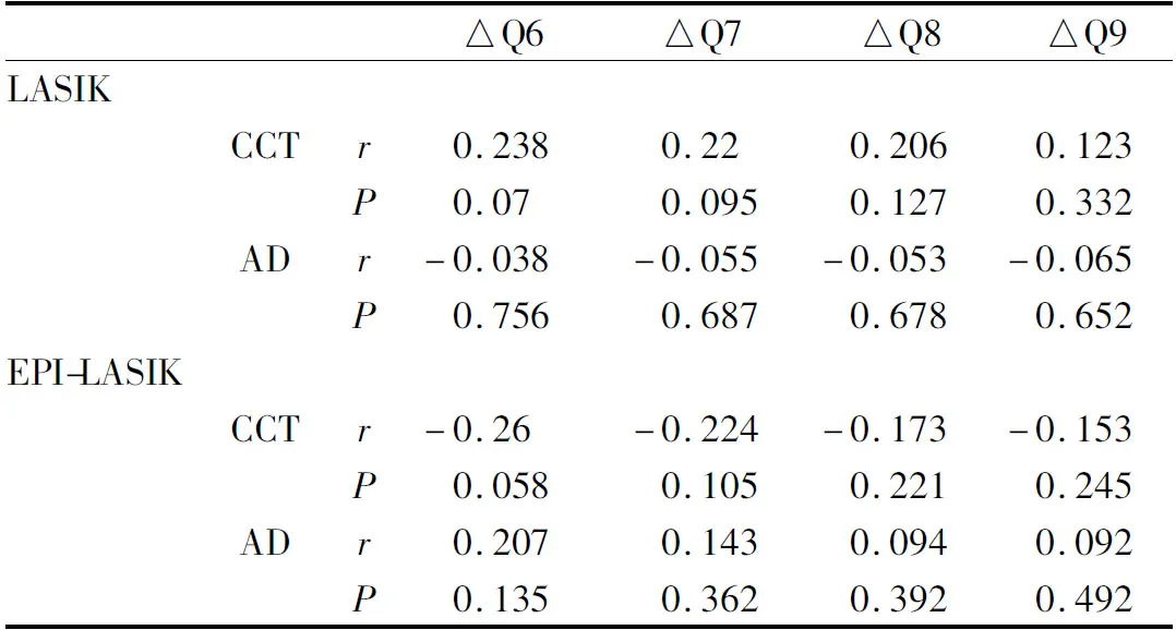
表3 手术前后角膜后表面Q值变化(△Q)与CCT、AD的相关性Table 3 Correlation between Q value changes before and after surgery (△Q)with CCT and AD
3 讨论
准分子激光角膜屈光手术对角膜组织进行适度、适量切削,改变角膜前表面的椭球形状,促使其转变为扁球形,从而降低角膜屈光力,矫正屈光不正[5]。但也正由于手术的原因,角膜组织受切削后,尤其是术中切削过多的情况下,角膜组织会不可逆转地变薄,再加上术后患者眼压或多或少的变化,都有可能会影响到角膜后表面非球面性的改变,如能早期了解术后患者角膜后表面形态变化,对于预测术后是否会出现继发性角膜扩张等有着积极的意义[6]。
Pentacam作为一种新型角膜地形图系统,能够更准确地描绘角膜后表面的真实形态。不同于传统的高度值等指标,用Q值来描述非球面系数,能对角膜后表面的特性进行更为整体化的非球面性分析[7]。屈光手术中,激光切削角膜组织以后,角膜基质的减少必然会使得剩余基质间的张力降低,继而板层必会受到周边组织(角膜基质)对其产生的牵拉力,再结合术后患者眼压的变化,最终的结果就是角膜后表面向扁球形发展,导致了患者角膜后表面的形态学改变[8]。本研究利用Pentacam系统对屈光手术后角膜后表面不同区域的非球面形态变化进行研究,结果显示,无论是实施LASIK手术或是Epi-LASIK手术,术后1个月角膜后表面的Q值均有所增加,即角膜后表面术后1个月较术前变得更扁平;而至术后6个月时,所有患者Q值均低于术前,但只有行LASIK患者Q9与术前比较,差异有统计学意义(P=0.017),其余与术前比较,差异均无统计学意义(均为P>0.05);术后6个月所有患者角膜后表面Q值均较术后1个月明显有所下降,差异均有统计学意义(均为P<0.05)。说明随着角膜手术所致创面的愈合,基质胶原纤维结构恢复术前的连续性和完整性,角膜后表面的Q值基本已降至或低于术前水平,提示患者术后6个月角膜后表面的形态稳定性已与术前差异不大,这也与Smadja等[9]研究结果基本一致。本研究结果还显示,行LASIK与EPI-LASIK患者手术前后角膜后表面Q值的变化与CCT、AD并无明显的相关性,提示屈光手术切削深度和剩余基质床厚度并未对角膜后表面Q值产生明显的影响[10]。
综上所述,准分子激光角膜屈光手术早期能够使得原本大致呈椭球形的角膜后表面形态向扁球形发展(即后表面Q值增加),而术后6个月角膜后表面的形态又会逐渐发生适应性改变,最终与术前水平保持一致。
1 Lai IA,Wang IJ,Hsieh YT.Persistent adherence of lens capsule fragment to posterior corneal surface after cataract surgery[J].CanJOphthalmol,2012,47(6):e51-52.
2 Yan P,Du Z,Wu N,Zhang Y,Xu YL.Minor influence of sub-bowman keratomileusis on the posterior corneal surface at early stage[J].CurrEyeRes,2013,38(8):871-879.
3 Vetter JM,Butsch C,Faust M,Schmidtmann I,Hoffmann EM,Sekundo W,etal.Irregularity of the posterior corneal surface after curved interface femtosecond laser-assisted versus microkeratome-assisted descemet stripping automated endothelial keratoplasty[J].Cornea,2013,32(2):118-124.
4 Sogutlu E,Pinero DP,Kubaloglu A,Alio JL,Cinar Y.Elevation changes of central posterior corneal surface after intracorneal ring segment implantation in keratoconus[J].Cornea,2012,31(4):387-395.
5 Cheng LS,Tsai CY,Tsai RJ,Liou SW,Ho JD.Estimation accuracy of surgically induced astigmatism on the cornea when neglecting the posterior corneal surface measurement[J].ActaOphthalmol,2011,89(5):417-422.
6 Lombardo M,De Santo MP,Lombardo G,Schiano Lomoriello D,Desiderio G,Ducoli P,etal.Surface quality of femtosecond dissected posterior human corneal stroma investigated with atomic force microscopy[J].Cornea,2012,31(12):1369-1375.
7 Mahmoud AM,Nunez MX,Blanco C,Koch DD,Wang L,Weikert MP,etal.Expanding the cone location and magnitude index to include corneal thickness and posterior surface information for the detection of keratoconus[J].AmJOphthalmol,2013,156(6):1102-1111.
8 Vetter JM,Holtz C,Vossmerbaeumer U,Pfeiffer N.Irregularity of the posterior corneal surface during applanation using a curved femtosecond laser interface and microkeratome cutting head[J].Jrefractsurg,2012,28(3):209-214.
9 Smadja D,Santhiago MR,Mello GR,Roberts CJ,Dupps WJ Jr,Krueger RR.Response of the posterior corneal surface to myopic laser in situ keratomileusis with different ablation depths[J].JCataractRefractSurg,2012,38(7):1222-1231.
10 Yamaguchi T,Ohnuma K,Tomida D,Konomi K,Satake Y,Negishi K,etal.The contribution of the posterior surface to the corneal aberrations in eyes after keratoplasty[J].InvestOphthalmolVisSci,2011,52(9):6222-6229.
date:Oct 27,2013
Changes of corneal posterior surface asphericity after excimer laser corneal refractive surgery
ZHANG Xiao-Lin,FAN Guang-Zhong,YE Shu-Bo
excimer laser corneal refractive surgery; corneal posterior surface; asphericity
Objective To analyze the changes of asphericity of corneal posterior surface after excimer laser corneal refractive surgery.Methods Clinical data of patients who received excimer laser corneal refractive surgery at our hospital from March 2012 to March 2013 were retrospectively analyzed, the right eyes of all patients were chosen for study, the Q values of corneal posterior surface under pupil diameter with 6 mm, 7 mm, 8 mm, 9 mm were examined by Pentacam at post-operation and postoperative 1 month, 6 months, the preoperative central corneal thickness (CCT)and intraoperative ablation depth (AD)were also detected.Results At postoperative 1 month, the Q values of corneal posterior surface in all patients were higher than pre-operation, there were statistical differences in Q6, Q7 and Q8 compared with pre-operation (allP<0.05), and no statistical difference in Q9 (P>0.05). At postoperative 6 months, the Q values of all patients were lower than pre-operation, but the difference was statistically significant only in Q9 (P=0.017). The Q values of all patients at postoperative 6 months were obviously lower than those at postoperative 1 month, there were statistical differences (allP<0.05).The changes of Q value before and after operation was not correlated with CCT and AD (allP<0.05).Conclusion The asphericity of corneal posterior surface is influenced by excimer laser corneal refractive surgery at the early time, the Q value increase, and then it return to its original state over time.
张晓琳,女,1974年5月出生,副主任医师。研究方向:准分子手术。联系电话:15096600539;E-mail:zhangxl74@163.com
AboutZHANGXiao-Lin:Female,born in May,1974.Associate chief physician.Tel:15096600539; E-mail:zhangxl74@163.com
2013-10-27
650231 云南省昆明市, 昆明普瑞眼科医院
张晓琳,范光忠,叶树波.准分子激光角膜屈光手术后角膜后表面非球面性的变化[J].眼科新进展,2014,34(4):364-365,368.
��
10.13389/j.cnki.rao.2014.0099
修回日期:2014-01-18
本文编辑:周志新
Accepteddate:Jan 18,2014
From thePuruiOphthalmologicHospitalofKunminCity,Kunming650231,YunnanProvince,China
[RecAdvOphthalmol,2014,34(4):364-365,368]
猜你喜欢
中医眼耳鼻喉杂志(2019年3期)2019-04-13
中国眼镜科技杂志(2017年13期)2017-08-16
中国眼镜科技杂志(2017年13期)2017-08-16
中国眼镜科技杂志(2017年13期)2017-08-16
航天制造技术(2016年6期)2016-05-09
中国继续医学教育(2015年4期)2016-01-07
中国塑料(2015年9期)2015-10-14
中国医疗美容(2015年1期)2015-07-12
中国医疗美容(2015年1期)2015-07-12
中国当代医药(2015年32期)2015-03-01