
Prevention of renal dysfunction by nutraceuticals prepared from oil rich plant foods
2014-03-22 13:01SaharAlOkbiDohaMohamedThanaaHamedRehamSHEsmailSouriaDonya
Sahar Y. Al-Okbi, Doha A. Mohamed*, Thanaa E. Hamed, Reham SH. Esmail, Souria M. Donya
1Food Sciences and Nutrition Department, National Research Centre, Dokki, Cairo, Egypt
2Pathology Department, National Research Centre, Dokki, Cairo, Egypt
3Cytogenetic Department, National Research Centre, Dokki, Cairo, Egypt
Prevention of renal dysfunction by nutraceuticals prepared from oil rich plant foods
Sahar Y. Al-Okbi1, Doha A. Mohamed1*, Thanaa E. Hamed1, Reham SH. Esmail2, Souria M. Donya3
1Food Sciences and Nutrition Department, National Research Centre, Dokki, Cairo, Egypt
2Pathology Department, National Research Centre, Dokki, Cairo, Egypt
3Cytogenetic Department, National Research Centre, Dokki, Cairo, Egypt
PEER REVIEW
Peer reviewer
Chang Gue Son, Professor, OMD, Ph.D. Liver & Immunology Research Center, Daejeon Oriental Hospital of Daejeon Univ, 22-5 Daeheung-dong, Junggu, Daejeon, Korea 301-724. Email: ckson@dju.kr
Ibrahim Hamed, Professor of Nutritional Biochemistry, Food Sciences and Nutrition Department, National Research Centre, Dokki, P.O. Box 12622, Cairo, Egypt. Tel: 0021224457158, Fax: +(202)33370931,
E-mail: hamed5858@yahoo.com
Co-reviewers: Prof. Nagwa M. Ammar, Cairo, Egypt. Prof. Khadiga Ibrahim, Cairo, Egypt.
Comments
This is an important research that showed the possible protection from kidney dysfunction by administration of avocado, walnut, flaxseed and E. sativa extracts.
Details on Page 625
Objective:To investigate the protective effect of extracts prepared from avocado, walnut, flaxseed and Eruca sativa seeds in a rat model of kidney dysfunction induced by intraperitoneal cisplatin.Methods:Ethanol and petroleum ether extracts mixture was prepared from each plant. Six groups of rats were conducted; control healthy, cisplatin group and four test groups where rats were given daily oral dose of each extract mixture before cisplatin injection. Different biochemical and cytogenetic parameters and kidney histopathology were determined. Acute toxicity was tested for the nutraceuticals. Total phenolic contents, fatty acids (FA) and unsaponifiable matter were assessed in the extracts.
Nutraceutical, Cisplatin, Kidney dysfunction, Rats, Oxidative stress
1. Introduction
Kidneys are dynamic organs and represent the major control system maintaining the body haemostasis; they are affected by many chemicals and drugs that may affect their function[1]. Changes in renal function are one of the most common manifestations of severe illness. Their importance is reflected in the routine physiological and biochemical monitoring of kidney function via urine output measures and blood laboratory measurements in critically ill patients[2]. Chronic kidney disease is associated with high morbidity and mortality[3]. Oxidative stress, an imbalance between the generation of reactive oxygen species (ROS) and antioxidant defense capacity of the body, is closely associated with the majority of chronic diseases[4,5]. Elevated ROS can cause oxidative damages to many vital components of the cells thereby expose the body to susceptibility to different diseases[5]. Oxidative stress mediates a wide range of renal impairments, ranging from acute renal failure, obstructive nephropathy and glomerular damage to chronic renalfailure associated with inflammation[6-9]. Increased levels of malondialdehyde and F2-isoprostanes, two products of lipid peroxidation, have been reported in various clinical settings associated with renal damage[10]. Oxidative stress may alter the structure and function of the glomerulus because of the effect of ROS on mesangial and endothelial cells[11]. The glomerulus is considerably more sensitive to oxidative injuries than other nephron parts[12]. Oxidative stress involved in inflammatory lesions caused by series of mediators, including cytokines and chemokines leading to leukocyte activation, production of ROS and increased glomerular damage[13]. Also, the molecules causing inflammation could be produced by the resident renal cells[14].
There is clear evidence that diet rich in fruits, vegetables, and some sorts of oil seeds and nuts are considered as chronic diseases preventive[15-18]. This is due to presence of different active constituents called nutraceuticals. Phytosterols, polyunsaturated fatty acids and phenolic compounds are important constituents of food (nutraceuticals) that have been proved previously to have health benefits such as antioxidant and anti-inflammatory effects, and can be utilized in form of nutraceuticals[19,20]. The aim of the present study was preparation and evaluation of the protective effect of different plants´ extract (nutraceuticals) towards kidney dysfunction. Total phenolic contents, fatty acids and unsaponifiable matter in the nutraceuticals were determined. Assessment of safety of nutraceuticals is among the aim of the present study.
2. Materials and methods
2.1. Plant materials
Avocado (Persea americana), walnut (Juglans regiaL.), flaxseed (Linum usitatissimumL.) andEruca sativa(E. sativa) seeds were purchased from local markets, Cairo, Egypt.
2.2. Main chemical
Cisplatin (platinol, 1 mg/mL) was purchased from Mayne Pharmaceuticals (Warwickshire, UK) to induce nephrotoxicity in rats according to the method of Prabhuet al[21]. The applied dose was 7.5 mg/kg rat body weight (intraperitoneal).
2.3. Animals
Male Sprague-Dawley rats weighing 140-160 g were used in the present study. Animals were obtained from animal house of National Research Centre, Cairo, Egypt. They were kept individually in metabolic stainless steel cages; water and food were givenad libitumall over the experiment. Male and female albino mice of 21-25 g body weight were used in acute toxicity test.
2.4. Methods
2.4.1. Preparation of plant materials
Fresh avocado (whole fruit including seed) was washed by tap water and cut into small pieces. Avocado, walnut, flaxseed andE. sativaseeds were dried separately in an air circulating oven at 40 °C till complete dryness, and then they were reduced into powder.
2.4.2. Preparation of plant extracts
The dried powder of all plants’ were separately placed in a continuous extraction apparatus and subjected to extraction by petroleum ether (40-60 °C) then ethanol. The solvent of each extract was completely removed by evaporation under reduced pressure at a temperature not exceeding 40 °C. All extracts were kept in deepfreeze till used.
2.4.3. Determination of total phenolic contents in extracts
Total phenolics were determined in the ethanol extracts using Folin-Ciocalteu reagent[22]. Absorbance was measured at 765 nm using UVPC spectrophotometer. The total phenolic content was expressed as gallic acid equivalent (GAE) in grams per 100 g.
2.4.4. Assessment of fatty acids, hydrocarbons and phytosterols contents in the petroleum ether extracts (oils)
The unsaponifiable fraction and fatty acid methyl esters of the studied extracts were prepared according to Association of Official Analytical Chemists[23], and were subjected to gas-liquid chromatographic (GLC) analysis of fatty acids, hydrocarbons and phytosterols.
The unsaponifiable fraction was analyzed by GLC adopting the following conditions: column: 10% OV-101 packed column; stationary phase: chromosorb W-HP; detector temperature: 290 °C; injector temperature: 28 °C; carrier gas: N2; flow-rate 30 mL/min; air flow-rate: 300 mL/ min; H2flow-rate 30 mL/min; detector: flame ionization detector; chart speed: 0.5 cm/min; oven program: initial temperature, 70 °C; final temperature: 270 °C; programmed 4 °C/min. The process lasted for 35 min at 270 °C, total time, 85 min. Identification of hydrocarbons and phytosterols contents of the unsaponifiable matter was carried out by comparison of their retention times with co-injected authentic reference compounds. Quantification was based on peak area integration.
Analysis of the methyl ester by GLC was carried out according to the following conditions: stationary phase: 10% diethylene glycosuccinate packed column; oven temperature: 170 °C; detector temperature: 300 °C; injector temperature: 250 °C; carrier gas: N2; flow-rate: 30 mL/min; air flow-rate: 350 mL/min; H2flow-rate: 350 mL/min; detector: flame ionization detector; chart speed: 2 cm/min. Identification of the fatty acid methyl ester was carried out by direct comparison of retention times of each of the separated compounds with authentic samples of the fatty acid methyl esters analyzed under the same conditions. Quantification was based on peak area integration.
2.4.5. Diets
A balanced diet composed of 10% casein, 10% corn oil, 23.5% sucrose, 47% maize starch, 5% fiber, 3.5% salt mixture[24], and 1% vitamin mixture[25] was prepared for feeding the rats all over the experimental period.
2.4.6. Preparation of nutraceuticals
Ethanol and petroleum ether extracts of each plant was mixed together in the ratio of their presence in the whole plant. Each plant extract mixture was dispersed separately in water using the same amount of gum acacia. For the control, the vehicle was prepared through dissolving the same amount of gum acacia in water.
2.5. Experimental procedures
Thirty-six male rats were divided into six groups, each comprised six rats. Four groups were served as the test groups. Rats of the test groups were given daily oral dose of nutraceuticals prepared from avocado,E. sativa, flaxseed or walnut as 250 mg/kg rat body weight. Two groups served as control where rats were given only daily equal amounts of the vehicle received by the test groups. All treatments were given by stomach tube. After 20 d of nutraceuticals´ dosing, rats of all groups except one group of control (served as normal control) were received a single intraperitoneal dose of cisplatin 7.5 mg/kg rat body weight[21]. Rats were maintained on the balanced diet all over the experiment. On the Day 5 after cisplatin administration, 24 h urine was collected from rats and blood samples were obtained from fasted animals. Heparin was used as an anticoagulant and plasma was separated by centrifugation at 3 500 r/min for 10 min and stored at -20 °C till being analyzed. Plasma malondialdehyde (MDA) was determined according to Satoh as indicator of lipid peroxidation and oxidative stress[26]. Plasma total antioxidant capacity and catalase were assessed according to Perez-Gutierrezet al.[27], and Winyardet al.[28], respectively as an antioxidant biomarker. Plasma adiponectin as inflammatory biomarker was determined using Quantikine enzyme-linked immunosorbent kit (R&D Systems, Minneapolis, MN). Plasma creatinine[29], urea[30], total protein[31], and albumin were estimated as indicator of kidney function[32]. After blood sampling, all rats were injected intraperitoneally by 2 mL colchicine (0.2 g/100 mL saline). After 2 h of injection, bone marrow was separated for studying chromosomal aberration. Also the epididymides were excised for assessing sperm-shape abnormalities. Kidney was immediately removed for histopathological examination. Creatinine was determined in the collected 24 h urine for calculation of creatinine clearance[29]. Animal procedures were performed in accordance with the Ethics Committee of the National Research Centre, Cairo, Egypt, and followed the recommendations of the National Institutes of Health Guide for Care and Use of Laboratory Animals (Publication No. 85-23, revised 1985).
2.5.1. Histopathological examination
Kidneys from rats of all the groups were fixed in 10% formaldehyde, dehydrated in graded alcohol and embedded in paraffin. Fine sections were obtained, mounted on glass slides and counterstained with hematoxylin and eosin for light microscopic analysis[33].
2.5.2. Assessment of cytogenetic parameters
For chromosomal aberrations, bone marrow metaphases were prepared following the method of Yosida and Amano[34], and stained with 7% Giemsa stain in phosphate buffer (pH 6.8). Then 100 well spread metaphases per animal were analyzed for chromosomal aberrations. The structural aberrations included gaps, breaks, fragments and deletions, numerical aberrations, tetraploidy and polyploidy.
For sperm shape abnormalities, the epididymides were excised and minced in isotonic sodium citrate solution (2.2%). Fixed in acetic acid:methanol (1:3) once a time. Smears were prepared and sperms were stained with Eosin Y[35]. At least 1 000 sperm per animal (5 000/group) were assessed for morphological abnormalities of the sperm shape.
Acute lethal toxicity test of nutraceuticals was carried out according to Goodmanet al[36]. The 24 h mortality counts among equal sized groups of mice (eight animals/group) receiving progressively increasing oral dose levels of the different extracts (up to 12 g/kg mice body weight) were recorded.
2.6. Statistical analysis
The results of animal experiments were expressed as the mean±SE and they were analyzed statistically using the One-way analysis of variance ANOVA followed by Duncan’s test. In all casesP<0.05 was used as the criterion of statistical significance. For statistical analysis of cytogenetic parametersChi-square test (2×2 contingency table) was applied.
3. Results
Total phenolic content of the ethanol extracts of different plants under study were present as 37.44, 6.34, 5.46 and 47.42 g GAE/100 g ethanol extract in avocado,E. sativa, flaxseed and walnut, respectively.
Tables 1 and 2 shows the fatty acids and unsaponifiable matter in petroleum ether extract of the studied plants, respectively. Avocado oil showed the highest content of linoleic acid (66.00%) followed by walnut oil (46.6%) then flaxseed oil (11.8%) andE. sativaoil (0.84%). Flaxseed oil showed the highest content of linolenic acid (48.4%). Total saturated fatty acids ranged from 9.0% in walnut oil to 10.86% in avocado oil. The highest content of unsaturated fatty acid was present in walnut oil (74.9%) followed by flaxseed oil 73.8% then avocado oil (70.71%) andE. sativaoil (50.04%) (Table 1). GLC investigation of the unsaponifiable matter showed the presence of campesterol, stigmasterol and β-sitosterol in all oils under investigation. The highest content of campesterol was present in avocado oil as 7.90%. Walnut oil showed the highest content of β-sitosterol (6.10%). Total phytosterol was 8.50% in flaxseed oil, 9.30% inE. sativaoil 11.03% in walnut oil and 16.2% in avocado oil. Total hydrocarbon was more or less equal in all plants; it ranges from 5.02% in avocado oil to 15.28% inE. sativaoil.

Table 1 Fatty acids’ content of the petroleum ether extracts of different studied plants (as percentage of total fatty acids).
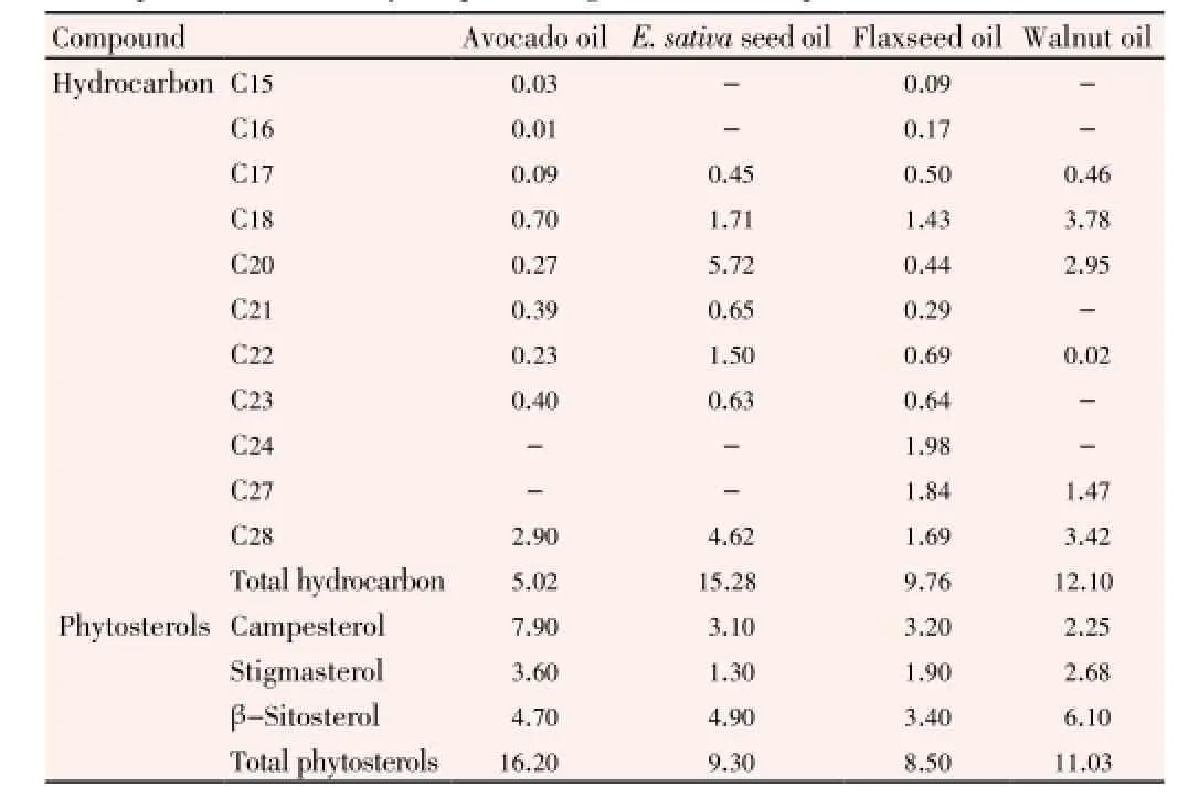
Table 2 GLC analysis of unsaponifiable matter of the different petroleum ether extracts of the plants under study (as percentage of total unsaponifiable matter).
The determined biochemical parameters of different experimental groups are shown in Table 3. Plasma levels of creatinine and urea as indicator of kidney function were significantly elevated in cisplatin control when compared with all groups. Administration of all studied nutraceuticals showed significant reduction in plasma levels of creatinine and urea compared to cisplatin control but still significantly higher than normal control except for avocado nutraceutical that showed non-significant change in creatinine compared to control normal. Plasma levels of total protein and albumin were reduced significantly in cisplatin control compared with control normal. Oral administration of different nutraceuticals showed significant elevation in plasma level of total protein and albumin. Total protein was comparable to control normal in case ofE. sativaand flaxseed groups. MDA level as indicator of lipid peroxidation showed significant increase in cisplatin group compared with normal group. Plants’ nutraceuticals administration showed significant reduction in MDA level with different degrees when compared with cisplatin control but the levels were still significantly higher than control normal group. Plasma levels of catalase and total antioxidant capacity as antioxidant indicator were reduced significantly in cisplatin control compared to control normal group. Administration of different nutraceuticals produced significant elevation of plasma catalase and total antioxidant capacity with different degrees. Catalase levels in the groups treated with flaxseed and walnut nutraceuticals were similar to that of normal rats. Plasma level of total antioxidant capacity ofE. sativaextract group was also normalized. Plasma level of adiponectin was similar among all groups.
Creatinine clearance of different experimental groups is present in Table 4. Creatinine clearance was reduced significantly in cisplatin group compared to normal group. Administration of avocado and flaxseed showed significant increase in creatinine clearance compared with both cisplatin group and normal rats while treatment withE. sativaand walnut produced significant increase in creatinine clearance compared to cisplatin control and which was comparable to normal control.
3.1. Cytogenetic Study
The antigenotoxic effect of different plant nutraceuticals against the genotoxic effect of cisplatin was assessed through studying chromosomal aberration in bone marrow cells and sperm shape abnormalities.
Table 5 illustrates number and percentage of different types of chromosomal aberration in different experimental groups. The percentage induced aberrations in cisplatin control group was found to be statistically highly significantP≤0.001 in including gaps andP≤0.01 after excluding gaps compared to normal control. Breaks and fragments recorded 5.4% of structural aberrations to be the highly affected onewhile numerical aberrations reached 1.2%, which is a marker of carcinogenic effect.

Table 3 Biochemical parameters of different experimental groups.

Table 4 Creatinine clearance of different experimental groups.

Table 5 Number and percentage of different types of chromosomal aberration in bone marrow in different experimental groups.
Pre-treatment of rats withE. sativa, flaxseed, walnut and avocado nutraceuticals significantly decreased the aberration percentage to be 4.6%, 4.6%, 4.2% and 5.4%, respectively, after excluding gaps compared to control cisplatin. Also, significant reduction was noticed in percentage including gaps on administration of different nutraceutical compared to cisplatin control. The previous improvement did not reach that of the control. Table 6 demonstrates the different types of sperm shape abnormalities. Cisplatin induced highly significant percentage of abnormal sperms, reached 7.29% in cisplatin control group compared to normal control. Pretreatment with nutraceuticals inhibited this percentage significantly, the efficiency of nutraceuticals was in the following order, flax seed>avocado>E. sativa>walnut.
3.2. Histopathology
Photomicrographs of kidney sections from various treatment groups are shown in Figure 1. Histopathological examination of sections from rat kidney treated with cisplatin (control rats given cisplatin without nutraceuticals) (Figure 1E and 1F) showed severe and generalized tubular epithelial cell necrosis associated with diffuse tubular lumina (hyalinized casts), diffuse intratubular ducts, foci of fibrosis and congested vessels. The normal control rats were free from any significant pathological changes (Figure 1G).
Administration of avocado nutraceutical afforded some protection against the nephro-toxic effect of cisplatin as compared with the unprotected treated control where rats showed only focal casts in about 30% of the medullary renal tubules, with congested vessels and scattered foci of fibrosis (Figure 1A).E. sativaextract provided complete protection against the nephrotoxic effect of the cisplatin in 70% of the rats while only 30% of them showed focal effect in the form of few casts in some tubules (Figure 1B). Flaxseed nutraceutical produced good protection aginst the nephrotoxic effect of the cisplatin with almost complete protection at histopatholgical level in about 50% of the rats. The changes are noted in halfof the cases in the form of few casts inside the medullary tubules (Figure 1C). Nutraceuticals prepared from walnut exerted partially protective effect against the nephrotoxicity of cisplatin and the kidneys were only focally affected in the form of intratubular castsin in some medullary tubules (Figure 1D).
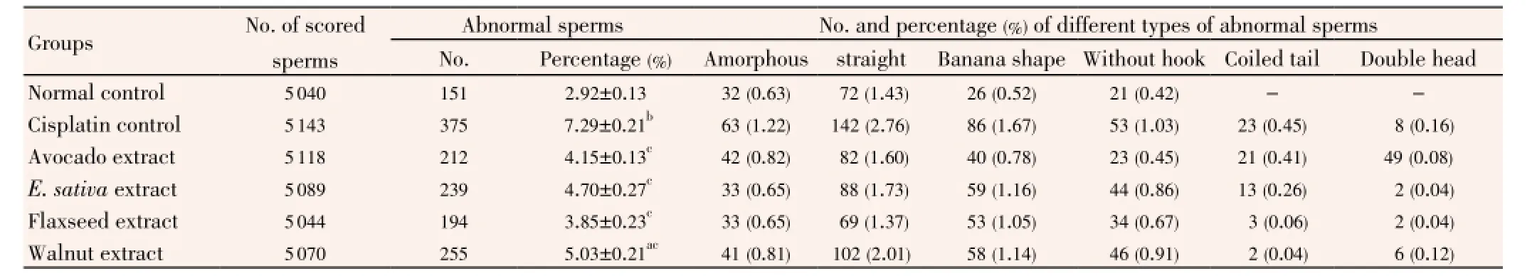
Table 6 Number and mean percentage of different types of sperm shape abnormalities in rat sperms in different experimental groups.
The acute lethal toxicity test revealed that all studied nutraceuticals were very safe up to the highest studied dose (12 g/kg mice body weight) which corresponds to 93 g/70 kg man body weight for human when the dose of mice was extrapolated to corresponding estimates in human adopting interspecies dosage conversion scheme[37]. This reflects the highest safety of the bioactive extracts.
4. Discussion
Induction of kidney dysfunction in experimental animals is important for studying new therapeutic agents including nutraceuticals that may possess therapeutic or protective effect towards kidney dysfunction. Experimental impairment of kidney function is induced through treatment by specific chemical or drugs or through surgical means. In the present research, kidney dysfunction was induced in rats by injection of cisplatin according to Prabhuet al[21]. Cisplatin accumulates in the renal tubular cells approximately five times its extracellular concentration[38]. Consequently, the kidney is considered to be the primary target organ for cisplatin toxicity[1,39]. The mechanism for cisplatin nephrotoxicity may involve decreased protein synthesis, membrane peroxidation, mitochondrial dysfunction, and/or DNA injury and thereby cause tubular injury. Cytochrome P450, a group of heme proteins, may serve as a significant source of catalytic iron in cisplatin-induced nephrotoxicity[40]. Cisplatin nephrotoxicity has also been demonstrated to be mediated by DNAaseI a highly reactive renal endonuclease[38,41]. The nephrotoxicity of the drug may also be due to complex metabolic pathways that activates the drug to become a potent kidney toxin[38,40]. Cisplatin is known to accumulate in the mitochondria of renal epithelial cells and induces ROS in these cells via decreasing the activity of antioxidant enzymes. Reactive oxygen molecules can trigger several apoptotic mechanisms activated by cisplatin. Cisplatin induces apoptotic process by initiating the cytochrome C release and then generating superoxide[42].
Searching new natural agents that might improve renal dysfunction is considered as an important issue nowadays due to increased renal dysfunction cases in Egypt. Natural agents that possess antioxidant, anti-inflammatory and hypotensive effects are expected to possess a renal protective effect.
In the present study, increased creatinine and urea in plasma along with decreased plasma albumin, plasma total protein and creatinine clearance were noticed on treatment with cisplatin. The serum albumin concentration may be directly altered, due to increased loss of albumin through damaged glomeruli in case of renal failure[43]. These results agreed with previous researches[44,45]. In the present study, pretreatment with different nutraceuticals provided a significant protection towards kidney dysfunction reflected in the significant reduction of the levels of plasma creatinine and urea and the increase in plasma albumin, plasma total protein and creatinine clearance.
Significant increase in lipid peroxides represented by malondialdehyde along with significant reduction of the antioxidant catalase enzymes and total antioxidant capacity reflected an elevated oxidative stress on cisplatin treatment in the present study. A significant decline in antioxidant enzymes and increase in free radicals in experimental models as well as in subjects is typical during cisplatin treatment[46]. Signs of injury including the decrease in kidney glutathione levels and increase in MDA levels in addition to decreased activities of kidney superoxide dismutase, catalase, glutathione peroxidase and glutathione s-transferase enzymes are a proof of the oxidative stress caused by cisplatin treatment and have been previously reported in a number of studies[1,44,47].
Thiobarbituric acid reactive substances are produced by lipid peroxidation and are considered as indicators of oxidative stress[48]. Lipid peroxidation is ascribed to a free radical-mediated chain reaction that damages cell membranes and inhibition of this process by different nutraceuticals in the present study is mainly attributed to their high ability of scavenging free radicals. Free radicals induced by cisplatin can evoke extensive tissue damage, reacting with macromolecules, such as membrane lipids, proteins and nucleic acids[49,50]. Such elevated oxidative stress was reflected in the significant reduction in total antioxidant capacity and catalase noticed in rats treated with cisplatin in the present study. Administration of different nutraceuticals produced elevation of total antioxidant capacity and catalase thereby reducing free radicals which may be reflected in improvement of kidney function through reduction of tissue damage and reduction of inflammatoryprocess. This effect can be noticed by the improvement of histopathological changes noticed in the present study. Also reduction in free radicals due to administration of nutraceuticals resulted in reduction of chromosomal aberration and inhibition of percentage of abnormal sperms appeared on treatment with cisplatin.
Nutraceuticals studied in the present research were prepared from avocado, walnut, flaxseed andE. sativaseeds. Avocado is a good source of bioactive compounds such as unsaturated fatty acids, vitamin E and sterols[51], and has been shown to possess antioxidants, anti-inflammatory and hypotensive effect[52,53]. Walnut is a good source of essential fatty acids (linoleic acid), tocopherols, tocotrienols, phytosterols, tannins and other polyphenols[54]. Most phenolic compounds commonly identified in walnut seeds are phenolic acids, namely gallic, ellagic, syringic, 5-O-caffeoylquinic, caffeic, p-coumaric, ferulic and sinapic acids, and tannins, such as glansrins A, B and C, casuarinin and stenophyllarin[55,56], thereby it is expected to possess antioxidant and anti-inflammatory effect[57]. Flaxseed has been reported to have antioxidant effect[58], and beneficial effect towards certain autoimmune kidney disease. Dietary flaxseed and flaxseed oil attenuated the decline in renal function and reduced glomerular injury[59]. It has also been shown that treatment with flaxseed oil caused a significant improvement in histopathological picture of the kidney as well as the kidney function and antioxidant status, against lead acetate-induced renal toxicity[60].E. sativaseeds have been reported to possess a potent antioxidant, antiinflammatory and renal protective activity in rats[61,62]. The renal protective effect of the studied nutraceuticals might be due to presence of phenolic compounds, polyunsaturated fatty acids, and phytosterols as shown from phytochemical analysis in the present study. Phenolic compounds possess several therapeutic properties including antioxidant and anti-inflammatory activity thereby they are free radicals scavengers, singlet oxygen quenchers, and metal chelators, in addition, a direct relationship between total phenolic content and antioxidant activity has been reported[63,64]. Phytosterols have been reported to possess antioxidant and anti-inflammatory activity[65]. Previous studies showed polyunsaturated fatty acids to possess anti-inflammatory activity[66].
Cisplatin induced chromosomal aberration and sperm shape abnormalities might be due to increased oxidative stress during cisplatin treatment. Reduction of such chromosomal aberration and sperm shape abnormalities on nutraceutical administration might be ascribed to their antioxidant activity due to presence of the previously mentioned bioactive compounds especially phenolic compounds. Many of the phenolic compounds have properties including antioxidant, anti-mutagenic, anticarcinogenic and anti-inflammatory effects that might potentially be beneficial in protecting the stability of the genome[67]. A significant cancer risk reduction by avocado extract,E. sativaand walnut extract has been reported which might be related to prevention of DNA damage[68-70].
Cisplatin-induced oxidative stress might be responsible for the induced kidney dysfunction reflected in biochemical parameters and histopathological changes. Cisplatin also resulted in chromosomal aberration and increased percentage of abnormal sperms. Administration of the studied nutraceuticals proved to possess protective role against cisplatin-induced nephrotoxicity, chromosomal aberration and abnormal sperms. All studied nutraceuticals showed complete safety.
Conflict of interest statement
We declare that we have no conflict of interest.
Acknowledgements
The authors thank the National Research Centre, Cairo, Egypt for financing this work. The present research is a part of a project entitled “Prevention and Management of Some Chronic Diseases (Renal and hepatic) by Nutraceuticals and Functional Foods” (2010-2013). This paper is granted and totally funded by National Research Centre, Cairo, Egypt (Ninth plan of National Research Centre) (Grant No. 9130103).
Comments
Background
Kidneys are vital organs in the body. They have an important role in body hemostasis. So natural agents that may guard against renal dysfunction are needed.
Research frontiers
The present research studied kidney protection by different plant food extracts through assessing different biomarkers of renal functions and examining the histopathology of the kidney.
Related reports
Renal dysfunction rat model were reported using cisplatin or by high adenine phosphate diet. These models were used to evaluate new agents that may protect from renal dysfunction.
Innovations and breakthroughs
Avocado, walnut, flaxseed andE. sativaare rich in antioxidant and anti-inflammatory bioactive constituents. In the present study, the others utilized these previous activities to evaluate their protective effect towards kidneys.
Applications
This study recommends the use of avocado, walnut, flaxseed andE. sativaas renal protective natural agent.
Peer review
This is an important research that showed the possible protection from kidney dysfunction by administration of avocado, walnut, flaxseed andE. sativaextracts.
[1] Maliakel DM, Kagiya TV, Nair CK. Prevention of cisplatininduced nephrotoxicity by glucosides of ascorbic acid and alphatocopherol. Exp Toxicol Pathol 2008; 60(6): 521-527.
[2] Hawkins R. New biomarkers of acute kidney injury and the cardio-renal syndrome. Korean J Lab Med 2011; 31: 72-80.
[3] Wen CP, Matsushita K, Coresh J, Iseki K, Islam M, Katz R, et al. Relative risks of chronic kidney disease for mortality and endstage renal diseaseacross races are similar. Kidney Int 2014; doi: 10.1038/ki.2013.553.
[4] Wenceslau CF, McCarthy CG, Szasz T, Spitler K, Goulopoulou S, Webb RC, et al. Mitochondrial damage-associated molecular patterns and vascular function. Eur Heart J 2014; 35: 1172-1177.
[5] Yazdanparast R, Bahramikia S, Ardestani A. Nasturtium officinale reduces oxidative stress and enhances antioxidant capacity in hypercholesterolaemic rats. Chem Biol Interact 2008; 172: 176-184.
[6] Bhandari S, Galanello R. Renal aspects of thalassaemia a changing paradigm. Eur J Haematol 2012; 89(3): 187-197.
[7] Ebrahimi B, Eirin A, Li Z, Zhu XY, Zhang X, Lerman A, et al. Mesenchymal stem cells improve medullary inflammation and fibrosis after revascularization of swine atherosclerotic renal artery stenosis. PLoS One 2013; 8(7): e67474.
[8] Park S, Kim CS, Lee J, Suk Kim J, Kim J. Effect of regular exercise on the histochemical changes of d-galactose-induced oxidative renal injury in high-fat diet-fed rats. Acta Histochem Cytochem 2013; 46(4): 111-119.
[9] Conner TA, McQuade C, Olp J, Pai AB. Effect of intravenous vitamin C on cytokine activation and oxidative stress in endstage renal disease patients receiving intravenous iron sucrose. Biometals 2012; 25(5): 961-969.
[10] Günal SY, Ustündağ B, Günal Aİ. The assessment of oxidative stress on patients with chronic renal failure at different stages and on dialysis patients receiving different hypertensive treatment. Indian J Clin Biochem 2013; 28(4): 390-395.
[11] Yadav AK, Kumar V, Jha V. Heat shock proteins 60 and 70 specific proinflammatory and cytotoxic response of CD4+CD28nullcells in chronic kidney disease. Mediators Inflamm 2013; doi: 10.1155/2013/384807.
[12] Yi X, Nickeleit V, James LR, Maeda N. α-Lipoic acid protects diabetic apolipoprotein E-deficient mice from nephropathy. J Diabetes Complications 2011; 25(3): 193-201.
[13] Ma H, Wu Y, Xu Y, Sun L, Zhang X. Human umbilical mesenchymal stem cells attenuate the progression of focal segmental glomerulosclerosis. Am J Med Sci 2013; 346(6): 486-493.
[14] Lin Q, Chen Y, Lv J, Zhang H, Tang J, Gunaratnam L, et al. Kidney injury molecule-1 expression in IgA nephropathy and its correlation with hypoxia and tubulointerstitial inflammation. Am J Physiol Renal Physiol 2014; 306: F885-F895.
[15] Piscopo S. The Mediterranean diet as a nutrition education, health promotion and disease prevention tool. Public Health Nutr 2009; 12: 1648-1655.
[16] Luciano RL. Acute kidney injury from cherry concentrate in a patient with CKD. Am J Kidney Dis 2014; 63(3): 503-505.
[17] Lu Y, Sun J, Petrova K, Yang X, Greenhaw J, Salminen WF, et al. Metabolomics evaluation of the effects of green tea extract on acetaminophen-induced hepatotoxicity in mice. Food Chem Toxicol 2013; 62: 707-721.
[18] Peterson J, Dwyer J, Adlercreutz H, Scalbert A, Jacques P, McCullough ML. Dietary lignans: physiology and potential for cardiovascular disease risk reduction. Nutr Rev 2010; 68: 571-603.
[19] Davis JM, Murphy EA, Carmichael MD. Effects of the dietary flavonoid quercetin upon performance and health. Curr Sports Med Rep 2009; 8: 206-213.
[20] Rosenbaum CC, O’Mathúna DP, Chavez M, Shields K. Antioxidants and antiinflammatory dietary supplements for osteoarthritis and rheumatoid arthritis. Altern Ther Health Med 2010; 16: 32-40.
[21] Prabhu VV, Kannan N, Guruvayoorappan C. 1,2-Diazole prevents cisplatin-induced nephrotoxicity in experimental rats. Pharmacol Rep 2013; 65(4): 980-990.
[22] Ainsworth EA, Gillespie KM. Estimation of total phenolic content and other oxidation substrates in plant tissues using Folin-Ciocalteu reagent. Nat Protoc 2007; 2(4): 875-877.
[23] Association of Official Analytical Chemists. Official methods of analysis of AOAC international. 17th ed. Gaithersburg, MD, USA: Association of Analytical Communities; 2000.
[24] Briggs GM, Williams MA. A new mineral mixture for experimental rat diets and evaluation of other mineral mixtures. Fed Proc 1963; 22: 261-266.
[25] Morcos SR. The effect of protein value of the diet on the neurological manifestations produced in rats by betaimmodipropionitrile. Br J Nutr 1967; 21: 269-274.
[26] Satoh K. Serum lipid peroxide in cerebrovascular disorders determined by a new colorimetric method. Clin Chim Acta 1978; 90: 37-43.
[27] Perez-Gutierrez RM, Muñiz-Ramirez A, Gomez YG, Ramírez EB. Antihyperglycemic, antihyperlipidemic and antiglycation effects of Byrsonima crassifolia fruit and seed in normal and streptozotocin-induced diabetic rats. Plant Foods Hum Nutr 2010; 65(4): 350-357.
[28] Winyard PG, Ryan B, Eggleton P, Nissim A, Taylor E, Lo Faro ML, et al. Measurement and meaning of markers of reactive species of oxygen, nitrogen and sulfur in healthy human subjects and patients with inflammatory joint disease. Biochem Soc Trans 2011; 39(5): 1226-1232.
[29] Houot O. Interpretation of clinical laboratory tests. Seal Beach: Biomedical publications; 1985.
[30] Fawcett JK, Scott JE. A rapid and precise method for the determination of urea. J Clin Pathol 1960; 13: 156-159.
[31] Rheinhold JG. Total protein, albumin and globulin in standard methods of clinical chemistry. New York: Academic Press Inc.; 1953, p. 88.
[32] Doumas BT, Watson WA, Biggs HG. Albumin standards and the measurement of serum albumin with bromocresol green.1971. Clin Chim Acta 1997; 258: 21-30.
[33] Ekor M, Emerole GO, Farombi EO. Phenolic extract of soybean (Glycine max) attenuates cisplatin-induced nephrotoxicity in rats. Food Chem Toxicol 2010; 48: 1005-1012.
[34] Yosida TH, Amano K. Autosomal polymorphism in laboratory bred and wild Norway rat, Rattus norvegicus. Chromosoma 1965; 16: 658-667.
[35] Wyrobek AJ, Bruce WR. The induction of sperm-shape abnormalities in mice and humans. In: Hollaender A, de Serres FJ, editors. Chemical mutagens: principles and methods for their detection. New York: Plenum Press; 1978, p. 255-285.
[36] Goodman AG, Goodman LS, Gilman A. Goodman and Gilman’s: the pharmacological basis of therapeutics. 6th ed. New York: Macmillan; 1980, p. 1602-1615.
[37] Paget P, Barnes T. Evaluation of drug activities pharmacometrics. New York: Academic Press; 1964, p. 135-140.
[38] Hussein A, Ahmed AA, Shouman SA, Sharawy S. Amelioratingeffect of DL-α-lipoic acid against cisplatin-induced nephrotoxicity and cardiotoxicity in experimental animals. Drug Discov Ther 2012; 6(3): 147-156.
[39] Shalby AB, Assaf N, Ahmed HH. Possible mechanisms for N-acetyl cysteine and taurine in ameliorating acute renal failure induced by cisplatin in rats. Toxicol Mech Methods 2011; 21(7): 538-546.
[40] Peres LA, da Cunha AD Jr. Acute nephrotoxicity of cisplatin: molecular mechanisms. J Bras Nefrol 2013; 35(4): 332-340.
[41] Hodeify R, Megyesi J, Tarcsafalvi A, Safirstein RL, Price PM. Protection of cisplatin cytotoxicity by an inactive cyclindependent kinase. Am J Physiol Renal Physiol 2010; 299(1): F112-F120.
[42] Naqshbandi A, Rizwan S, Khan MW, Khan F. Dietary flaxseed oil supplementation ameliorates the effect of cisplatin on brush border membrane enzymes and antioxidant system in rat intestine. Hum Exp Toxicol 2013; 32(4): 385-394.
[43] Cekmen M, Ilbey YO, Ozbek E, Simsek A, Somay A, Ersoz C. Curcumin prevents oxidative renal damage induced by acetaminophen in rats. Food Chem Toxicol 2009; 47(7): 1480-1484.
[44] Atasayar S, Gürer-Orhan H, Orhan H, Gürel B, Girgin G, Ozgüneş H. Preventive effect of aminoguanidine compared to vitamin E and C on cisplatin-induced nephrotoxicity in rats. Exp Toxicol Pathol 2009; 61(1): 23-32.
[45] Saad AA, Youssef MI, El-Shennawy LK. Cisplatin induced damage in kidney genomic DNA and nephrotoxicity in male rats: the protective effect of grape seed proanthocyanidin extract. Food Chem Toxicol 2009; 47(7): 1499-1506.
[46] Longo V, Gervasi PG, Lubrano V. Cisplatin induced toxicity in rat tissues: the protective effect of Lisosan G. Food Chem Toxicol 2011; 49(1): 233-237.
[47] Zhao M, Isami K, Nakamura S, Shirakawa H, Nakagawa T, Kaneko S. Acute cold hypersensitivity characteristically induced by oxaliplatin is caused by the enhanced responsiveness of TRPA1 in mice. Mol Pain 2012; 8: 55.
[48] Boghdady NA. Antioxidant and antiapoptotic effects of proanthocyanidin and ginkgo biloba extract against doxorubicininduced cardiac injury in rats. Cell Biochem Funct 2013; 31(4): 344-351.
[49] Sindhu ER, Kuttan R. Carotenoid lutein protects the kidney against cisplatin-induced acute renal failure. J Environ Pathol Toxicol Oncol 2013; 32(1): 21-28.
[50] Nicolson GL, Conklin KA. Molecular replacement in cancer therapy: reversing cancer metabolic and mitochondrial dysfunction, fatigue and the adverse effects of cancer therapy. Cancer Genomics Proteomics 2006; 3: 159-168.
[51] Plaza L, Sánchez-Moreno C, de Pascual-Teresa S, de Ancos B, Cano MP. Fatty acids, sterols, and antioxidant activity in minimally processed avocados during refrigerated storage. J Agric Food Chem 2009; 57: 3204-3209.
[52] Gorinstein S, Haruenkit R, Poovarodom S, Vearasilp S, Ruamsuke P, Namiesnik J, et al. Some analytical assays for the determination of bioactivity of exotic fruits. Phytochem Anal 2010; 21: 355-362.
[53] Heinecke LF, Grzanna MW, Au AY, Mochal CA, Rashmir-Raven A, Frondoza CG. Inhibition of cyclooxygenase-2 expression and prostaglandin E2 production in chondrocytes by avocado soybean unsaponifiables and epigallocatechin gallate. Osteoarthritis Cartilage 2010; 18: 220-227.
[54] Pereira JA, Oliveira I, Sousa A, Ferreira I, Bento A, Estevinho L. Bioactive properties and chemical composition of six walnut (Juglans regia L.) cultivars. Food Chem Toxicol 2008; 46: 2103-2111.
[55] Pribis P, Bailey RN, Russell AA, Kilsby MA, Hernandez M, Craig WJ, et al. Effects of walnut consumption on cognitive performance in young adults. Br J Nutr 2011; 107(9): 1393-1401.
[56] Bolling BW, Chen CY, McKay DL, Blumberg JB. Tree nut phytochemicals: composition, antioxidant capacity, bioactivity, impact factors. A systematic review of almonds, Brazils, cashews, hazelnuts, macadamias, pecans, pine nuts, pistachios and walnuts. Nutr Res Rev 2011; 24(2): 244-275.
[57] Muthaiyah B, Essa MM, Chauhan V, Chauhan A. Protective effects of walnut extract against amyloid beta peptide-induced cell death and oxidative stress in PC12 cells. Neurochem Res 2011; 36: 2096-2103.
[58] Upadhyay R, Mohan Rao LJ. An outlook on chlorogenic acidsoccurrence, chemistry, technology, and biological activities. Crit Rev Food Sci Nutr 2013; 53(9): 968-984.
[59] Caligiuri SP, Love K, Winter T, Gauthier J, Taylor CG, Blydt-Hansen T, et al. Dietary linoleic acid and α-linolenic acid differentially affect renal oxylipins and phospholipid fatty acids in diet-induced obese rats. J Nutr 2013; 143(9): 1421-1431.
[60] Abdel Moneima AE, Dkhila MA, Al-Quraishy S. The protective effect of flaxseed oil on lead acetate-induced renal toxicity in rats. J Hazard Mater 2011; 194: 250-255.
[61] Sarwar AM, Kaur G, Jabbar Z, Javed K, Athar M. Eruca sativa seeds possess antioxidant activity and exert a protective effect on mercuric chloride induced renal toxicity. Food Chem Toxicol 2007; 45: 910-920.
[62] Yehuda H, Khatib S, Sussan I, Musa R, Vaya J, Tamir S. Potential skin antiinflammatory effects of 4-methylthiobutylisothiocyanate (MTBI) isolated from rocket (Eruca sativa) seeds. Biofactors 2009; 35: 295-305.
[63] Costa RM, Magalhães AS, Pereira JA, Andrade PB, Valentão P, Carvalho M, et al. Evaluation of free radical scavenging and antihemolyticactivities of Cydonia oblonga leaf: a comparative study with green tea (Camellia sinensis). Food Chem Toxicol 2009; 47: 860-865.
[64] Yang J, Liu R, Halim L. Antioxidant and antiproliferative activities of common edible nut seeds. LWT-Food Sci Technol 2009; 42: 1-8.
[65] Dreher ML. Pistachio nuts: composition and potential health benefits. Nutr Rev 2012; 70(4): 234-240.
[66] Wall R, Ross RP, Fitzgerald GF, Stanton C. Fatty acids from fish: the anti-inflammatory potential of long-chain omega-3 fatty acids. Nutr Rev 2010; 68: 280-289.
[67] Xie C, Wang C, Wang X, Yang X. Two modified RNA extraction methods compatible with transcript profiling and gene expression analysis for cotton roots. Prep Biochem Biotechnol 2013; 43(5): 500-511.
[68] Jackson MD, Walker SP, Simpson-Smith CM, Lindsay CM, Smith G, McFarlane-Anderson N, et al. Associations of whole-blood fatty acids and dietary intakes with prostate cancer in Jamaica. Cancer Causes Control 2012; 23(1): 23-33.
[69] Lamy E, Schröder J, Paulus S, Brenk P, Stahl T, Mersch-Sundermann V. Antigenotoxic properties of Eruca sativa (rocket plant), erucin and erysolin in human hepatoma (HepG2) cells towards benzo(a)pyrene and their mode of action. Food Chem Toxicol 2008; 46: 2415-2421.
[70] Aithal BK, Kumar SM, Rao BN, Udupa N, Rao BS. Juglone, a naphthoquinone from walnut exerts cytotoxic and genotoxic effects against cultured melanoma tumor cells. Cell Biol Int 2009; 33: 1039-1049.
10.12980/APJTB.4.201414B66
*Corresponding author: Doha A. Mohamed, Professor of Nutritional Biochemistry, Ph.D., Food Sciences and Nutrition Department, National Research Centre, Dokki, Cairo, Egypt.
Tel: 00201222357571
Fax: +(202)33370931
E-mail: dohamohamed@yahoo.com
Foundation Project: Supported by National Research Centre, Cairo, Egypt (Ninth plan of National Research Centre) (Grant No. 9130103).
Article history:
Received 26 Jun 2014
Received in revised form 4 Jul, 2nd revised form 13 Jul, 3rd revised form 20 Jul 2014
Accepted 5 Aug 2014
Available online 28 Aug 2014
Results:Walnut ethanol extract showed the highest content of total phenolic. FA analysis revealed that all the studied plants were rich in unsaturated FA. Gas-liquid chromatographic investigation of the unsaponifiable matter showed the presence of campesterol, stigmasterol and β-sitosterol in all the studied plants. Cisplatin treatment induced significant increase in plasma urea, creatinine and malondialdehyde along with significant reduction of plasma albumin, total protein, catalase and total antioxidant as well as reduction in creatinine clearance. Histopathological examination proved the induction of kidney dysfunction. Some sorts of chromosomal aberration and spermshape abnormalities were noticed after cisplatin treatment. Administration of extracts mixtures produced improvements in biochemical, histopathological and cytogenetic parameters.
Conclusions:Administration of the studied nutraceuticals proved to possess protective role against cisplatin-induced nephrotoxicity, chromosomal aberration and abnormal sperms. All studied nutraceuticals showed complete safety.
 Asian Pacific Journal of Tropical Biomedicine2014年8期
- Asian Pacific Journal of Tropical Biomedicine的其它文章
- An overview of travel-associated central nervous system infectious diseases: risk assessment, general considerations and future directions
- Genetic diversity of the msp-1, msp-2, and glurp genes of Plasmodium falciparum isolates along the Thai-Myanmar borders
- Impact of antibacterial drugs on human serum paraoxonase-1 (hPON1) activity: an in vitro study
- High levels of Zinc-α-2-Glycoprotein among Omani AIDS patients on combined antiretroviral therapy
- Toxicity effects of water extracts of Holothuria atra Jaeger in mice
- Polyketide and benzopyran compounds of an endophytic fungus isolated from Cinnamomum mollissimum: biological activity and structure