
The effect of m inimal shoes on arch structure and intrinsic foot muscle strength
2014-03-21 02:33ElizethillerKtherineWhitomeDnielLieermnHetherNortonRhelDyer
Elizeth E.M iller,Ktherine K.Whitome,*,Dniel E.Lieermn, Hether L.Norton,Rhel E.Dyer
aDepartment of Anthropology,University of Cincinnati,Cincinnati,OH 45221-0380,USA
bDepartment of Human Evolutionary Biology,Harvard University,Cambridge,MA 02138,USA
cDepartment of Biomedical,Chemical and Environmental Engineering,University of Cincinnati,Cincinnati,OH 45221-0380,USA
The effect of m inimal shoes on arch structure and intrinsic foot muscle strength
Elizabeth E.M illera,Katherine K.Whitcomea,*,Daniel E.Liebermanb, Heather L.Nortona,Rachael E.Dyerc
aDepartment of Anthropology,University of Cincinnati,Cincinnati,OH 45221-0380,USA
bDepartment of Human Evolutionary Biology,Harvard University,Cambridge,MA 02138,USA
cDepartment of Biomedical,Chemical and Environmental Engineering,University of Cincinnati,Cincinnati,OH 45221-0380,USA
Background:This prospective study explored the effects of endurance running(ER)in m inimal versus standard running shoes on the foot’s superficial layer intrinsic muscles and the function of the longitudinal arch.Our hypothesis was that running in m inimal shoes would cause hypertrophy in these muscles and lead to higher,stronger,stiffer arches.
Methods:The hypothesis was tested using a sample of 33 healthy runners random ized into two groups,a control group shod in traditional running footwear and an experimentalgroup shod in minimal support footwear,whose feetwere scanned in an MRIbefore and after a 12-week training regime.Running kinematics as well as arch stiffness and height were also assessed before and after the treatment period.
Results:Analysis of anatom ical cross-sectionalareas and muscle volumes indicate that the flexor digitorum brevis muscle became larger in both groups by 11%and 21%,respectively,butonly the m inimally shod runners had significantarealand volumetric increases of the abductor digiti m inim i of 18%and 22%,respectively,and significantly increased longitudinal arch stiffness(60%).
Conclusion:These results suggest thatendurance running in m inimal support footwear w ith 4 mm offsetor less makes greater use of the springlike function of the longitudinal arch,thus leading to greater demands on the intrinsic muscles that support the arch,thereby strengthening the foot.
CopyrightⒸ2014,Shanghai University of Sport.Production and hosting by Elsevier B.V.A ll rights reserved.
Endurance running;Foot strength;Foot strike;Intrinsic foot muscles;Longitudinal arch;M inimal support footwear
1.Introduction
Approximately 10%of the U.S.population regularly participates in endurance running(ER).1A lmost all of them run in highly cushioned shoes w ith elevated heels,stiff soles,and arch supports,designed to increase running com fort,especially on hard substrates.2However,throughout much of human evolution humans ran barefootor in m inimal footwear, whose earliest direct evidence is approximately 10,000 years old.3M inimal footwear design today differs markedly from conventional running shoes.M inimal shoes became popular in the 1970s,by featuring smaller heels,little to no cushioning, more flexible soles,and no built-in arch supports.4Despite perceived benefi ts of modern conventional running shoes, several aspects of their design likely affect the spring-like function of the longitudinal arch during stance.5During the fi rst half of stance,the arch deflects inferiorly,stretching the many muscles,ligaments and other connective tissues that hold the arch together.It subsequently allows these tissues torecoilduring the second half of stance,releasing elastic energy to help raise the body’s center of mass.6—9Conventional running shoes have several features,notably rigid arch supports,which enhance com fort but potentially restrict this motion.In addition,most shoes have stiffened soles and toesprings that lessen how much work the intrinsic muscles have to do.10
A lthough conventional shoes are built w ith features which reduce the workload of the foot’s intrinsic muscles,these features potentially interfere w ith the normal function and development of the arch.If shoes weaken the intrinsic muscles,they could increase the likelihood of a low or collapsed arch(pes planus),which not only lessens the arch’s ability to act as a spring and a shock absorber but also promotes excessive pronation.11Over pronation is linked with a greater risk of injury due to increased rearfoot motion,tibial accommodation and other components of the lower extrem ity kinetic chain.3,11,12In addition,weak intrinsic foot muscles likely increase the load that must be borne by the plantar fascia, increasing the possibility and severity of plantar fasciitis.12,13
The hypothesis thatstandard running shoes may contribute to atrophy of the intrinsic foot muscles is conjectural,in part because of the challenges of measuring the force production of these muscles.The few studies that have addressed this issue have various limitations.Robbins and Hanna14reported that subjects who spent 4 months in various unspecified barefoot weight-bearing activities shortened the long axis of the medial arch increasing arch height.Robbins and Hanna,14however, did notassess variation in the treatmentand control conditions relevant to how the arch was loaded,they did not control for activity,and they assessed the effects of being barefootusing only radiographs to quantify arch heighton a self-constructed wooden board atop a spring.More recently,Bru¨ggemann and colleagues15compared cross-sectional muscle area from 25 subjects who used Nike Frees to warm up(but not run)for 5 months compared with 25 controls who used traditional training shoes for the same program.This study,published as a conference abstract,found thatwarm ing up in a non-structured m inimal shoe(the Nike Free;Nike,Inc.,Beaverton,OR, USA),was associated w ith an increase in the anatom ical cross-sectional area(ACSA)and strength of four plantar muscles of the metatarsophalangeal joints.This study,however,did not directly examine the strength effect of minimal shoes among habitual endurance runners,test the accuracy of the magnetic resonance imaging(MRI)measurements,or consider(self-reported or otherw ise)variation in the type of warm up activities or amount of time spent in m inimal footwear.Thus,the effect of running w ith minimal support footwear on foot strength associated with ER remains poorly understood.
Another factor to considerwhen assessing the effectofshoes on arch conformation is kinematic variation.Whereas most shod runners use a rearfootstrike(RFS),which leads to a large impactpeak in the verticalground reaction force,barefootand m inimally shod runners are more likely to land w ith a forefoot strike(FFS)orm idfootstrike(MFS).16—21An FFS generates no discernable impactpeak and also loads the arch differently than RFS.Perl et al.9showed that the arch in an RFS is not loaded until foot flat,and undergoes less deformation than in an FFS, which loads the arch from the momentof contactin three-point bending.However,the effectof these different loading patterns on arch conformation has notbeen tested.
Therefore,there are several reasons to hypothesize that minimal shoes engage the intrinsic muscles of the foot to a greater extent than conventional running shoes,since they lack built-in arch support and have lower heels and more flexible midsoles.Therefore,runners who transition to m inimal footwear are predicted to increase foot strength by increasing the CSA and volume of the intrinsic plantar musculature.However,this hypothesis needs to be tested more thoroughly.This study therefore used a random ized controlled study design to test three hypotheses about the effects of running in m inimal shoes on the arch and intrinsic muscles of the foot.First,we tested if runners who transitioned from standard running shoes to m inimal footwear landed w ith more of an MFS or FFS. Second,we tested if runners who adapt to a m inimalist shoe increased the ACSA and muscle volume(MV)of the three main intrinsic muscles of the longitudinalarch.These include the abductor hallucis(ABH),flexor digitorum brevis(FDB), and abductor digiti minim i(ADM),all of which run like longitudinal bowstrings from the calcaneus to the metatarsals or phalanges.22These most superficial intrinsic plantar muscles span much of the long axis of the footand are easiest to measure using MRIas itdistinguishes wellbetween bone and soft tissues.Finally,we tested the hypothesis that runners who transitioned to m inimal support footwear developed higher, stronger arches.
2.M ethods
2.1.Subjects
Thirty-three healthy adults(17 males,16 females)were solicited from the Cincinnati area.Inclusion criteria required an average of 30 running m iles per week(48.3 km/week)in standard running shoes for no less than 12 months.Exclusion resulted from minimal shoe running,barefootactivities,orany lower limb injury w ithin the previous year that restricted running for more than 5 consecutive days.Subjects were random ly assigned to one of two study groups(Table 1).The controlgroup(n=16)ran only in conventional footwearw ith plastic arch supports and a cushioned heel offset approximately 12 mm from the m idsole heightat forefoot to midsole height at heel.Footwear among control subjects was selfselected,and all shoes met the standard design requirement. Shoe brand and modelwere individually assessed according to the criteria and recorded for each participant.Subjects assigned to the experimentalgroup(n=17)transitioned from standard running footwear to m inimal support footwear that lacked built-in arch support,provided reduced cushioning,and had a forefoot-heel offset of 4 mm or less.M inimal models included the New BalanceⓇRoad M inimus 10(4 mm offset; New BalanceⓇ,Boston,MA,USA)or MerrellⓇPace/Trail Glove(0 mm offset;MerrellⓇ,Rockford,M I,USA).Subjectand minimal shoe model were random ly paired.All participants were asked to follow one of two custom designed training programs.Those who ran only in conventional shoes maintained a weekly regimen of 30 shod m iles(Appendix 1). Those transitioning to m inimal shoes matched weekly m ileage w ith the control group while gradually increasing the percentage of m inimally shod m iles(Appendix 2).In an attempt to preventinjuries associated w ith abrupt transition to m inimal support footwear4,23our transitioning protocol eased runners into greater minimal footwear mileage across a longitudinal 12-week study.Transitional runners were encouraged to maintain vertical trunk posture,use a high cadence and avoid overstriding,4but they were not instructed on foot strike.A ll subjects were advised to reportoccurrence of running pain and injury.The Institutional Review Board of the University of Cincinnati approved the study,and all participants gave w ritten consent.

Table 1 General descriptive statistics of study groups(experimental and control)(mean±SD).
2.2.Kinematic footstrike data collection and processing
Running kinematics were captured for each subject on a standard treadm ill(Smooth Fitness 76HRPRO,King of Prussia,PA,USA)using an eight-camera Vicon MX T10 3D motion capture system(Vicon Nexus,Centennial,CO,USA) at 120 fps and a Basler Pilot pia640 monochrome high-speed digital camera(Balser AG,Ahrensburg,Germany).Video recording occurred at 200 fps w ith the lens set perpendicular to the long axis of the treadm illatdistance of 1.0 m and 0.5 m above the lab floor.A ll trials were of 10-s duration follow ing a brief period of treadm ill acclimation:two quiet stance trials (recorded before and after gait trials),two walking trials at 1.25 m/s and three at 1.75 m/s,and self-determ ined running speeds for seven trials at half of race pace and seven at half marathon race pace.During the initial baseline session,all participants wore standard running shoes.Subsequently,the experimental group began transitioning to m inimal footwear. For the concluding post-treatment session,the control group ran in standard shoes and experimental group in minimal footwear.The foot strike event was identified visually(by EEM)on synchronized high-speed digital video and 2D Vicon reconstruction run at 1/8th speed.Video based foot contact was assessed relative to the treadm ill deck.Vicon 2D contact was then identified by fi rst foot marker deceleration to zero, either the heel or metatarsal head marker.Vicon frame numbers associated w ith foot contact were recorded and later processed w ith custom MATLAB(Math Works Inc.,Natick, MA,USA)scripts fi ltered through a 4th order zero-lag lowpass Butterworth fi lter w ith a cut-off frequency of 10 Hz.
Follow ing Lieberman et al.,21we calculated the right foot angle of incidence(AOI)at foot strike as the angle between the footsegmentdefined by 14-mm markers overlying the left lateral malleolus and the fi fth metatarsal head(LMT5),and a global horizontal through the LMT5.The running AOI was standardized to the angle obtained in quiet stance(Table 2). We identified foot strike type by AOI as an angle greater than 0°indicating forefoot contact(FFS),less than 0°heel strike (RFS),and an angle equal to 0°indicating m idfoot contact (MFS)21(Table 2).
2.3.MRI data acquisition and processing
Because there is no direct method to measure force production of the ABH,FDB,and ADM,we used muscle CSA and MV to assess strength of the intrinsic muscles based on correlations between maximal force production and muscle area and volume.24—29In order to quantify CSA and MV,we performed same-day MRI scans matched to the kinematic session schedule.Five 1.5 T scans of the left foot were performed at each session follow ing methods of Recht and Donley30(Siemens Magnatom Espree;Siemens AG,Erlangen, Germany).With the body supine and the medial malleolus centered w ithin the scanner coil,the foot assumed plantarflexion of 10°—20°and external rotation of 10°—30°.To suppress fat tissue from appearing brighter,as it does in turbo spin echo(TSE),both the axial and sagittal tests were performed w ith a fatsaturation scan to reduce the contribution of the fatty acids to the MR signal.30Coronal,sagittal,and axial scans were later viewed to identify muscle length,shape,and attachments.Axial scans only allowed reliable measurement of CSA and MV.
Each muscle was measured from the T2 TSE fatsaturation axial scan along its full length.CSA was obtained by tracing muscle belly perimeters of each MRI slice using a Wacom Intuos 3 66-square inch pen tablet(www.wacom.com)25(Table 2).DICOM images of the muscles were then imported into ImageJplanimetric software(v1.44,http://rsb.info. nih.gov/nih-image/)where they were outlined and areal dimensions were quantified foreach scan slice.We validated the MRI protocol by comparing the ImageJ acquired maximum CSAs to direct sliding caliper measurements taken on the maximum CSAs of the ABH,FDB,and ADM ofa leftcadaver foot obtained from an anonymous adult male.Five independent ImageJ measurements on each muscle were taken over multiple days(single observer:EEM).Mean measurement relative error was 4.3%for the ABH,1.9%for the FDB,and 0.2%for the ADM.
The MRI acquired CSAs of all axial scan slices for each intrinsic muscle were averaged to obtain the ACSA28(Table 2). The MRIbased CSA was further used to calculate MV(Table 2).Relationships between the muscle size variables andmeasures of both body mass and foot length were examined. Differences in body mass explained only a small portion of muscle size variation in oursample as indicated by low Pearsonr2values(0.12—0.23).Correlations w ith foot length were similarly low for the ACSA variables(0.09—0.15)buthigher for the MV variables(0.16—0.26).Thus forallanalyses of relative muscle size,raw ACSA and MV variates were log normalized to foot length(lnACSA/lnFL).
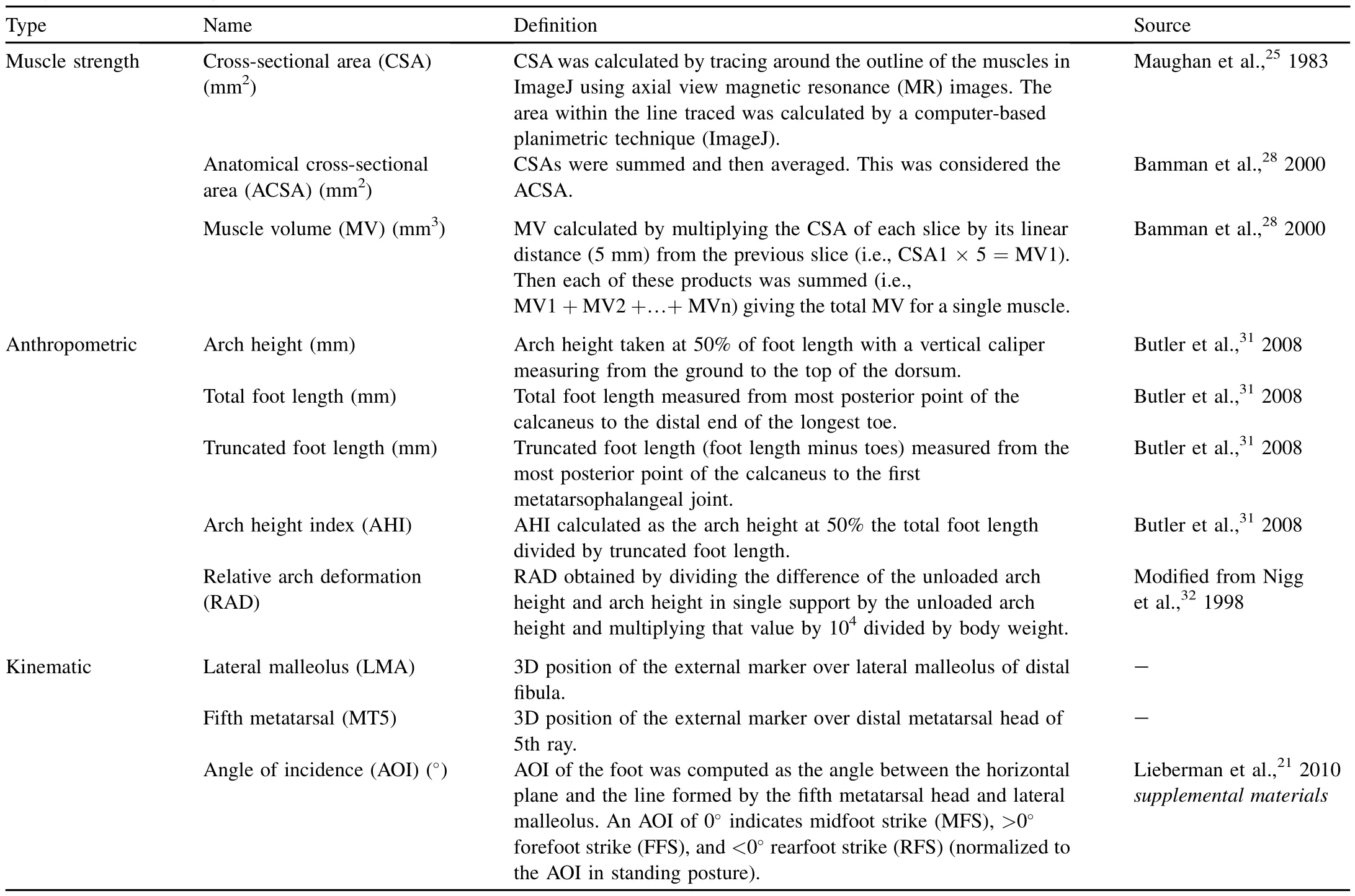
Table 2 Study variables including abbreviations,definitions and source.
2.4.Arch height and deformation data
We defined total foot length,truncated foot length,and arch height follow ing Butler etal.31(Table 2,Fig.1).W ith subjects seated,we measured linear dimensions of the unloaded left foot resting on an osteometric board using sliding calipers. Measurements were repeated w ith subjects standing to obtain loaded footdimensions in both single limb supportand double limb support.From these measurements we derived an arch height index(AHI)and quantified relative arch deformation (RAD),which assesses stiffness32(Table 2).We defined AHI as the arch height at 50%the total foot length divided by truncated foot length31,33—35(Fig.1).Given independent loading of the two limbs during running,we used the AHI in single stance(AHIss)for our measure of arch height.To calculate RAD we used sitting AHI(AHsit)and AHIss in the equation:
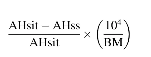
modified from Nigg et al.32(Table 2).
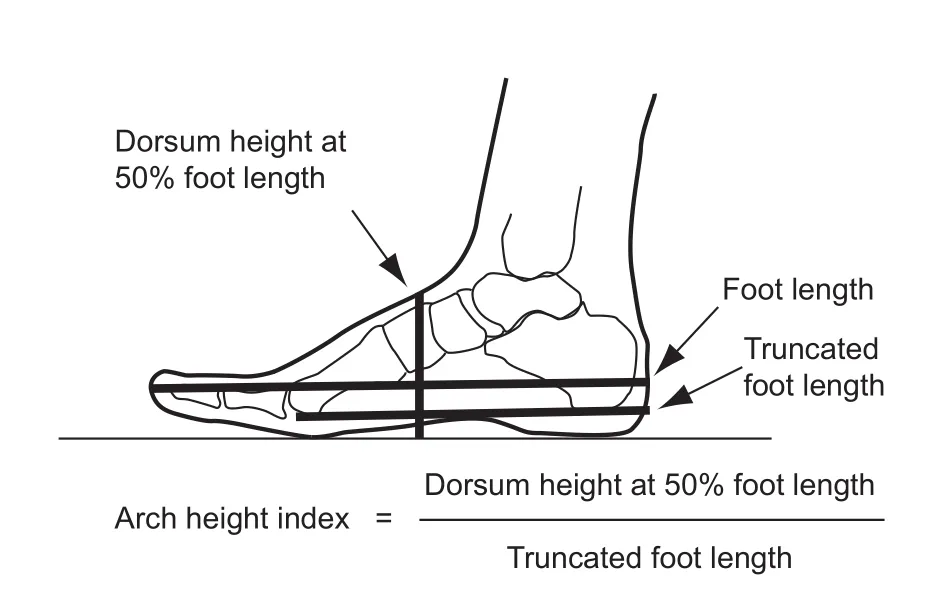
Fig.1.The arch height index formula w ith visual depiction of the three measurements,total foot length,truncated foot length,and arch height.
2.5.Analyses
M ixed w ithin and between subjects designs were used to test for experimental effects of minimal shoe running on the ASCA and MV of the ABH,FDB,and ADM muscles and the foot AHIss and RAD.All statistical analyses were performed in JMP(version 9.0;SAS Institute Inc.,Cary,NC,USA). Normality of data was assessed w ith the Shapiro—W ilkWTest and variance homogeneity using Bartlett’s Test.To identify stochastic differences between the random ly assigned groups at intake,we performed the nonparametric Wilcoxon Rank Sums Test comparing control and experimental runners. Data collected in the term inal session were exam ined as baseline—term inal comparisons using a nested repeatedmeasures multivariate analysis of variance(MANOVA)for time and time×treatment(standard shoesvs.minimal shoes) effects between-groups and w ithin-subjects.Where w ithinsubject differences were significant,we also performed w ithin-group pairedttests.For all statistical tests,we usedα 0.05 to determ ine significance.If no significant changes were found,the Cohen’sdeffect size(ES)was calculated36,37and reported and reviewed according to Cohen’s effect scale36as small ES(0.2—0.5),medium ES(>0.5 and≤0.8),and large ES(> 0.8).Researchers were blind to all subjects during analyses.Of the four participants who withdrew prior to the term inal session,three control subjects variably reported insertional Achilles tendonitis,plantar fascia tear,and lower back pain.One experimental subject w ithdrew for non-study related reasons.
3.Results
3.1.Foot strike
All subjects ran in conventional footwear during the baseline pre-treatment trials.Foot strike pattern varied among subjects w ithin the pooled sample(n=33)at baseline. A lthough forefoot and m idfoot landings were infrequent, four subjects routinely ran FFS and one MFS.The remaining 28 subjects,comprising 85%of the overall sample,ran RFS.Between-group tests of the AOI showed there was no statistical difference in contact angle at baseline between the control and experimental groups(p=0.310,d=0.27, Table 3).
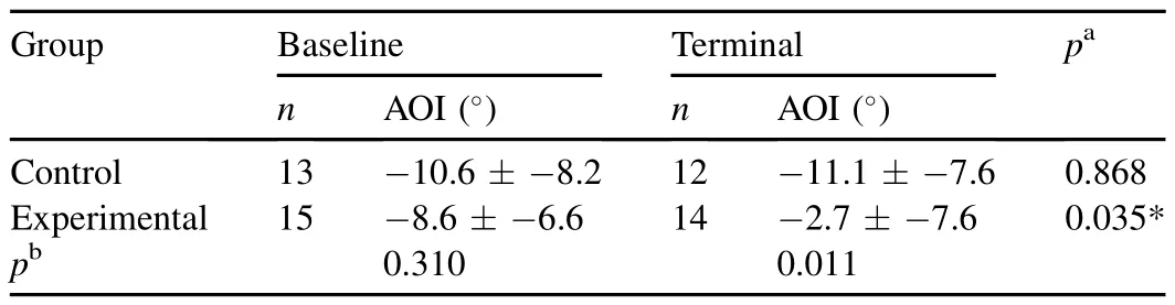
Table 3 Foot strike pattern reported as the angle of incidence(AOI)by group (mean±SD).
Term inal session comparison of the AOI revealed a significant post-treatment difference between-groups (p=0.011).Upon completion of the experimental protocol, the m inimally shod group had a significant 8°mean decrease in dorsiflexion at foot contact(p=0.035).Over the same study period of standard shod running,the contact AOI comparison of pre-and post-treatment w ithin the control group was not significant(p=0.868,d=0.06).In other words, from baseline to term inal testing,distribution of controlgroup foot strike pattern did not change.However,w ithin the experimental group there was a shift from runners using predominately RFS atbaseline to a more MFS or FFS at terminal session.
3.2.ACSA and MV
Baseline tests of the relative ACSA and relative MV of the ADM,ABH,and FDB muscles showed no significant pretreatment difference between the control and experimental groups(Figs.2A and 3A).Term inal testing revealed significant differences between the relative ACSA and relative MV of the ADM and ABH muscles of the two study groups(Figs. 2B and 3B).W ith 12 weeks of standard shod running,the control group signifi cantly increased only MV of the FDB (p=0.03,Table 4,Fig.4).Follow ing the same 12-week period,the experimental group having transitioned to m inimal shod running increased not only MV of the FDB (p=0.03,Table 4,Fig.5)but also MV and ACSA of the ADM(p=0.009 andp=0.007,respectively,Table 4, Fig.5).Neither group significantly increased MV or ACSA of the ABH muscle.
3.3.AHI and RAD
Prior to treatment,conformation of the longitudinal arch did notdiffer between the random ly assigned groups(Table 5). The AHIss index of mean arch height in single limb support was equivalent at 0.36 in the two groups.Similarly,group comparison of the mean RAD,our measure of stiffness, showed no initial difference between the control and experimental groups(p=0.33,d=0.33).
Neither group experienced a significant change in AHIss over the 12-week study period(Table 5).Sim ilarly,posttreatment test of RAD showed no significant change in arch stiffness w ithin either group(p=0.21,d=0.37).However, we identified an outlier among experimental runners at3.5 SD from the group mean.Anad hoctest after outlier deletion yielded a significant effect of time by group(p=0.04).A follow-up pairedttest of experimental runners showed significant change in post-treatment RAD(p=0.013)suggesting a stiffening of the arch w ith m inimally shod running (Table 5).
4.Discussion
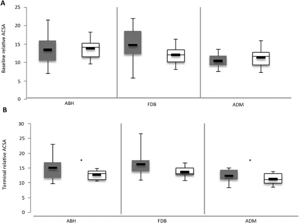
Fig.2.Between-group tests of relative ACSA(lnACSA/lnFL)of the ABH,FDB,and ADM.Experimentalgroup gray and controlgroup white.Heavy black line indicates group mean.(A)Baseline session tests showed no significant group difference in relative ACSA(ABH:p=0.6647,FDB:p=0.0579,ADM:p=0.2206).(B)Terminal session tests indicated significantgroup differences over time for the ABH(p=0.0016)and ADM(p=0.0001)and no significant difference for FDB(p=0.9881).ACSA=anatom ical cross-sectional area;ABH=abductor hallucis;FDB=flexor digitorum brevis;ADM=abductor digiti m inim i.*indicates signifi cant result.
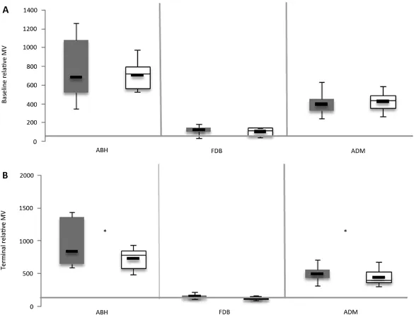
Fig.3.Between-group tests of relative MV(lnMV/lnFL)of the ABH,FDB,and ADM.Experimental group gray and control group white.Heavy black line indicates group mean.(A)Baseline session tests showed no signifi cant difference in relative MV(ABH:p=0.5847,FDB:p=0.3,ADM:p=0.2067).(B) Terminalsession tests indicated significantgroup differences in the ABH(p=0.0011)and ADM(p=0.0256)and no significantdifference in FDB(p=0.5542). MV=muscle volume;ABH=abductor hallucis;FDB=fl exor digitorum brevis;ADM=abductor digiti m inim i.*indicates significant result.
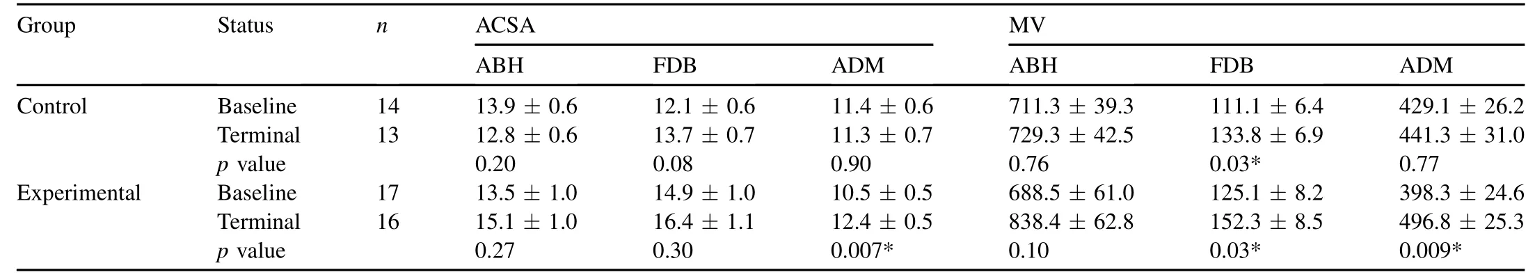
Table 4 Anatomical cross-sectionalarea(ACSA)and muscle volume(MV)of abductor hallucis(ABH),flexor digitorum brevis(FDB),and abductor digitim inim i(ADM) relative to foot length(mean±SD).
The results of this 12-week longitudinal study suggest that endurance running in m inimal support footwear stimulates changes in arch function and the intrinsic foot muscles of runners who previously used conventional running shoes.The experimental runners who transitioned from conventional running shoes to m inimal footwear experienced multiple changes in their landing kinematics, foot musculature and arch conformation as hypothesized. No such changes were observed in the controlgroup w ith the exception of an increase in flexor digitorum brevis volume. Volume appears to be a more sensitive and robust measure than CSA as the majority of signifi cant findings were in the volume of the muscles over time.A lthough foot strength was not directly measured,the results of this prospective experimental study suggest that runners who transition to m inimal footwear can develop a significant increase in foot strength.
At the start of the study,85%of our subjects were RFS,a proportion well w ithin the range of previous reports3,4,21and one that suggests an RFS is typical of conventional shod running at endurance speeds.As predicted,runners who adapted to a m inimalist shoe over the 12-week study period shifted from a predom inantly rearfoot strike pattern to more MFS or FFS landings.We found a significant decrease in dorsiflexion angle at foot contactamong experimental runners in minimalistshoes and notamong control runners in standard footwear.The controlgroup remained unchanged w ith runners landing mainly w ith an RFS.Our findings agree w ith several other studies that have shown runners who transition from standard to m inimalist or barefoot running change from RFS to more MFS/FFS,16,17,38just as most habitual barefoot runners often land more MFS/FFS.18,20,21
This change in foot strike pattern may lead to greater muscle recruitment and therefore increased work performed by muscles of the foot.38Runners w ithin the experimental group who transitioned to a more MFS/FFS increased the ACSA and MV of the ADM muscle.The increase was similar for the MFS and the FFS transitions.However,ACSA and MV of the abductor muscles remained unchanged in runners who consistently used RFS.During the fi rst half of stance the longitudinal arch deforms inferiorly in MFS and FFS,6—9and the intrinsic muscles spanning the arch stretch sim ilarly to mechanicalsprings under tension.These muscles subsequently contract,stiffening the longitudinalarch as load shifts from the m idfoot onto the ball of the foot,pulling the calcaneus and metatarsals closer.This w indlass sequence does not characterize RFS because the arch stretches later in stance only during flatfoot21when arch stiffening and muscle stabilization are less likely.
The intrinsic ADM,ABH,and FDB muscles provide structural integrity to the medial longitudinal arch by their origins on the medialcalcaneal tubercle and insertionsdistal to the metatarsal-phalangeal joints(MPJ).39As the runner’s center of mass shifts to the forefoot the heel rises and the toes dorsiflex.The ground reaction force in response to MPJ rotation generates a dorsiflexion moment ranging in magnitude from 20 to 40 Nm.40Activation of the long and short toe flexors(ADM,ABH,and FDB)counteract the external MPJ dorsiflexion moments.40A lthough this heel rise—MPJ dorsiflexion event occurs regardless of foot strike pattern,those runners whose initial footcontact is either MFS or FFS clearly position the MPJ in greater dorsiflexion at foot contact than otherwise occurs in RFS.9Such repetitive contact events in which the impact force occurs during high MPJ dorsiflexion may lead to increase in both MV and CSA as the short flexors act to m itigate the high MPJ dorsiflexion moment associated w ith MFS and FFS contact.
We predicted that the runners transitioning from conventional running shoes to m inimalist footwearwould increase the MV and ASCA of the intrinsic ABH,FDB,and ADM muscles. Notably,the experimentalgroup significantly increased both the MVand ACSAsof the ADM muscle(p=0.009 andp=0.007, respectively).This muscle,attaching proximally at the calcaneus and distally to the lateral base of the lateral proximal phalanx,flexesonly the fi fth digitand supports the longitudinal arch.39Evident in its volumetric and areal increase,greater force production in the ADM suggests increased recruitmentof itand the longitudinalarch among m inimalshoe runners.Justas barefoot running has been shown to increase work of the leg compartment triceps surae in association w ith increased plantarflexion moments,9,38our results for m inimally shod running show increase in work of the foot compartment muscles in association with plantarflexion at foot strike.Importantly,volumetric and areal increases of the ADM in minimally shod runners only suggest that the mean 8°decrease in dorsiflexion (i.e.,increase in plantarflexion)at footstrike affects the work ofthe ADM muscle more so than the other intrinsic muscles we exam ined.Of the three intrinsic musclesstudied,only the ADM lies entirely within the midfoot region.Thus,routine MFS may recruit the ADM more heavily than either the FDB or the ABH and explain why it significantly increased in both volume and CSA.
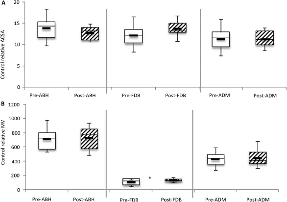
Fig.4.W ithin-group tests of relative ACSA and relative MV between baseline and term inalsession for the ABH,FDB,and ADM in control group.Baseline/pretreatmentsession(white)and terminal/post-treatmentsession(striped).Heavy black line indicates group mean.(A)No significantdifference in relative ACSA for all three muscles(ABH:p=0.2,FDB:p=0.08,ADM:p=0.9).(B)Significantdifference in relative MV of the FDB(p=0.03).No significantdifference in the ABH(p=0.76)and the ADM(p=0.77).ACSA=anatom ical cross-sectional area;MV=muscle volume;ABH=abductor hallucis;FDB=flexor digitorum brevis;ADM=abductor digiti m inimi.*indicates significant result.
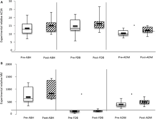
Fig.5.Within-group tests of relative ACSA and relative MV between baseline and term inalsession for the ABH,FDB,and ADM in experimentalgroup.Baseline/ pre-treatmentsession(white)and terminal/post-treatmentsession(striped).Heavy black line indicates group mean.(A)Significantdifference in relative ACSA of the ADM(p=0.007).No significantdifference the ABH(p=0.27)and FDB(p=0.3).(B)Signifi cantdifference in the relative MVof the ADM(p=0.007)and the FDB(p=0.03)over time.No significantdifference in the ABH(p=0.1).ACSA=anatomicalcross-sectionalarea;ABH=abductorhallucis;FDB=flexor digitorum brevis;ADM=abductor digiti m inimi;MV=muscle volume.*indicates significant result.
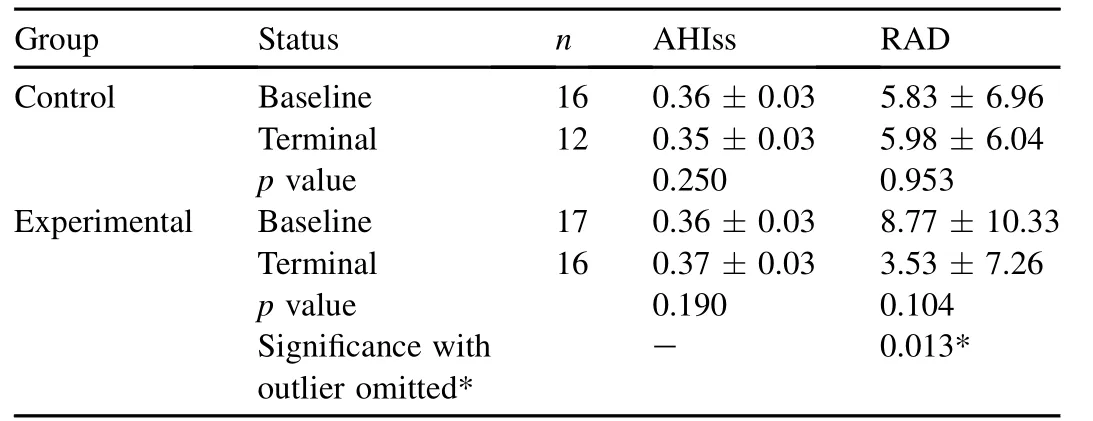
Table 5 Mean arch height index in single support(AHIss)and relative arch deformation(RAD)(mean±SD).
A lthough the FDB muscle increased in relative size (MV),unlike the ADM it did so in both running groups. This suggests that sustained running,whether in standard or m inimal footwear recruits the centrally positioned muscle underlying superfi cial plantar facsia.We suspect that endurance running,regardless of preferred foot strike pattern,heavily recruits the m idline FDB.Furthermore,it appears that running w ithout heel-cushioned and stiff m idsole shoes,as in m inimal footwear running,increases the work of the central FDB as well as the lateral ADM. Because m inimal shoes are constructed w ith a low heel and have no built-in arch support,they may recruit the ADM differently than standard running shoes.Previous work has shown the occurrence of a second peak in center of pressure (COP)follow ing initial foot contact pronation.41This second trajectory peak occurs more laterally in barefoot runners than in standard shod runners.41,42Lateral deflection and laterally oriented velocity peak of COP in the absence of built-in arch support,whether barefoot or in m inimal shoe, may lead to greater demand on the ADM.
Foot muscles appear to respond quickly to increased mechanical stimuli.In a recent study of resistance training, Goldman and colleagues40found that an effort of 90% maximum voluntary isometric contraction repeated over 7 weeks increased intrinsic toe flexor strength 40%.Over the course of our 12-week study,running in conventional and m inimal footwear led to an increase in FDB size and m inimal shoe running only led to additional increase in ADM size.We interpret size change measured as increase in muscle CSA and volume to indicate greater muscle strength.24Increased muscle strength was likely induced by recruitment of the intrinsic group for arch stabilization during toe-off.6,22,43,44Whereas arch stabilization in standard footwear is largely provided by the extrinsic arch support of rigid shoe design,stability in m inimal footwear is contingent only on intrinsic factors of foot conformation, including the tensile and contractile properties of muscle. Furthermore,although both groups saw an increase in MV of the FDB,in maintaining RFS throughout the longitudinal study,the control group showed no change in both abductor size and arch stiffness.In contrast,the m inimal footwear group additionally increased ADM abductor size and increased arch stiffness.
We found the most robustdifference between conventional shod and m inimally shod groups in the variable of longitudinal arch stiffness(RAD),which increased approximately 60%in the m inimally shod runners butunderwentno change in the control group.Our random ly assigned groups entered the study w ith no significant difference in RAD and AHI in single limb support(AHIss).The pre-treatment AHIss of 0.36 for both groups was consistent w ith values previously reported for the habitually shod (conventional running shoe).31,35Most conventional running shoes place a relatively stiff support below the longitudinal arch.This support combined w ith a relatively stiff m idsole likely reduce the extent of stretch in soft tissues during loading,and effectively replace or inhibit the natural spring mechanism of the arch.6,9It is reasonable to infer that these soft tissues are able to function more naturally as a spring in a m inimal shoe.The abductors,which fl ex the hallucal and fi fth digit metatarsalphalangeal joints,also enhance the w indlass mechanism of the plantar aponeurosis.45Thus,volumetric increase of the ADM in the m inimally shod runners suggests notonly greater stiffness in the m inimally shod foot but also greater capacity for force production when the arch deforms and recoils. Further,MFS/FFS may heavily recruit the ADM more than the highly dorsiflexed RFS as this abductor stabilizes the longitudinal arch during initial foot strike and is held in tension until toe-off.
The results of this study suggest the need for several additional experiments.Future research on the effects of barefoot and m inimal shoe running on foot strength would benefi t from a larger sample size and a longer treatment period.Although the ADM and FDB responded quickly in this study and others,40,45a longer treatment period m ight be hypothesized to yield arch height differences between treatment groups.Another area for future study would be to improve the ability to delineate deep intrinsic muscles in MRI scans.We examined only superficial plantar musculature of the foot, om itting the quadratus plantae muscle that lies deep w ithin the second layer.Finer differentiation of the interdigitating fibers of the quadratus plantae muscle would capture more of the intrinsic musculature’s response to different running conditions.
Our study design aimed to vary only footwear among control and experimental subjects in order to assess effects of m inimal footwear on arch structure and intrinsic foot muscle strength.However,our protocol for the experimental runners included a brief discussion on safe practice(posture and cadence)in m inimal shoe running in order to prevent injury. Subjects were not instructed on which foot strike pattern to use.Nonetheless,these instructions may have led to other changes in form in the experimental group.
A potential weakness of this study was the variation of footwear worn by the both groups.The shoes worn by control subjects varied w idely by model and make,but met all construction criteria.Although the experimental group used just two models of m inimal footwear,which also meta prioricriteria,the drop offset of minimal shoe models differed by 4 mm.The Merrell Pace/TrailGlove w ith its 0 mm differential is a more m inimal shoe than the New Balance M inimus.Post hoctests of experimental runners accounting for the two m inimal shoe models showed a significant difference in the RAD(p=0.0009),w ith a stiffer arch among the New Balance model runners.Thus,it is likely that the New Balance shoe required the intrinsic muscles to do more work.Nonetheless,both m inimal shoes were shown to recruit the plantar intrinsic musculature of the foot more than highly cushioned standard running shoes.However,in vivoelectromyography analyses are necessary to test this hypothesis.
To conclude,these findings support earlier studies,which suggested that running barefoot or in m inimal shoes increases the overall area and volume of the plantar intrinsic musculature,makes greater use of the spring-like function of the longitudinal arch and its associated muscles,and promotes stiffer arches.9,15,16These results suggest that runners can adapt successfully to using m inimal shoes w ithout increased risk of injury if they do so gradually and carefully,but future studies w ith larger samples sizes are clearly necessary to test this hypothesis more carefully.
Acknow ledgm ent
This research was supported by the Charles Phelps Taft Research Center at the University of Cincinnati.We thank Randy Cox M.S.S.for the training plans.
Appendix 1.
Control group training plan
Included in this packet are your training programs for the next 3 months and a key explaining the various workouts. Please make sure to read over it carefully to ensure you understand it correctly.If you have any questions do nothesitate to contact the Principal Investigator for the study,Liz M iller.
Key
VE(very easy)=not faster than 70%of current5000 pace/ mile.For instance,if you can run a 5000 at6:00/mile,VE pace would be no faster than 8:30/m ile.Calculated as 6 divided by 0.70. This is near effortless pace. AC (aerobic conditioning)=75%—82%of current 5000 pace/m ile.A medium effort run.
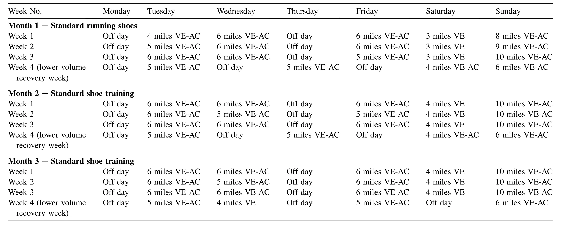
Week No. Monday Tuesday Wednesday Thursday Friday Saturday Sunday M onth 1—Standard running shoes Week 1 Off day 4 m iles VE-AC 6 m iles VE-AC Off day 6 m iles VE-AC 3 m iles VE 8 m iles VE-AC Week 2 Off day 5 m iles VE-AC 6 m iles VE-AC Off day 6 m iles VE-AC 3 m iles VE 9 m iles VE-AC Week 3 Off day 6 miles VE-AC 6 miles VE-AC Off day 5 miles VE-AC 3 miles VE 10 miles VE-AC Week 4(lower volume recovery week) Off day 5 m iles VE-AC Off day 5 m iles VE-AC Off day 4 m iles VE-AC 6 m iles VE-AC M onth 2—Standard shoe training Week 1 Off day 6 m iles VE-AC 6 m iles VE-AC Off day 6 m iles VE-AC 4 m iles VE 10 m iles VE-AC Week 2 Off day 6 m iles VE-AC 5 m iles VE-AC Off day 5 m iles VE-AC 4 m iles VE 10 m iles VE-AC Week 3 Off day 6 m iles VE-AC 6 m iles VE-AC Off day 6 m iles VE-AC 4 m iles VE 10 m iles VE-AC Week 4(lower volume recovery week) Off day 5 m iles VE-AC Off day 5 m iles VE-AC Off day 4 m iles VE-AC 6 m iles VE-AC M onth 3—Standard shoe training Week 1 Off day 6 m iles VE-AC 6 m iles VE-AC Off day 6 m iles VE-AC 4 m iles VE 10 m iles VE-AC Week 2 Off day 6 miles VE-AC 5 miles VE-AC Off day 6 miles VE-AC 4 miles VE 10 miles VE-AC Week 3 Off day 6 m iles VE-AC 6 m iles VE-AC Off day 6 m iles VE-AC 4 m iles VE 10 m iles VE-AC Week 4(lower volume recovery week) Off day 5 m iles VE-AC 4 m iles VE Off day 5 m iles VE-AC Off day 6 m iles VE-AC
Appendix 2.
Transition group training plan
Included in this packetare your training programs for the next 3 months and a key explaining the various workouts. Please make sure to read over it carefully to ensure you understand it correctly.If you have any questions do not hesitate to contact the Principal Investigator for this study, Liz M iller.You w ill be running in your standard running shoes while transitioning into the minimalist shoes.Please make sure you note which workouts are to be done in which shoes.
Key
VE(very easy)=not faster than 70%of current5000 pace/ m ile.For instance,if you can run 5000 at 6:00/m ile,VE pace would be no faster than 8:30/m ile.Calculated as 6 divided by 0.70. This is near effortless pace. AC (aerobic conditioning)=75%—82%of current 5000 pace/m ile.A medium effort run.
IMS=in m inimalist shoes;ISS=in standard shoes.
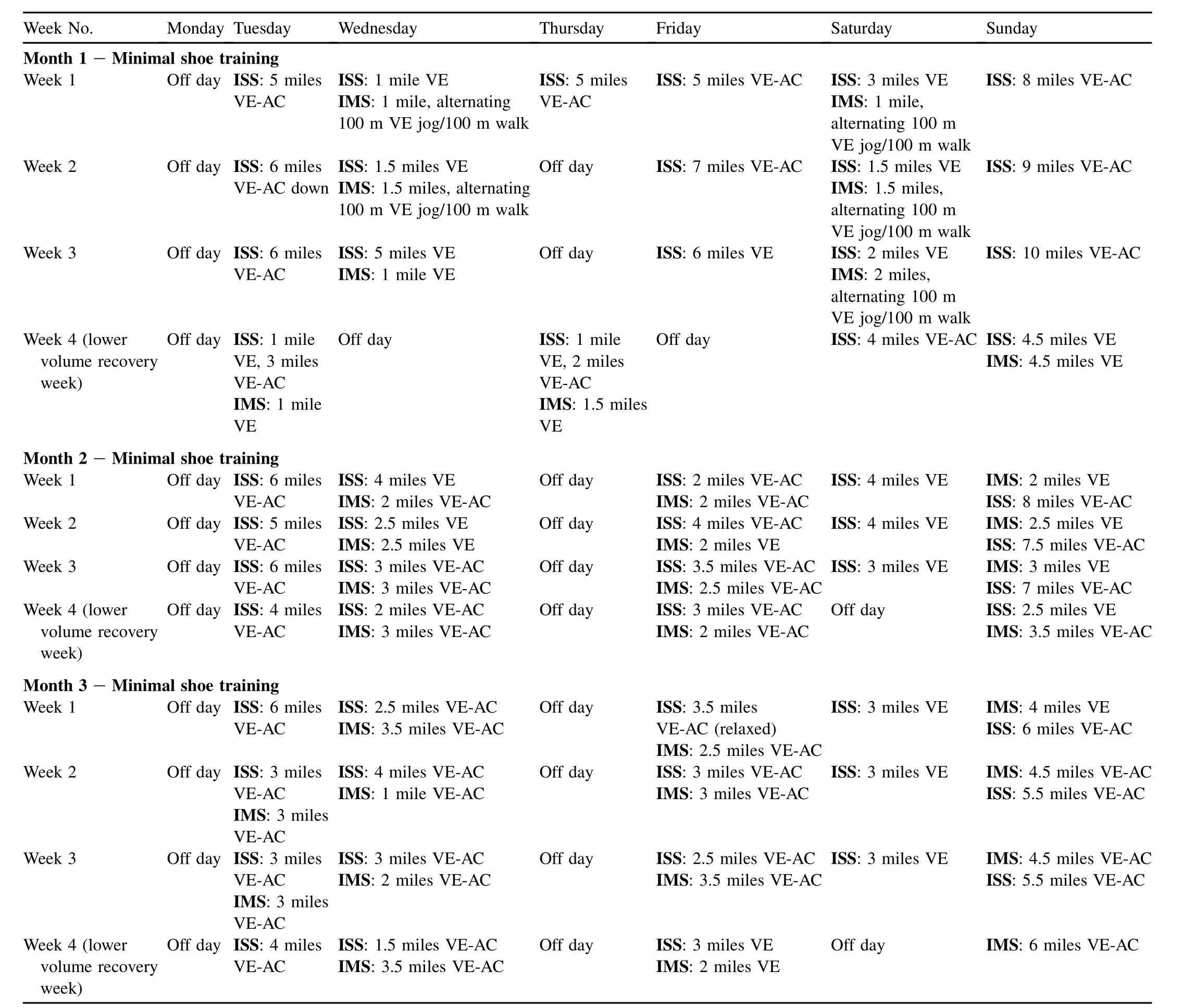
Week No. Monday Tuesday Wednesday Thursday Friday Saturday Sunday M onth 1—M inimal shoe training Week 1 Off day ISS:5 m iles VE-AC ISS:1 m ile VE IMS:1 m ile,alternating 100 m VE jog/100 m walk ISS:5 miles VE-AC ISS:5 m iles VE-AC ISS:3 m iles VE IMS:1 m ile, alternating 100 m VE jog/100 m walk ISS:8 m iles VE-AC Week 2 Off day ISS:6 m iles VE-AC down ISS:1.5 m iles VE IMS:1.5 m iles,alternating 100 m VE jog/100 m walk Off day ISS:7 m iles VE-AC ISS:1.5 m iles VE IMS:1.5 m iles, alternating 100 m VE jog/100 m walk ISS:9 m iles VE-AC Week 3 Off day ISS:6 m iles VE-AC ISS:5 m iles VE IMS:1 m ile VE Off day ISS:6 m iles VE ISS:2 m iles VE IMS:2 m iles, alternating 100 m VE jog/100 m walk ISS:10 m iles VE-AC Week 4(lower volume recovery week) Off day ISS:1 m ile VE,3 m iles VE-AC IMS:1 m ile VE Off day ISS:1 mile VE,2 m iles VE-AC IMS:1.5 m iles VE Off day ISS:4 miles VE-AC ISS:4.5 miles VE IMS:4.5 m iles VE M onth 2—M inim al shoe training Week 1 Off day ISS:6 m iles VE-AC ISS:4 m iles VE IMS:2 m iles VE ISS:8 m iles VE-AC Week 2 Off day ISS:5 m iles VE-AC ISS:4 m iles VE IMS:2 m iles VE-AC Off day ISS:2 m iles VE-AC IMS:2 m iles VE-AC ISS:4 m iles VE IMS:2.5 m iles VE ISS:7.5 m iles VE-AC Week 3 Off day ISS:6 m iles VE-AC ISS:2.5 m iles VE IMS:2.5 m iles VE Off day ISS:4 m iles VE-AC IMS:2 m iles VE ISS:3 m iles VE-AC IMS:3 m iles VE-AC Off day ISS:3.5 m iles VE-AC IMS:2.5 m iles VE-AC ISS:3 m iles VE IMS:3 m iles VE ISS:7 m iles VE-AC Week 4(lower volume recovery week) Off day ISS:4 miles VE-AC ISS:2 miles VE-AC IMS:3 m iles VE-AC Off day ISS:3 miles VE-AC IMS:2 m iles VE-AC Off day ISS:2.5 miles VE IMS:3.5 m iles VE-AC M onth 3—M inim al shoe training Week 1 Off day ISS:6 m iles VE-AC ISS:2.5 m iles VE-AC IMS:3.5 m iles VE-AC Off day ISS:3.5 m iles VE-AC(relaxed) IMS:2.5 m iles VE-AC ISS:3 miles VE IMS:4 m iles VE ISS:6 m iles VE-AC Week 2 Off day ISS:3 m iles VE-AC IMS:3 m iles VE-AC ISS:4 m iles VE-AC IMS:1 m ile VE-AC Off day ISS:3 m iles VE-AC IMS:3 m iles VE-AC ISS:3 m iles VE IMS:4.5 m iles VE-AC ISS:5.5 m iles VE-AC Week 3 Off day ISS:3 m iles VE-AC IMS:3 m iles VE-AC ISS:3 m iles VE-AC IMS:2 m iles VE-AC Off day ISS:2.5 m iles VE-AC IMS:3.5 m iles VE-AC ISS:3 m iles VE IMS:4.5 m iles VE-AC ISS:5.5 m iles VE-AC Week 4(lower volume recovery week) Off day ISS:4 m iles VE-AC ISS:1.5 m iles VE-AC IMS:3.5 m iles VE-AC Off day ISS:3 m iles VE IMS:2 m iles VE Off day IMS:6 m iles VE-AC
1.National Sporting Goods Association.Sports participation in 2009:series I.National Sporting Goods Association;2009.
2.Nigg B,Cole GK,Bruggemann GP.Impact forces during heel-toe running.J Appl Biomech1995;11:407—32.
3.A ltman A,Davis I.Barefoot running:biomechanics and implications for running injuries.Curr Sports Med Rep2012;11:244—50.
4.Lieberman DE.What we can learn about running from barefoot running: an evolutionary medicalperspective.Exerc SportSciRev2012;40:63—72.
5.D’Aout K,Pataky TC,De Clercq D,Aerts P.The effects of habitual footwear use:foot shape and function in native barefoot walkers.Footwear Sci2009;1:81—94.
6.Ker RF,Bennett MB,Bibby SR,Kester RC,A lexander RM.The spring in the arch of the human foot.Nature1987;325:147—9.
7.Bandholm T,Boysen L,Haugaard S,Kreutzfeldt Zebis M,Bencke J.Foot medial longitudinal-arch deformation during quiet standing and gait in subjects w ith medial tibial stress syndrome.J Foot Ankle Surg2008;47:89—95.
8.Fukano M,Fukubayashi T.Motion characteristics of the medial and lateral longitudinal arch during landing.EurJApplPhysiol2009;105:387—92.
9.Perl DP,Daoud AI,Lieberman DE.Effects of footwear and strike type on running economy.Med Sci Sports Exerc2012;44:1335—43.
10.Nigg B.Biomechanical considerations on barefoot movement and barefoot shoe concepts.Footwear Sci2009;1:73—9.
11.Kaufman KR,Brodine SK,Shaffer RA,Johnson CW,Cullison TR.The effect of foot structure and range of motion on musculoskeletal overuse injuries.Am J Sports Med1999;27:585—93.
12.Bishop M,Fiolkowski P,Conrad B,Brunt D,Horodyski M.Athletic footwear,leg stiffness,and running kinematics.JAthl Train2006;41:387—92.
13.Pohl MB,Hamill J,Davis IS.Biomechanical and anatomical factors associated with a history ofplantar fasciitis in female runners.Clin J Sport Med2009;19:372—6.
14.Robbins SE,Hanna AM.Running-related injury prevention through barefoot adaptations.Med Sci Sports Exerc1987;19:148—56.
15.Bru¨ggemann GP,Potthast W,Braunstein B,Niehoff A.Effectof increased mechanical stimuli on foot muscles functional capacity.ISB XXth Congress—ASB 29th annual meeting,July 31—August 5,2005,Cleveland,OH,USA.
16.De Wit B,De Clercq D,Aerts P.Biomechanical analysis of the stance phase during barefoot and shod running.J Biomech2000;33:269—78.
17.Divert C,Mornieux G,Freychat P,Baly L,Mayer F,Belli A.Barefootshod running differences:shoe or mass effect?Int J Sports Med2008;29:512—8.
18.Squadrone R,Gallozi C.Biomechanicaland physiological comparison of barefoot and two shod conditions in experienced barefoot runners.J Sports Med Phys Fitness2009;49:6—13.
19.Jenkins DW,Cauthon DJ.Barefoot running claims and controversies:a review of the literature.J Am Podiatr Med Assoc2011;101:231—46.
20.Hatala KG,DingwallHL,Wunderlich RE,Richmond BG.Variation in foot strike patterns during running among habitually barefoot populations.PLoS One2013;8:e52548.http://dx.doi.org/10.1371/journal.pone.0052548.
21.Lieberman DE,Venkadesan M,Werbel WA,Daoud AI,D’Andrea S, Davis IS,et al.Foot strike patterns and collision forces in habitually barefoot versus shod runners.Nature2010;463:531—5.
22.Mann R,Inman VT.Phasic activity of intrinsic muscles of the foot.J Bone Jt Surg Am1964;46-A:469—81.
23.Rooney BD,Derrick TR.Joint contact loading in forefoot and rearfoot strike patterns during running.J Biomech2013;46:2201—6.
24.Fukunaga T,Roy RR,Shellock FG,Hodgson JA,Edgerton VR.Specific tension of human plantar fl exors and dorsiflexors.J Appl Phys1996;80:158—65.
25.Maughan RJ,Watson JS,Weir J.Strength and cross-sectional area of human skeletal muscle.J Physiol1983;338:37—49.
26.Lieber RL.Skeletal muscle structure,function,and plasticity.Baltimore, MD:Lippincott W illiams&W ilkins;2002.
27.Narici MV,Landoni L,M inetti AE.Assessmentof human knee extensor muscle stress fromin vivophysiological cross-sectional area and strength measurements.Eur J Appl Physiol1992;65:438—44.
28.Bamman MM,New comer BR,Larson-Meyer DE,Weinsier RL, Hunter GR.Evaluation of the strength-size relationshipin vivousing various muscle size indices.Med Sci Sports Exerc2000;32:1307—13.
29.A lbrachtK,Arampatzis A,Baltzopoulos V.Assessmentofmuscle volume and physiological cross-sectional area of the human triceps surae musclein vivo.J Biomech2008;41:2211—8.
30.Recht MP,Donley BG.Magnetic resonance imaging of the footand ankle.J Am Acad Orthop Surg2001;9:187—99.
31.Butler RJ,Davis IS,Ham ill J.Interaction of arch type and footwear on running mechanics.Am J Sports Med2006;34:1998—2005.
32.Nigg B,Khan A,Fisher V,Stefanyshyn D.Effect of shoe insert construction on foot and leg movement.MedSci Sports Exerc1998;30:550—5.
33.Williams IIIDS,McClay IS.Measurements used to characterize the foot and the medial longitudinal arch:reliability and validity.Phys Ther2000;80:864—71.
34.Richards CJ,Card K,Song J,Hillstrom H.A novel arch height index measurement system(AHIMS):intra-and inter-rater reliability.Proceedings of the American Society of Biomechanics meeting Toledo,OH; 2003.
35.Zifchock RA,Davis I,Hillstrom H,Song J.The effectof gender,age,and lateral dominance on arch height and arch stiffness.Foot Ankle Int2006;27:367—72.
36.Cohen J.Statistical power analysis for the behavioral sciences.2nd ed. Hillsdale,NJ:Law rence Erlbaum Associates;1988.
37.Enders H,von Tscharner V,Nigg B.The effects of preferred and nonpreferred running strike patterns on tissue vibration properties.J Sci Med Sport2014;17:218—22.
38.Bonacci J,Saunders P,Hicks A,Rantalainen T,Vicenzino B, Spratford W.Running in a m inimalist and lightweight shoe is not the same as running barefoot:a biomechanical study.Br J Sports Med2013;47:387—92.
39.Augur AMR,Lee MJ.Grant’s atlas of anatomy,10th ed.Baltimore,MD: Lippincott Williams&Wilkins;1999.
40.Goldman JP,Sanno M,W illwacher S,Heinrich K,Bruggemann GP.The potential of toe flexor muscles to enhance performance.J Sports Sci2013;31:424—33.
41.De Cock A,Varenterghem J,Willems T,Witvrouw E,De Clercq D.The trajectory of the centre of pressure during barefoot running as a potential measure for foot function.Gait Posture2008;27:669—75.
42.Hampson J,Sinclair J,Greenhalgh A.The trajectory of the centre of pressure during running in barefoot,m inimalist footwear and traditional running shoe conditions in females.BASES annual conference of the British Association of Sport and Exercise Sciences;2013.Preston,UK.
43.Aiello LC,Dean C.An introduction to human evolutionary anatomy.San Jose,CA:California Academ ic Press;2002.
44.Kelly L,Cresswell A,Racinais S,Whiteley R,Lichtwark G.Intrinsic foot muscles have the capacity to controldeformation of the longitudinal arch.J R Soc Interface2014;11:93.
45.Hicks JH.The mechanics of the foot,III:the foot as support.Acta Anat1955;25:34—45.
Received 2 September 2013;revised 9 February 2014;accepted 18 March 2014
*Corresponding author.
E-mail address:katherine.whitcome@uc.edu(K.K.Whitcome)
Peer review under responsibility of Shanghai University of Sport
2095-2546/$-see front matter CopyrightⒸ2014,Shanghai University of Sport.Production and hosting by Elsevier B.V.A ll rights reserved. http://dx.doi.org/10.1016/j.jshs.2014.03.011
 Journal of Sport and Health Science2014年2期
- Journal of Sport and Health Science的其它文章
- Can m inimal running shoes im itate barefoot heel-toe running patterns? A comparison of lower leg kinematics
- Strike type variation among Tarahumara Indians in m inimal sandals versus conventional running shoes
- Foot strike patterns and hind limb joint angles during running in Hadza hunter-gatherers
- Muscle activity and kinematics of forefoot and rearfoot strike runners
- Impact shock frequency components and attenuation in rearfoot and forefoot running
- The effectof shoe type on gait in forefootstrike runners during a 50-km run