Mathematical Models for Teenager’s Living Age Evaluation Based on CT Image of Medial Clavicular Epiphysis
WEI Hua,WAN Lei,YING Chong-liang,WANG Ya-hui,ZHU Guang-you
(1.East China University of Political Science and Law,Shanghai 200042,China;2.Shanghai Key Laboratory of Forensic Medicine,Institute of Forensic Science,Ministry of Justice,P.R.China,Shanghai 200063,China)
Mathematical Models for Teenager’s Living Age Evaluation Based on CT Image of Medial Clavicular Epiphysis
WEI Hua1,2,WAN Lei2,YING Chong-liang2,WANG Ya-hui2,ZHU Guang-you2
(1.East China University of Political Science and Law,Shanghai 200042,China;2.Shanghai Key Laboratory of Forensic Medicine,Institute of Forensic Science,Ministry of Justice,P.R.China,Shanghai 200063,China)
ObjectiveTo explore the correlation between volume rendering(VR)statistics of medial clavicular epiphysis and living age,and establish the mathematical models for living age evaluation using the CT image of medial clavicular epiphysis based on the growth rules of osteoepiphysis of medial clavicle. Methods The CT images of the medial clavicles from 795 teenagers aged 15-25,387 males and 408 females,were collected in East and South China.VR 3D images were reconstructed from 0.60mm-thick slice CT images.The epiphyseal diameter,sternal end diameter,and their respective diameter ratio(the left:x1;the right:x3);epiphyseal area,sternal end area,and their respective area ratio(the left:x2;the right:x4),were measured and calculated.All these observations were analyzed using SPSS 19.0 statistical software.The statistical differences in gender and age were analyzed by Mann-Whitney U test.The mathematical models were established using least square.Sixty trained subjects,30 males and 30 females,were tested to verify the accuracy of the established mathematical models.ResultsIn the group of same age,x1showed significant difference in gender;the same results were observed in x2,x3,and x4, which suggested that the growth rules of osteoepiphysis of medial clavicle were highly correlated with living age.The accuracy of these mathematical models were all above 67.6%(±1.0 year)and 78.5%(±1.5 year).ConclusionThe mathematical models with reasonable accuracy could be manageable in practice to confirm the conclusion of the atlas method.The current study can contribute to the single skeletal age evaluation.
forensic anthropology;living skeletal age;medial clavicular epiphysis;volume rendering; mathematical model
Article IC:1004-5619(2013)04-0248-04
Introduction
In the past investigations,comparative studies have been conducted on living age and big joints, with the mathematical models established[1-5].These models can be readily put into practice so as to confirm the conclusion of the atlas method and improve the accuracy of living bone age evaluation[1].Medial clavicular epiphysis has been investigated for decades because it is one of the most important indicators in the living age evaluation of old teenagers[2-4].
The current study was inspired by the research results of Schmeling[4].The subjects were scanned by 2.0 mm,the images reconstructed to the thickness of 0.06mm.Measured and calculated were the epiphyseal diameter,sternal end diameter,and their respective diameter ratio;epiphysealarea,sternal end area,and their respective area ratio as well as other data using volume rendering(VR)images. The results of the atlas method had been obtained by the VR statistics,and the accuracy and practicability,testified using several experimental data in our previous study[3].These statistics were used as theobservationsforclassificationoftheatlas method,which covered the shortage of 2D statistics in Schmeling’s method[4].Meanwhile,these indexes were all found to be sequential quantitative variables,having high correlation with living age in our previous study[3].Thus it is necessary that mathematical models of the living bone age evaluation should be established to make further investigation on these statistics[3].
Materials and methods
Subjects
The CTimages of medialclavicles of 795 teenagers aged 15-25,387 males and 408 females, were collected in East and South China(Table 1).
To be eligible for the current study,the subjects matched the following criteria:their physical health and nutrition situation were in good condition;and their heights and weights,measured by Ma Erding metal gauge and beam balance,were in the specialized range according to the national criteria[5].Those were excluded who had received special recreation and sports training,taken the medications which could influence bone development,or had a history of bone injury or bone developmentdisorder.All the subjects signed informed consents. The investigation complied with the related regulations of medical ethics.

Table 1Age and gender distribution
CT scanning
The 40 ranks’SOMATOM Definition AS MSCT(Siemens Health Group,Germany)was employed in the experiment.Scanning parameters were set as follows:the slice 2.0 mm thick,slice internal 1.0 mm thick,tube voltage 120kV,and tube current 80mA. Upon their measurements of height and weight,the subjects received CT scanning in the supine position[6-8].
Data collection
The thin layer CT images were ranked according to the age ascending.The images(slice thickness 0.6mm and slice internal 0.5 mm)reconstructed and transferred to Syngo workstation,the tomographic images were reconstructed as VR images. The images of secondary ossification center or medial clavicular epiphysis were selected for the current study[4-7].With the MIWORK S5.0.0.6 PACS bone age reading software[Pnoenist(Shanghai)Information Technology Co.,Ltd.],epiphyseal growth were analyzed in thin slice CTimages including transverse and coronal.
The observation was made on the appearance of the secondary ossification center.If it appeared, the position and characteristics were observed.The epiphyseal diameter,sternal end diameter,and their respective diameter ratio(the left:x1;the right:x3); epiphyseal area,sternal end area,and their respective area ratio(the left:x2;the right:x4),were measured and calculated on VR images[1].
Statistical analysis
All data were analyzed using SPSS 19.0 statistical software.The dependent variable of living age was assumed as y.The statisticaldifferences in genderandagewererespectivelyanalyzedby Mann-Whitney U test.The level of significance was set at P<0.05.The correlation between y and x was analyzed by least square for establishing the mathematicalmodels.Sixty trained subjects,30 males and 30 females,were tested to verify the accuracy of the established models[3-5].
Results
Statistical differences between male and female subjects
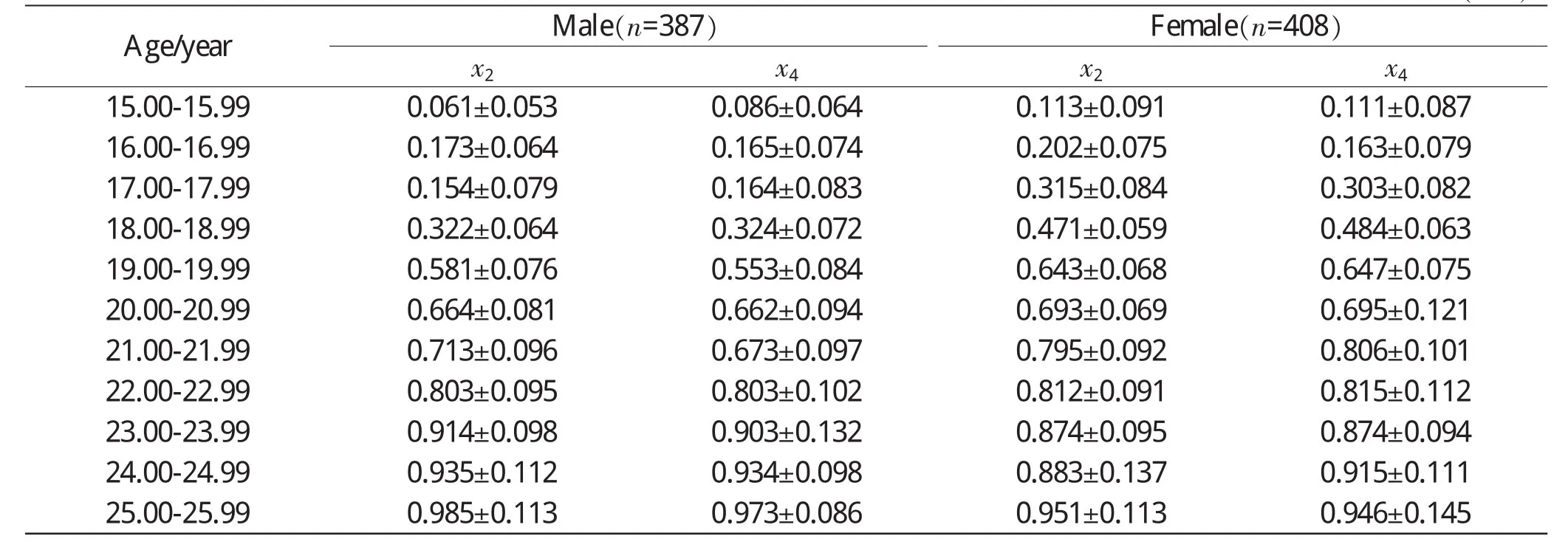
The results showed that x1,x3,x2,and x4had significant differences in gender(P<0.05).Since the variables of x2and x4had a higher correlation with ages than those of x1and x3,which had been reported in our previous study[3],the mean values of x2and x4were respectively analyzed by Mann-Whitney U test.In the female group,the mean value of x2was 0.693±1.473,and the mean value of x4was 0.694±1.476.In the male group,the mean value of x2was 0.653±1.289,and the mean value of x4was 0.644±1.322.The differences of x2and x4in gender were observed in each age group(Table 2).
Table 2Mean values of x2and x4of both genders in each age group(±s)

Table 2Mean values of x2and x4of both genders in each age group(±s)
Age/yearMale(n=387)Female(n=408)x2x4x2x415.00-15.990.061±0.0530.086±0.0640.113±0.0910.111±0.087 16.00-16.990.173±0.0640.165±0.0740.202±0.0750.163±0.079 17.00-17.990.154±0.0790.164±0.0830.315±0.0840.303±0.082 18.00-18.990.322±0.0640.324±0.0720.471±0.0590.484±0.063 19.00-19.990.581±0.0760.553±0.0840.643±0.0680.647±0.075 20.00-20.990.664±0.0810.662±0.0940.693±0.0690.695±0.121 21.00-21.990.713±0.0960.673±0.0970.795±0.0920.806±0.101 22.00-22.990.803±0.0950.803±0.1020.812±0.0910.815±0.112 23.00-23.990.914±0.0980.903±0.1320.874±0.0950.874±0.094 24.00-24.990.935±0.1120.934±0.0980.883±0.1370.915±0.111 25.00-25.990.985±0.1130.973±0.0860.951±0.1130.946±0.145
The mathematical models
It was found that there was no statistical difference between the left and right side of the variables;therefore,those variables were divided into three groups to analyze their adjusted R2with y,which were x1,x3group,x2,x4group,and x1,x3,x2,x4group.
The correlation between x and y were analyzed by least square to establish the mathematical models.The adjusted R2of the variables of x1,x3,x2, and x4was calculated,the results of which were that the adjusted R2of x1and x3was 0.521,low enough to be rejected.The variables of x2and x4and those of x1,x2,x3,and x4were adopted to form the mathematical models.Furthermore,the 60 subjects were examined to verify the accuracy of established mathematical models(Table 3).

Table 3The mathematical models of medial clavicular epiphysis of teenagers
Discussion
Mathematical model method is also known as multivariate regression method.Through statistical software,multiple regression equation is used to analyze the statistics of observations on bones.The best correlation between living age and related observations is explored to establish the mathematical model[1].As the technological development of teenager’s living bone age evaluation,severalresearch methods have been reported,of which the mathematical model and Atlas method,whose accuracy have been testified by experimental data,are regarded as the precise inference methods.Because of their excellent operability,the two methods have received growing attention.In 2000,Tian et al.[9]established the mathematical model of 27 X-ray observations from 360 male and female subjects.In 2005,Wang etal.[1]made a statisticalanalysis about 24 indexes of teenager’s bone,analyzing the relationship between height,weight and living age. Using multiple stepwise regression,clustering methodology,Fisher’s two kinds of discriminate analysis,a series of mathematical models were established to deduce living age[1-6].
It has been found that the medial clavicular epiphysis is highly correlated to living age in the previous study[3].Because the medial clavicular epiphysis is located under the tissues of breast,it is difficult to obtain the clear images using conventional radiography.It can reduce the effect of artifactandobtainclearimageswithtomographic imaging technology.In the current study,the conventional X-ray imaging technology was substituted with CTscanning.The tomographic images were reconstructed to 0.6 mm-thick slice images,since it has been testified that the thickness below 2.0 mm can reduce the partial volume effect at mostin Schmeling’s study[4].The VR images were recombined with the 0.6 mm-thick slice images on the Syngo workstation.The observations could be readily and precisely measured in these clear VR images.
Compared with those of the previous study,the observations of the current study were sequential instead of categorical variables.The observations of categorical variables should be classified at first and then the correlation analysis could be done.After measuring and calculating the quantitative observations,the correlation analysis could be done in a direct way.Therefore,it could avoid the artificial judgment which might lead to some artificial error in classifying the observations.
In the current study,x1had significant difference in gender of the same age group.The same results were obtained from the analysis of x2,x3, and x4.Because the variables of x2and x4had a higher correlation with living age than those of x1and x3,the variables of x2and x4were made to conduct further research.Compared with the mean value of x2and x4in gender of the same age group,it showed that female’s development was earlier than male’s,especially from 15 to 20.
In the mathematical models,the adjusted R2of x1,x3group was lower than 0.521,so the group was rejected from the correlation with living age. The adjusted R2of the other groups were all higher than 0.641,so they were adopted for the performance of analysis by least square.
It was shown that the male’s adjusted R2was higherthanthefemale’sinthetwoadopted groups.After being testified by the 60 trained subjects,the accuracy of these models were all above 67.6%(±1.0 year)and 78.5%(±1.5 year),suggesting that medial clavicular can have significant reference in teenager’s skeletal age evaluation.The male accuracy was higher than the female in these mathematical models.The mathematical model of x2,x4group was more accurate than x1,x3,x2,x4group,which might have resulted from the area data superior to the linear data in the correlation with living age.
In the current study,the operability and accuracy of these established models were reasonable; therefore it was worth to make the further research in the other single bone.The mathematical model method,which could confirm Atlas method of medial clavicular,can improve the accuracy of skeletal age evaluation.
Acknowledgments
This study was funded by the Natural Science Fund Projects(81102305)and the 12th five-year National Science and Technology Support Project(2012BAK16B01)and Fund Projects of Institute of ForensicScience,MinistryofJustice,P.R.China(2011-1)and the Council of Shanghai Key Laboratory of Forensic Medicine(13DZ2271500).
[1]Wang YH,Zhu GY,Wang P,et al.Mathematical models of forensic bone age assessment of living subjects in Chinese Han female teenagers[J].Fa Yi Xue Za Zhi, 2008,24(2):110-113.
[2]Wang YH,Wei H,Ying CL,et al.Progress in thin layer CT scan technology in estimating skeletal age of sternal end of clavicle[J].Fa Yi Xue Za Zhi,2013, 29(2):130-133.
[3]Wang YH,Wei H,Ying CL,et al.The staging method of sternal end of clavicle epiphyseal growth by thin layer CT scan and imaging reconstruction[J].Fa Yi Xue Za Zhi,2013,29(3):168-171,179.
[4]Schmeling A,Grundmann C,Fuhrmann A,et al.Criteria for age estimation in living individuals[J].Int J Legal Med,2008,122(6):457-460.
[5]Zhu GY,Fan LH,Zhang GZ,et al.Staging methods of skeletal growth by X-ray in teenagers[J].Fa Yi Xue Za Zhi,2008,24(1):18-24.
[6]李果珍,张德苓,高润泉,等.中国人的骨发育研究:上肢骨发育的初步研究[J].中华放射学杂志,1979,13(1):19-23.
[7]Schulz R,Mühler M,Reisinger W,et al.Radiographic staging of ossification of the medial clavicular epiphysis[J].Int J Legal Med,2008,122(1):55-58.
[8]Kellinghaus M,Schulz R,Vieth V,et al.Enhanced possibilities to make statements on the ossification status of the medial clavicular epiphysis using an amplified staging scheme in evaluating thin-slice CT scans[J].Int J Legal Med,2010,124(4):321-325.
[9]田雪梅,张继宗,闵建雄,等.青少年骨关节X线片的骨龄研究[J].刑事技术,2001,(2):6-10.
(Received date:2013-06-14)
(Editor:ZHOU Xiao-rong)
DF795.1Document code:A
10.3969/j.issn.1004-5619.2013.04.003
Author:WEI Hua(1988—),postgraduate in forensic medicine; E-mail:mustweihua@hotmail.com
WANG Ya-hui,assistant research fellow,postgraduate in forensic medicine;E-mail:wangyh@ssfjd.cn
Corresponding author:ZHU Guang-you,research fellow,postgraduate tutor in forensic medicine;E-mail:zhugy@ssfjd.cn

