改良鼻-上颌-硬腭截骨联合鼻充填治疗Binder综合征
王衡健 袁捷 张英 韦敏
改良鼻-上颌-硬腭截骨联合鼻充填治疗Binder综合征
王衡健 袁捷 张英 韦敏
目的 探索一种新的截骨方法以纠正Binder综合征患者的中面部凹陷和鼻畸形。方法 选取上海第九人民医院8例Binder综合征患者。采用改良鼻-上颌-硬腭截骨联合肋软骨充填,纠正面中部凹陷及鼻畸形。结果 术后6个月随访,患者面中部及鼻突度维持正常。骨块及鼻假体固位良好,无明显吸收。定位测量结果S-N距离增加约5 mm,SNA角度增加约10°;软组织测量面角增加约8°,鼻唇角增加约10°。鼻尖前移约1 cm。患者对术后结果均满意,无严重并发症出现。结论 改良鼻-上颌-硬腭截骨联合肋软骨或鼻假体充填,可以纠正Binder综合征患者面中部凹陷畸形;同时,肋软骨隆鼻可用于纠正该综合症患者的鼻畸形,术后效果良好。
鼻颌畸形 Binder综合征 截骨 鼻充填 肋软骨
Binder综合征是常见的遗传性发育畸形。1962年,Binder[1]详细报道其特征性畸形表现为面中部发育不全和鼻畸形。鼻畸形主要表现为扁平鼻,鼻小柱过短,鼻唇角过小等。之后的报道补充并完善了Binder综合征畸形及病因学等相关研究,现在普遍认为其发病受多因素的影响,包括环境和基因等[2-5]。
Binder综合征的治疗主要以手术为主。轻度的畸形患者行单纯的鼻充填或者隆鼻手术,即可获得满意的效果;伴有面中部发育不全的严重患者,往往需要行面中部截骨手术,而传统的Le FortⅡ截骨手术需要将牙槽骨一并前移[12],但是梨状孔周围的凹陷畸形无法得到纠正,且不能纠正鼻畸形。现在的治疗措施是将这两种手术联合施行[6-11]。
我们采用改良的鼻-上颌-硬腭截骨联合鼻充填的方法,纠正Binder综合征患者的面中部及鼻畸形,术后效果满意。
1 资料和方法
1.1 临床资料
2003年至2008年,在我科就诊的Binder综合征患者共8例,男性3例,女性5例,年龄17-28岁。所有患者均无手术史。本组患者术前均呈现面中部及鼻畸形,表现为:鼻短小;鼻背塌陷(部分患者伴有驼峰样鼻背);鼻小柱短小,偏斜;鼻孔呈现特殊的“半月“”型,鼻唇角变锐;鼻翼周围扁平。上唇成凸状,人中嵴宽,咬颌关系可正常。全部患者术前行物理检查、X线定位测量、颅面部CT和三维重建。
1.2 手术方法
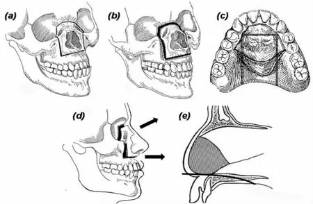
截骨包括鼻-上颌截骨和硬腭截骨。鼻部充填材料使用肋软骨和硅胶假体。改良截骨采用标准冠状切口,沿骨膜向下剥离暴露眶缘及鼻背,注意保护泪囊及内眦韧带。口内切口采用标准Le FortⅠ切口,剥离软组织及骨膜,充分暴露梨状孔。截骨从鼻额缝开始,水平向两侧经眶内壁,沿泪囊窝后方向下,转向眶下壁,在眶下神经孔内侧转向前,向下沿上颌窦前壁至梨状孔水平,然后截骨线在尖牙牙根上方直角水平转向内侧沿梨状孔下缘在中线位置汇合(图1a)。硬腭部位沿牙龈缘1 cm位置行小切口,使用剥离子沿硬腭表面剥离至硬腭后缘,充分松解。沿上颌窦前壁截骨线向后向下锯开硬腭侧缘并向前延伸,沿梨状孔下缘水平截骨线向后方锯开硬腭前缘(图1b,e)。充分游离骨块,并向前上方推进,钛板钛钉固定(图1d)。
沿第7肋表面切开皮肤及肌肉,暴露第7肋,根据患者鼻长度截取一定长度肋骨,保留约3 cm肋软骨,雕刻成“L”形[13]。上端与额骨固定,肋骨支撑鼻背,肋软骨充填鼻尖。截骨遗留缝隙使用多余的肋骨进行充填。对于2例因瘢痕原因而拒绝使用肋骨带肋软骨行鼻充填的患者,我们采用了简单的硅胶隆鼻。

图1 截骨示意图Fig.1 Schematic diagram of our modified nasal-maxillary-hard palatine osteotomy technique
2 结果
本组患者术后无坏死、感染等严重并发症出现,无泪囊及内眦韧带损伤。骨块固位良好,肋骨无明显吸收。
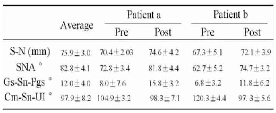
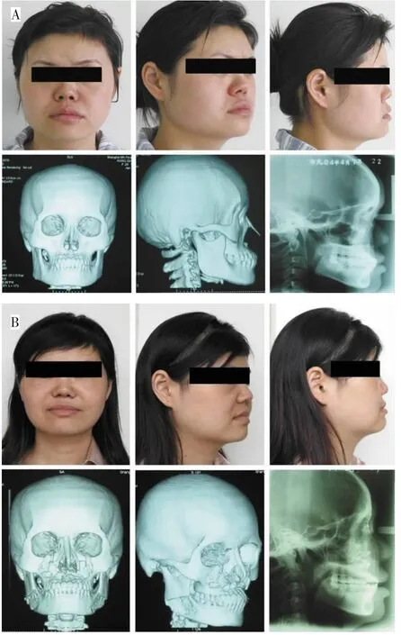
术后定位测量结果发现,患者S-N点距离平均增加5.0 mm,SNA角增大约10°,上颌后缩得到明显矫正。软组织测量显示,术后面角(Gs-Sn-Pgs)增加大约 8.0°,鼻唇角(Cm-Sn-UI)约在 95.0°左右(正常值 97.93 °±8.24 °)。 鼻尖前移约 10 mm,冠状位下移3.0 mm,(表1,图2)。本组患者的手术结果提示,使用本方法,术后患者的面中部及鼻外形都得到了前移,同时鼻小柱也得到了延长。

表1 两例患者术前术后测量结果Table 1 Anthropometric Measurements and Analysis for two cases

图2 典型病例(A:术前;B:术后)Fig.2 Typical case(A:Pre-operation;B:Post-operation)
3 讨论
Binder综合征是以面中部凹陷和鼻畸形为特点的先天畸形。该畸形是由于面中部和鼻部的骨发育异常,而不是软组织缺乏引起。因此,手术治疗应以纠正后缩的上颌骨,并使鼻部软组织复位为目的。
对于症状较轻的患者,单纯的鼻充填基本可以满足要求。常用的充填物有肋骨和硅胶假体等,采用骨条行鼻充填亦可获得满意效果[14-16]。然而由于鼻部皮肤覆盖非常紧密,植入的骨块往往会出现吸收。单纯的软骨重填术后鼻外形通常较好,但会出现软骨移位或者卷曲[17-18]。我们采用肋骨带软骨头进行鼻充填,术后没有明显的吸收且固位良好。说明肋骨带软骨头用于Binder综合征鼻畸形矫正具有良好效果。但是,对于较严重患者,单纯的鼻充填并不能纠正面中部因发育不全而造成的畸形,必须同时加以截骨手术以纠正面中部的凹陷。
严重面中部凹陷畸形可以采用Le Fort截骨[16]。不但可以纠正面中部凹陷,同时可以增加面中部的垂直高度。然而传统的截骨术不能很好地解决鼻畸形问题,因为鼻基底的塌陷后缩和鼻背支撑问题都未得到解决。Le FortⅡ和“金字塔”型截骨都需要移动上颌前牙,但许多患者并不伴有错颌畸形[2]。所以,我们对传统的截骨术式进行改良,既可以保留咬颌关系,而且可以达到同样的面中部前移效果。
尽管冠状切口手术时间较长,创伤较大,但是我们认为它还是该手术的最佳入路。相比之下,Converse等[16]介绍的手术入路对术区暴露稍显不足,而且瘢痕比较明显。Le FortⅠ型截骨可以充分将面中部前移,但是鼻基底部的塌陷仍然存在。“金字塔”型截骨能够很好地解决面中部凹陷的问题,然而需要前移上颌前牙,我们认为该方法可能更适用于伴有安氏Ⅲ错颌的患者。
我们的截骨区域下缘局限于鼻基底,不仅可以前移面中部,还可以使得鼻基底及梨状孔周围凹陷得到良好的纠正。同时,骨块不仅前移还伴有向上的旋转,可以增加面中部的高度。
综上所述,改良鼻-上颌-硬腭截骨联合肋软骨充填,可以有效地纠正Binder综合征患者的面中部凹陷及鼻畸形。
[1]Binder KH.Dysostosis maxillo-nasalis:ein archinencephaler Missbildungskomplex[J].Deutsche Zahnarztuche Zeitschift,1962,17:438-444.
[2]Holmstrom H:Clinical and pathologic features of maxillonasal dysplasia(Binder’s Syndrome)significance of the prenasal fossa on etiology[J].Plast Reconstr Surg,1986,78(5):559-567.
[3]Horswell BB,Holmes AD,Barnett JS,et al.Maxillonasal dysplasia(Binder’s syndrome):a critical review and case study[J].J Oral Maxillofac Surg,1987,45(2):114-122.
[4]Olow-Nordenram M,Valentin J.An etiologic study of maxillonasal dysplasia—Binder’s syndrome[J].Scand J Dent Res,1988,96(1):69-74.
[5]Roy-Doray B,Geraudel A,Alembik Y,et al.Binder syndrome in a mother and her son[J].Genet Couns,1997,8(3):227-233.
[6]Cronin TD.Lengthening columella by use of skin from nasal floor and alae[J].Plast Reconstr Surg Transplant Bull,1958,21(6):417-426.
[7]Dingman RO,Walter C.Use of composite ear grafts in correction of the short nose[J].Plast Reconstr Surg,1969,43(2):117-124.
[8]Hopkins GB.Hypoplasia of the middle third of the face associated with congenital absence of the anterior nasal spine,depression of the nasal bones,and angle class III malocclusion[J].Br J Plast Surg,1963,16:146-153.
[9]Meyer R,Flemming I.Congenital flat nose and its correction[J].Z Laryngol Rhinol Otol,1969,48(11):808-811.
[10]Obwegeser HL.Surgical correction of small or retrodisplaced maxillae.The"dish-face"deformity[J].Plast Reconstr Surg,1969,43(4):351-365.
[11]Henderson D,Jackson IT.Naso-maxillary hypoplasis--the Le Fort II osteotomy[J].Br J Oral Surg,1973,11(2):77-93.
[12]Jackson IT,Moos KF,Sharpe DT.Total surgical management of Binder’s Syndrome[J].Ann Plast Surg,1981,7(1):25-34.
[13]Monasterio FO,Molina F,McClintock JS:Nasal correction in Binder's syndrome:the evolution of a treatment plan[J].Aesthetic Plast Surg,1997,21(5):299-308.
[14]Ragnell A.A simple method of reconstruction in some cases of dish-face deformity[J].Plast Reconstr Surg,1952,10(4):227-237.
[15]Converse JM:Technique of bone grafting for contour restoration of the face[J].Plast Reconstr Surg,1954,14(5):332-346.
[16]Converse JM,Horowitz SL,Valauri AJ,et al.The treatment of nasomaxillary hypoplasia.A new pyramidal naso-orbital mazillary osteotomy[J].Plast Reconstr Surg,1970,45(6):527-535.
[17]Fry H:Cartilage and cartilage grafts:the basic properties of the tissue and the components responsible for them[J].Plast Reconstr Surg,1967,40(5):526-539.
[18]Gibson T,Davis WB.The distortion of autogenous cartilage grafts:Its cause and prevention[J].Br J Plast Surg,1957,10:257-274.
Modified Nasomaxillary and Hard Palatine Osteotomy Combined Nasal Implantation for Binder's Syndrome
WANG Hengjian,YUAN Jie,ZHANG Ying,WEI Min.Department of Plastic and Reconstructive Surgery,Shanghai Ninth People's Hospital,Shanghai Jiaotong University School of Medicine,Shanghai 200011,China.
WEI Min(E-mail:wmdoctor@yahoo.com).
ObjectiveTo explore a new alternative modified osteotomy method for correcting the midfacial defects of Binder's syndrome.MethodsEight patients with maxillonasal dysplasia were treated by modified naso-maxillary complex and hard palatine “box” osteotomy,combined with chondrocostal bone grafts or nasal implant to correct the retruded nasal deformity.ResultsAll patients were satisfied with outcome of operation without severe complications.Six-months follow-up evaluation showed good correction of the midfacial profile and nasal projection,the advancement of midface and chondrocostal bone graft was fixed perfectly.The lateral cephalometric analysis and superimposition results showed S-N distance had been increased approximately 5 mm and SNA angle increased about 10°.Soft tissue measurement showed the facial convexity angle had increased 8 °;the nasolabial angle increased about 10 °.The nasal tip had moved 10.0 mm anteriorly and 3.0 mm coronally.ConslusionModified naso-maxillary complex and hard palatine osteotomy can be a good alternative for correction of midfacial hypoplasia and nasal deformity of Binder's syndrome.
Maxillonasal dysplasia;Binder's syndrome;osteotomy;nasal implant;chondrocostal bone graft
R682.1+1
A
1673-0364(2012)05-0272-03
10.3969/j.issn.1673-0364.2012.05.007
200011 上海市 上海交通大学医学院附属第九人民医院整复外科。
韦敏(E-mail:wmdoctor@yahoo.com)。
2012年8月21日;
2012年9月18日)

