差异表达基因CDCA8、CDC20与肾母细胞瘤患者预后及免疫细胞浸润的关系
杨迪,黄业华,黄和松,蒙菲,黄靓,岑琴,覃花杏,张东虎


摘要:目的 探討差异表达基因CDCA8及CDC20在肾母细胞瘤中的表达,以及与肾母细胞瘤患者预后及免疫细胞浸润的关系。方法 登录并下载GEO数据库及Target数据库中与肾母细胞瘤相关的基因表达谱数据;利用R语言筛选出差异表达基因,筛选出与肾母细胞瘤预后有关的基因;利用R语言中的CIBERSORT计算方法计算出肾母细胞瘤中免疫细胞浸润的表达;利用SPSS分析肾母细胞瘤预后有关的基因与免疫细胞浸润的相关性。结果 GEO表达谱数据GSE66405、GSE73209、GSE11151筛选出共同差异表达基因1260个,其中高表达398个,低表达862个;蛋白互作网络筛选出15个核心蛋白,包括CDK1、CCNA2 、TOP2A、CCNB2、KIF11、BUB1、BUB1B、ASPM、NUSAP1、CDC20、CDCA8、KIF20A、DLGAP5、TPX2、CENPF,其中CDCA8及CDC20与肾母细胞瘤患者预后有关;T cells CD4 memory activated(P=0.026)、T cells follicular helper(P=0.031)、Macrophages M0(P=0.005)、Dendritic cells activated(P=0.025)的表达高于正常对照组,而Plasma cells(P=0.023)、T cells CD4 memory resting(P=0.002)、NK cells resting(P=0.045)、Monocytes(P=0.013)在肾母细胞瘤中低表达。相关性分析显示,CDCA8与6个免疫细胞浸润具有相关性,CDC20与5个免疫细胞浸润具有相关性。结论 CDCA8及CDC20有可能成为肾母细胞瘤患者治疗的新潜在靶点。
关键词:肾母细胞瘤;CDCA8;CDC20;免疫细胞浸润
中图分类号:R737.11 文献标识码:A DOI:10.3969/j.issn.1006-1959.2024.08.001
文章编号:1006-1959(2024)08-0001-09
Relationship Between Differentially Expressed Genes CDCA8, CDC20 and Prognosis,
Immune Cell Infiltration in Patients with Nephroblastoma
YANG Di1,HUANG Ye-hua2,HUANG He-song3,MENG Fei1,HUANG Liang1,CEN Qin4,QIN Hua-xing4,ZHANG Dong-hu1
(1.Department of Pediatric Surgery,Affiliated Hospital of Youjiang Medical University for Nationalities,Baise 533000,Guangxi,China;
2.Department of Emergency,Affiliated Hospital of Youjiang Medical University for Nationalities,Baise 533000,Guangxi,China;
3.Department of Respiratory Intensive Care,Affiliated Hospital of Youjiang Medical University for Nationalities,
Baise 533000,Guangxi,China;
4.Department of General Surgery,Baidong Hospital of Affiliated Hospital of Youjiang Medical University for Nationalities,Baise 533000,Guangxi,China)
Abstract:Objective To investigate the expression of differentially expressed genes CDCA8 and CDC20 in nephroblastoma and their relationship with prognosis and immune cell infiltration in patients with nephroblastoma.Methods The gene expression profile data related to nephroblastoma in GEO database and Target database were logged in and downloaded. The differentially expressed genes were screened by R language, and the genes related to the prognosis of nephroblastoma were screened. The expression of immune cell infiltration in nephroblastoma was calculated by CIBERSORT calculation method in R language. SPSS was used to analyze the correlation between genes related to the prognosis of nephroblastoma and immune cell infiltration.Results A total of 1260 common differentially expressed genes were screened out from GEO expression profile data GSE66405, GSE73209 and GSE11151, of which 398 were highly expressed and 862 were lowly expressed. The protein interaction network screened 15 core proteins, including CDK1, CCNA2, TOP2 A, CCNB2, KIF11, BUB1, BUB1B, ASPM, NUSAP1, CDC20, CDCA8, KIF20 A, DLGAP5, TPX2, CENPF, among which CDCA8 and CDC20 were related to the prognosis of nephroblastoma patients. The expression of T cells CD4 memory activated (P=0.026), T cells follicular helper (P=0.031), Macrophages M0 (P=0.005) and Dendritic cells activated (P=0.025) in nephroblastoma was higher than that in normal control group. Plasma cells (P=0.023), T cells CD4 memory resting (P=0.002), NK cells resting (P=0.045) and Monocytes (P=0.013) were lowly expressed in nephroblastoma. Correlation analysis showed that CDCA8 was correlated with 6 immune cell infiltration, while CDC20 was correlated with 5 immune cell infiltration.Conclusion CDCA8 and CDC20 may be new potential targets for the treatment of patients with nephroblastoma.
Key words:Nephroblastoma;CDCA8;CDC20;Immune cell infiltration
肾母细胞瘤(nephroblastoma)是儿童第二大最常见的腹内癌症,也是儿童第五大最常见的恶性肿瘤[1,2]。在儿童中,有95%以上的肾脏肿瘤是肾母细胞瘤,在2~3岁时肾母细胞瘤的发病率达到高峰[3]。随着手术、化疗、放疗的联合治疗,肾母细胞瘤的远期生存率由过去的30%提高到了90%以上,然而复发率仍然保持在15%~50%[1,2,4]。由此可见,肾母细胞瘤的治疗仍然有必要进一步提高,探索新的治疗肾母细胞瘤的靶点及方法具有一定的临床意义。人类细胞分裂周期相关基因8(cell division cycle associated 8, CDCA8)是人类细胞分裂周期相关基因家族中一个重要成员,是细胞有丝分裂周期调节基因,参与多种肿瘤细胞的增殖进程。研究发现[5],CDCA8基因在肝癌中高表达,且Kaplan-Meier生存分析中发现,高表达的CDCA8与肝癌低预后相关。在膀胱癌中,敲除CDCA8可抑制膀胱癌细胞的增殖并且促进其细胞凋亡[6]。研究发现[7],miR-133a-3p能够靶向调控CDCA8的表达,从而影响食管癌细胞的增殖、迁移和侵袭,进一步影响食管癌的进展。在肺腺癌的研究中[8],miR-133b可靶向调控CDCA8的表达从而抑制肺腺癌细胞的增殖、迁移及侵袭。细胞周期蛋白20(cell-division cycle protein 20, CDC20)是纺锤体组装检查点(spindle assembly checkpoint, SAC)的靶标,在有丝分裂中发挥重要作用。Zhang X[9]研究发现,CDC20在乳腺癌中高表达,并且高表达的CDC20提示乳腺癌患者较差的总生存时间。另有研究发现[10],在人胰腺癌细胞中CDC20呈高表达,沉默CDC20可以提高放射治疗对胰腺癌细胞的敏感性。在结直肠癌中,通过下调CDC20可通过调控Mcl-1/p-Chk1介导结直肠癌细胞的DNA损伤及细胞凋亡,进而增加结直肠癌细胞的放射敏感性[11]。目前关于CDCA8及CDC20基因在肾母细胞瘤中表达情况报道较少,本研究通过筛选出差异基因并分析CDCA8及CDC20在肾母细胞瘤中的表达情况,探讨CDCA8及CDC20与肾母细胞瘤患者总生存时间的关系,分析CDCA8及CDC20与肾母细胞瘤免疫细胞浸润的相关性,为寻找肾母细胞瘤新治疗靶点提供一定的理论基础。
1资料与方法
1.1数据来源及基本信息 登录美国国立生物信息技术中心(National Center for Biotechnology Information, NCBI)建立的GEO(Gene Expression Omnibus)数据库官网(https://www.ncbi.nlm.nih.gov/gds/?term=),以“wilms tumor”為关键词搜索并筛选与肾母细胞瘤相关的基因表达谱数据。登录儿童肿瘤数据库Target数据库(https://www.cancer.gov/ccg/)下载与肾母细胞瘤相关mRNA表达谱数据。其中,Target数据库中包含130例肿瘤患者以及6例对应正常癌旁组织,130例肾母细胞瘤患者均包含了相关的临床资料及生存资料。
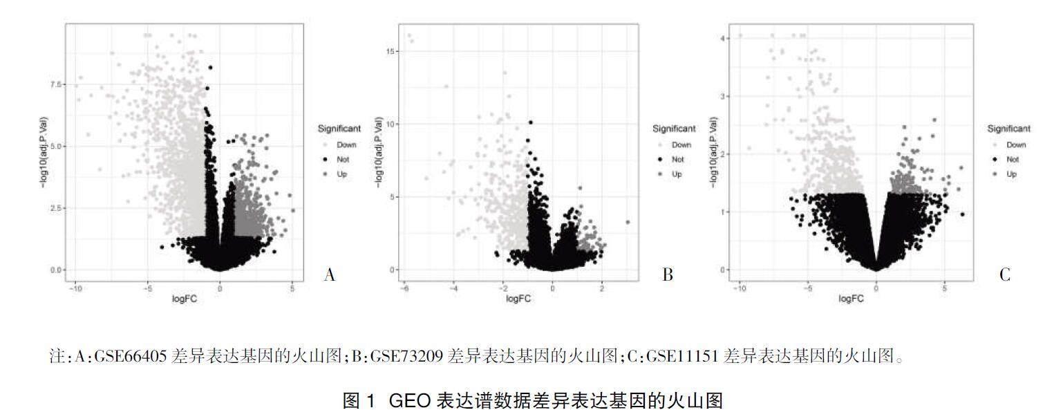
1.2差异表达基因的筛选 利用R语言中的limma包以logFCfilter=1及adj.P.Val.Filter=0.05为标准,分别筛选GEO表达谱数据GSE66405、GSE73209、GSE11151中的差异表达基因,并绘制出差异表达基因热图及火山图。在分别得到3个GEO表达谱数据的差异表达基因后,利用R语言中的RobustRankAggreg包筛选出3组差异表达基因的共同差异表达基因,并利用R语言中的pheatmap包绘制出共同差异表达基因的热图。
1.3共同差异表达基因的KEGG通路分析及GO富集分析 在得到3组GEO表达谱数据的共同差异表达基因后,利用R语言中的clusterProfiler、org.Hs.eg.db、enrichplot、ggplot2包分别计算出共同差异表达基因的KEGG通路分析及GO富集分析。
1.4蛋白互作网络的构建及核心蛋白的筛选 登录蛋白互作网络官网(https://string-db.org/),输入差异表达基因,在minimum required interaction score选择highest confidence(0.900),得到蛋白互作网络的结果,选择下载string_hires_image.png图片和string_interactions.tsv文件,文件继续用于下一步分析。将上述蛋白互作网络结果的string_interactions.tsv文件导入Cytoscape3.7.2软件。利用Cytoscape软件中的CytoHubba工具筛选出核心蛋白。
1.5 CDCA8及CDC20基因在肾母细胞瘤中的表达及与预后的关系 Target数据库中的数据包含有肾母细胞瘤患者的生存时间,利用R语言中的limma包及beeswarm包分别分析CDCA8及CDC20在肾母细胞瘤与正常组织中表达的差异。利用R语言中的survival包分别分析CDCA8基因及CDC20基因表达量与肾母细胞瘤患者总生存时间的关系。
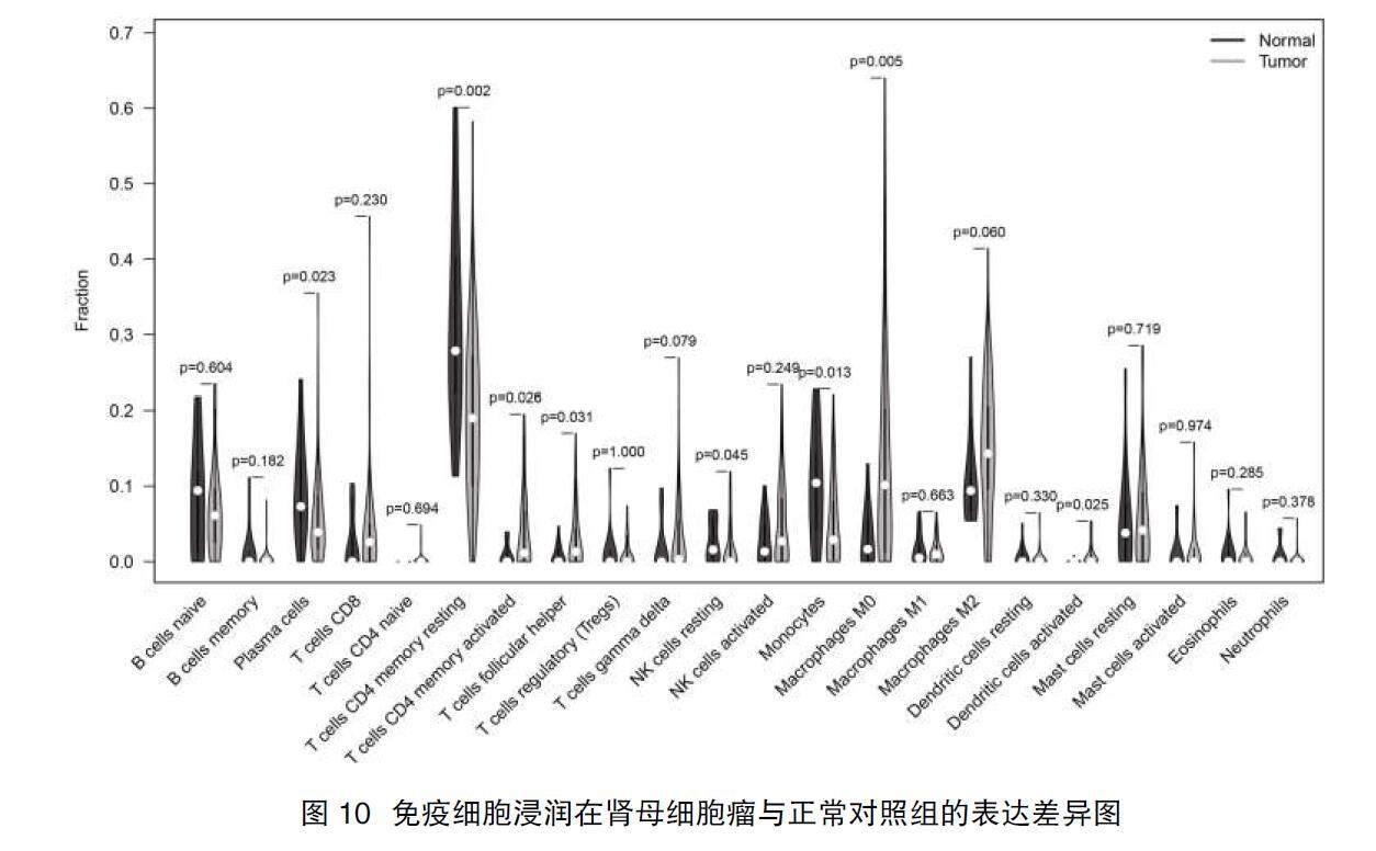
1.6免疫细胞浸润在肾母细胞瘤中的表达 登录CIBERSORT官网下载22种免疫细胞的CIBERSPRT.R代码,运用R语言中e1071包及preprocessCore数据包进行运算,最终得到22种免疫细胞在每例样品中的表达情况。运用R语言,绘制出22种免疫细胞在每例样品表达百分比以及免疫细胞浸润表达的热图;在R语言中下载vioplot包,分析免疫细胞浸润在肾母细胞瘤及正常组织中差异表达,绘制出小提琴图。
1.7 CDCA8、CDC20与免疫细胞浸润的关系 在得到免疫细胞浸润在肾母细胞瘤中的表达量后,将CDCA8、CDC20及22种免疫细胞浸润的表达结果导入SPSS 23.0,点击SPSS软件,选择栏目中的分析,点击双变量相关分析,统计学方法选择Spearman统计学方法分别分析CDCA8与22种免疫细胞浸润的关系以及CDC20与22种免疫细胞浸润的关系。
1.8统计学方法 本研究运用R语言(4.0.2)内的多个统计数据包将数据导入R软件并进行统计学分析及结果图片绘制。差异基因的筛选是利用R语言limma包,以logFCfilter=1及adj.P.Val.Filter=0.05为标准。共同差异表达基因的筛选利用R语言的RobustRankAggreg包筛选,P<0.05纳入共同差异表达基因。KEGG及GO富集分析利用R语言中的clusterProfiler、org.Hs.eg.db、enrichplot、ggplot2包,P<0.05认为被纳入的条件。核心蛋白的筛选利用Cytoscape3.7.2软件中的CytoHubba工具,选择MCC计算方法。免疫细胞浸润的计算是利用22种免疫细胞浸润的基因CIBERSPRT.R代码,运用R语言中e1071包及preprocessCore数据包进行运算。其他计量资料运用t检验,计数资料运用?字2检验或秩和检验,CDCA8及CDC20基因的生存分析在R语言中采用Kaplan-Meier法Log-rank检验,CDCA8、CDC20基因与免疫细胞浸润的相关性分析在SPSS 23.0中采用Spearman统计学方法,对于所有统计学计算,最终结果以P<0.05表示差异有统计学意义。
2结果
2.1差异表达基因的筛选 在GEO数据库中收集GSE66405、GSE73209、GSE11151表达谱数据,GSE66405包含肾母细胞瘤组织28例,正常组织4例;GSE73209包含肿瘤组织32例,正常组织6例;GSE11151表达谱数据包含4例肿瘤组织及5例正常组织。在Target数据库中收集到130例肾母细胞瘤,另有6例正常對照组。在GSE66405表达谱数据中筛选出2349个差异表达基因,包括上调差异表达基因745个,下调差异表达基因1604个。在GSE73209表达谱数据中,筛选出508个差异表达基因,其中54个高表达差异表达基因,454个低表达差异表达基因。在GSE11151表达谱数据中,筛选出684个差异表达基因,其中166高表达,518个低表达。利用R语言limm包,最终在3组GEO表达谱数据的差异表达基因中筛选出1260个共同差异表达基因,其中高表达398个,低表达862个。利用R语言绘制出差异表达基因的热图及火山图,见图1、图2。
2.2共同差异表达基因的KEGG基因通路分析及GO基因富集分析 在得到1260个共同差异表达基因后,利用R语言中的clusterProfiler、org.Hs.eg.db、enrichplot、ggplot2包分析1260个共同差异表达基因的KEGG通路分析及GO富集分析,其中GO富集分析包含BP、CC、MF共3部分,利用R语言绘制出前30个KEGG基因通路及GO富集分析结果,见图3。
2.3蛋白互作网络的构建及核心蛋白的筛选 将1260个共同差异表达基因导入String网页中,在minimum required interaction score选择highest confidence(0.900),最终构建出共同差异表达基因的蛋白互作网络,见图4。利用Cytoscape3.7.2软件中的CytoHubba工具,选择MCC计算方法筛选出15个核心蛋白,包括CDK1、CCNA2、TOP2A、CCNB2、KIF11、BUB1、BUB1B、ASPM、NUSAP1、CDC20、CDCA8、KIF20A、DLGAP5、TPX2、CENPF,见图5。
2.4 CDCA8及CDC20在肾母细胞瘤中的表达及与总生存时间的关系 得到15个核心蛋白后,利用Target数据库分析核心蛋白在肾母细胞瘤中的表达以及与总生存时间的关系,最终得到CDCA8在肾母细胞瘤中高表达(P=3.666e-05),并且与肾母细胞瘤患者总生存时间有关(P=0.008);CDC20在肾母细胞瘤中高表达(P=3.667e-05),与肾母细胞瘤患者总生存时间有关(P=0.036),其余核心蛋白与肾母细胞瘤患者总生存时间无关(P>0.05),见图6、图7。
2.5免疫细胞浸润在肾母细胞瘤中的表达情况 通过利用R语言计算出22种免疫细胞浸润在肾母细胞瘤的表达情况,绘制百分比图及热图见图8、图9。利用R语言中的vioplot包对比肾母细胞瘤患者与正常组免疫细胞浸润情况,结果发现T cells CD4 memory activated(P=0.026)、T cells follicular helper(P=0.031)、Macrophages M0(P=0.005)、Dendritic cells activated(P=0.025)的表达高于正常对照组,而Plasma cells(P=0.023)、T cells CD4 memory resting(P=0.002)、NK cells resting(P=0.045)、Monocytes(P=0.013)在肾母细胞瘤中低表达,见图10。
2.6 CDCA8及CDC20在肾母细胞瘤中的表达与免疫细胞浸润的相关性 将CDCA8基因、CDC20基因及22种免疫细胞浸润的表达数据导入SPSS 23.0,分析CDCA8与免疫细胞浸润的相关性及CDC20与免疫细胞浸润的相关性,结果得到在肾母细胞瘤中6种免疫细胞浸润与CDCA8基因的表达具有相关性,其中与NK cells activated(P=0.000,r=0.561)、Macrophages M0(P=0.000,r=0.432)呈正相关,与T cells CD4 memory resting(P=0.002,r=-3.840)、NK cells resting(P=0.002,r=-3.820)、Monocytes(P=0.003,r=-0.364)、Mast cells resting(P=0.043,r=-0.254)呈负相关。在CDC20与免疫细胞浸润相关性分析中,有5种免疫细胞浸润与CDC20的表达具有相关性,其中正相关免疫细胞浸润包括B cells naive(P=0.010,r=0.322)、T cells follicular helper(P=0.000,r=0.490)、NK cells resting(P=0.008,r=0.326);而T cells CD8(P=0.025,r=-0.280)、NK cells activated(P=0.008,r=-0.330)呈负相关,见表1、表2。
3讨论
肾母细胞瘤早期症状主要以无意中发现腹部包块为主,虽然目前治疗生存率较高,但是治疗后复发及远期较差的生存质量仍然是临床上需解决的问题。本研究通过下载GEO数据库中肾母细胞瘤相关基因表达谱数据,筛选出差异表达基因,通过对差异表达基因进行蛋白互作网络构建,筛选出15个核心蛋白。在Target数据库中,发现核心蛋白中的CDCA8及CDC20在肾母细胞瘤中的表达高于正常组织,并且与肾母细胞瘤患者预后的相关。目前CDCA8及CDC20在多个肿瘤中已有研究发表。Wu B等[12]研究发现,CDCA家族基因中的CDCA2、CDCA3、CDCA5及CDCA8在肝癌中高表达,并且均与肝癌患者的总生存时间具有相关性。Phan NN等[13]研究发现CDCA3、CDCA5和CDCA8在乳腺癌人体标本及实验室癌细胞的表达均高于正常对照组,并且高表达CDCA8提示较差的无复发生存率。Shi Q等[14]研究发现,通过Western blot 等检测发现在60例肾母细胞瘤中的CDC20蛋白呈高表达,并且利用Kaplan-Meier生存分析统计发现高表达CDC20蛋白提示较差的总生存时间。
目前多个研究已有关于免疫细胞浸润的分析,其中Ding J等[15]探讨了免疫细胞浸润在乳腺癌中的表达,同时分析SKA家族基因SKA1/2/3在乳腺癌中的表达,结果发现SKA1/2/3的表达与免疫细胞浸润具有相关性。Zhang H等[16]在肝癌中探讨免疫细胞浸润在肝癌中的表达情况,结果发现免疫细胞Mast Cells Resting在肝癌中表达较正常组织高,并且与肝癌患者的预后有关。Yang ML等[17]在头颈部鳞状细胞癌中分析了22种免疫细胞浸润的表达,并且发现SLC13A4与Monocytes、M1 macrophages、M2 macrophages、resting CD4+ memory T cells等呈负相关,而与plasma cells、T follicular helper cells、naive B cells呈正相关。Tian Z等[18]研究发现,TPM家族基因TPM1-4在肝癌中均高表达,TPM3是肝癌的独立预后因子,并且TPM1-4的表达均与肝癌中的免疫细胞浸润具有相关性。Lin Z等[19]研究发现,LncRNA ADAMTS9-AS2在肺癌中低表达,并且该LncRNA的表达量与免疫细胞NK CD56 bright cells、Th2 cells及TReg具有负相关性,与NK cells、T cells、T helper cells、Macrophages等免疫细胞具有正相关性。研究发现[20],CCL8基因在皮肤黑色素瘤中的表达与患者预后相关,并且发现CCL8的表达与免疫细胞M1 macrophages的表达具有相关性。本研究通过利用R语言计算出GEO表达谱数据中的免疫细胞浸润的表达,并且发现在肾母细胞瘤中T cells CD4 memory activated、T cells follicular helper、Macrophages M0、Dendritic cells activated免疫细胞呈高表达,而Plasma cells、T cells CD4 memory resting、NK cells resting、Monocytes在肾母细胞瘤中低表达。此外,本研究结果显示,CDCA8基因的表达与NK cells activated、 Macrophages M0、T cells CD4 memory resting、NK cells resting、Monocytes、Mast cells resting具有相关性;而CDC20的表达与B cells naive、T cells follicular helper、NK cells resting、T cells CD8、NK cells activated具有相关性。这些结果提示CDCA8及CDC20有可能通过参与调控肿瘤免疫细胞影响肾母细胞瘤的进展。
综上所述,CDCA8及CDC20有可能成为肾母细胞瘤患者治疗的新潜在靶点。但本研究目前仅在数据库中分析得出結果,需进一步行实验验证。
参考文献:
[1]Davidoff AM.Wilms tumor[J].Adv Pediatr,2012,59(1):247-267.
[2]de Sá Pereira BM,Montalv?觔o de Azevedo R,da Silva Guerra JV,et al.Non-coding RNAs in Wilms' tumor: biological function, mechanism, and clinical implications[J].J Mol Med (Berl),2021,99(8):1043-1055.
[3]Pastore G,Znaor A,Spreafico F,et al.Malignant renal tumours incidence and survival in European children (1978-1997):report from the Automated Childhood Cancer Information System project[J].Eur J Cancer,2006,42(13):2103-2114.
[4]Spreafico F,Pritchard Jones K,Malogolowkin MH,et al.Treatment of relapsed Wilms tumors:lessons learned[J].Expert Rev Anticancer Ther,2009,9(12):1807-1815.
[5]Shuai Y,Fan E,Zhong Q,et al.CDCA8 as an independent predictor for a poor prognosis in liver cancer[J].Cancer Cell Int,2021,21(1):159.
[6]Gao X,Wen X,He H,et al.Knockdown of CDCA8 inhibits the proliferation and enhances the apoptosis of bladder cancer cells[J].PeerJ,2020,8:e9078.
[7]Wang X,Zhu L,Lin X,et al.MiR-133a-3p inhibits the malignant progression of oesophageal cancer by targeting CDCA8[J].J Biochem,2022,170(6):689-698.
[8]Hu C,Wu J,Wang L,et al.miR-133b inhibits cell proliferation,migration,and invasion of lung adenocarcinoma by targeting CDCA8[J].Pathol Res Pract,2021,223:153459.
[9]Zhang X.Identification of potential prognostic markers associated with lung metastasis in breast cancer by weighted gene co-expression network analysis[J].Cancer Biomark,2022,33(3):299-310.
[10]Taniguchi K,Momiyama N,Ueda M,et al.Targeting of CDC20 via small interfering RNA causes enhancement of the cytotoxicity of chemoradiation[J].Anticancer Res,2008,28(3a):1559-1563.
[11]Gao Y,Wen P,Chen B,et al.Downregulation of CDC20 Increases Radiosensitivity through Mcl-1/p-Chk1-Mediated DNA Damage and Apoptosis in Tumor Cells[J].Int J Mol Sci,2020,21(18):6692.
[12]Wu B,Huang Y,Luo Y,et al.The diagnostic and prognostic value of cell division cycle associated gene family in Hepatocellular Carcinoma[J].J Cancer,2020,11(19):5727-5737.
[13]Phan NN,Wang CY,Li KL,et al.Distinct expression of CDCA3,CDCA5,and CDCA8 leads to shorter relapse free survival in breast cancer patient[J].Oncotarget,2018,9(6):6977-6992.
[14]Shi Q,Tang B,Li Y,et al.Identification of CDC20 as a Novel Biomarker in Diagnosis and Treatment of Wilms Tumor[J].Front Pediatr,2021,9:663054.
[15]Ding J,He X,Wang J,et al.Integrative analysis of prognostic value and immune infiltration of spindle and kinetochore-associated family members in breast cancer[J].Bioengineered,2021,12(2):10905-10923.
[16]Zhang H,Sun L,Hu X.Mast Cells Resting-Related Prognostic Signature in Hepatocellular Carcinoma[J].J Oncol,2021,2021:4614257.
[17]Yang ML,Zhang JH,Li S,et al.SLC13A4 Might Serve as a Prognostic Biomarker and be Correlated with Immune Infiltration into Head and Neck Squamous Cell Carcinoma[J].Pathol Oncol Res,2021,27:1609967.
[18]Tian Z,Zhao J,Wang Y.The prognostic value of TPM1-4 in hepatocellular carcinoma[J].Cancer Med,2022,11(2):433-446.
[19]Lin Z,Huang W,Yi Y,et al.LncRNA ADAMTS9-AS2 is a Prognostic Biomarker and Correlated with Immune Infiltrates in Lung Adenocarcinoma[J].Int J Gen Med,2021,14:8541-8555.
[20]Yang P,Chen W,Xu H,et al.Correlation of CCL8 expression with immune cell infiltration of skin cutaneous melanoma:potential as a prognostic indicator and therapeutic pathway[J].Cancer Cell Int,2021,21(1):635.
收稿日期:2023-01-09;修回日期:2023-06-16
編辑/杜帆

