
Comprehensive treatment and a rare presentation of Cronkhite-Canada syndrome: Two case reports and review of literature
2023-12-10 02:24:28YanQingLvMeiLanWangTongYuTangYuQinLi
Yan-Qing Lv,Mei-Lan Wang,Tong-Yu Tang,Yu-Qin Li
Abstract BACKGROUND Cronkhite-Canada syndrome (CCS) is a rare sporadic polyposis syndrome that presents with gastrointestinal and ectodermal symptoms in addition to nutritional deficiencies.CCS combined with hypothyroidism is an even rarer condition,with no standard treatment guidelines.CASE SUMMARY The present study described 2 patients with CCS: A 67-year-old woman with concomitant hypothyroidism and 68-year-old man treated with endoscopic mucosal resection (EMR).Both patients had multiple gastrointestinal symptoms and ectodermal changes,along with multiple gastrointestinal polyps.Microscopic examination showed that the mucosa in both patients was hyperemic and edematous,with pathologic examination showing distorted,atrophic,and dilated glands.Patient 1 had concomitant hypothyroidism and was treated with levothyroxine.Due to her self-reduction of hormone dose,her disease relapsed.Patient 2 underwent EMR,but refused further hormonal or biological treatments.Subsequently,he was treated with an oral Chinese medical preparation.CONCLUSION Pharmacotherapy can induce and maintain remission in CCS patients,with adjuvant EMR,long-term follow-up,and endoscopic surveillance being necessary.
Key Words: Cronkhite-Canada syndrome;Clinical features;Gastrointestinal polyps;Hypothyroidism;Case report
INTRODUCTION
Cronkhite-Canada syndrome (CCS),or polyposis hyperpigmentation alopecia nail dystrophy syndrome,is a rare,nongenetic syndrome characterized by ectodermal changes and gastrointestinal symptoms.Patients with CCS have a poor prognosis,with a 5-year mortality rate of 55%[1].There are no standardized treatments,as the etiology and pathogenesis of CCS are unknown.At present,glucocorticoid is the primary treatment,either alone or combined with immunosuppressive or biological agents.Other treatments include nutritional support,anti-Helicobacter pylori treatment,tumor necrosis factor-(TNF-) inhibitors,and traditional Chinese medicines.This report describes the clinical characteristics of 2 patients diagnosed with CCS at the First Bethune Hospital of Jilin University and presents a review of the literature.Patient 1 was a 67-year-old woman diagnosed with CCS,with concomitant hypothyroidism,whereas Patient 2 was a 68-year-old man diagnosed with CCS,who underwent endoscopic mucosal resection (EMR).The aim of this study was to provide the basis for basic and clinical research on CCS.
CASE PRESENTATION
Chief complaints
Case 1:A 67-year-old woman was admitted to our hospital in August 2016,due to intermittent diarrhea for 2 mo,aggravated with bloody stool for 1 mo.
Case 2:A 68-year-old man was admitted to our hospital in May 2023,due to hypogeusia and diarrhea for 1 mo.
History of present illness
Case 1:Two months prior to admission,the patient had developed yellowish watery diarrhea (3-5 times per day).One month later,the frequency of defecation had increased to 5-8 times per day,accompanied by a small amount of dark red bloody stool,abdominal pain,and tenesmus.The patient experienced nausea and vomited her gastric contents occasionally.She also experienced fatigue,apathy,loss of appetite,hypogeusia,and alopecia,with a weight loss of 5 kg.
Case 2:One month prior to admission,the patient had developed hypogeusia,loss of appetite,abdominal pain,and yellowish watery diarrhea at a frequency of 4-10 times per day,after eating and without obvious inducement.One week prior to admission,colonoscopy at a local hospital showed colonic mucosal lesions and multiple polypoid mucosal bulges throughout the rectum.He also experienced alopecia,exfoliation of the skin on the hands and face,poor diet and sleep,and a weight loss of 15.5 kg during the previous month.
History of past illness
Case 1:The patient denied a specific previous medical history.
Case 2:The patient denied a specific previous medical history.
Personal and family history
Case 1:The patient denied any personal history or family history related to the disease.
Case 2:The patient denied any personal history or family history related to the disease.
Physical examination
Case 1:On physical examination,the vital signs were as follows: Body temperature,36.5 °C;blood pressure,107/63 mmHg;heart rate,78 beats per min;respiratory rate,16 breaths per min.Furthermore,examination showed malnutrition,hyperpigmentation of the skin on the hands and feet,alopecia,and nail dystrophy (Figure 1).
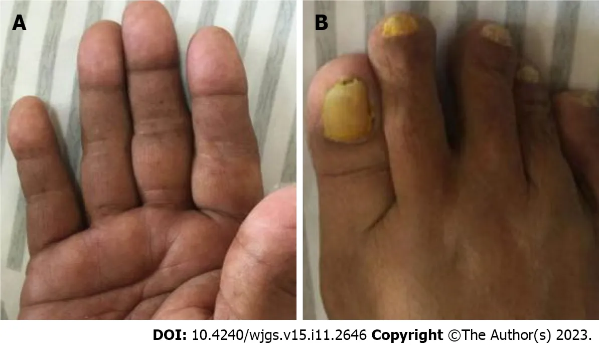
Figure 1 Cutaneous hyperpigmentation of Patient 1. A: Hands;B: Feet (with nail dystrophy).
Case 2:The vital signs were as follows: Body temperature,36.6 °C;blood pressure,96/61 mmHg;heart rate,90 beats per min;respiratory rate,18 breaths per min.Physical examination showed malnutrition,exfoliated skin on the face and hands,hyperpigmentation on the palms and backs of the hands (Figure 2A),alopecia (Figure 2B),and thickened and fragile nails on both hands and feet (Figure 2C).No edema was noted in either lower limb.
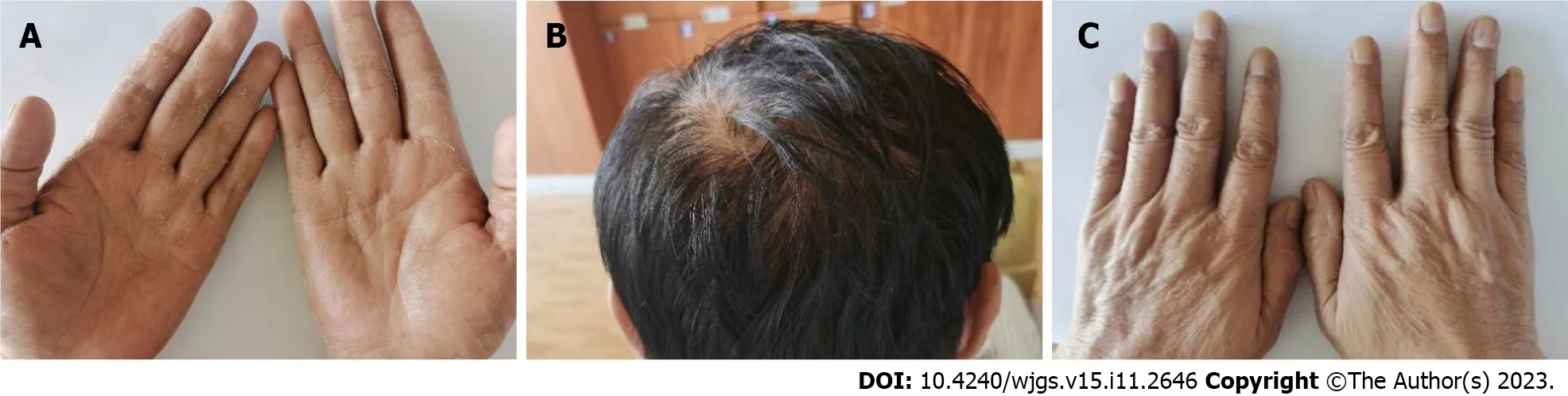
Figure 2 Physical examination findings of Patient 2. A and C: Hyperpigmentation on the (A) palms and (C) back of the hands;B: Alopecia.
Laboratory examinations
Case 1:Laboratory examination showed that the C-reactive protein level in this patient was 8.34 mg/L.Evaluation of thyroid function showed that her thyroid stimulating hormone (TSH) concentration was 26.080 µIU/mL (normal range:0.27-4.2 µIU/mL) and her free thyroxine (FT4) concentration was 10.63 pmol/L (normal range: 12.0-22.0 pmol/L).Rheumatic and immune-related results were normal.Other laboratory results are summarized in Table 1.
Table 1 Results of laboratory examinations of Patients 1 and 2
Case 2:Laboratory examinations showed that the patient had a cytokeratin 19 concentration of 8.63 ng/mL,a carcinoembryonic antigen of 9.26 ng/mL,a carbohydrate antigen 242 concentration of 40.05 U/mL,and a carbohydrate antigen 199 concentration of 55.83 U/mL.Routine blood tests,liver and kidney function tests,and blood lipids showed no significant abnormalities,and he was negative for IgG4,IgG9,and anti-mitochondrial antibody M2.Other laboratory results are summarized in Table 1.
Imaging examinations
Case 1:The second phase of contrast-enhanced computed tomography (CT) colonography in this patient showed that the gastric wall of the antrum and angle was thickened,with nodular protrusions and partial enhancement.The partial small intestinal wall was also thickened and heterogeneously enhanced.Diffuse wall thickening and polypoid masses with heterogeneous enhancements were observed throughout the colon,especially in the left colon.
The patient also underwent endoscopic examinations.Gastroscopy showed a thickened edematous mucosa with extensive congestion.The gastric antrum showed scattered nodular-like mucosal uplift.The duodenal bulb and descending segment showed multiple mucosal protrusions of different shapes and sizes (Figure 3).Colonoscopy showed that the mucosa of the large intestine was rough,with nodular and polypoid protuberances of different shapes and sizes.While the mucosa at the protuberances was hyperemic and edematous,the vascular texture of the intervening intestinal wall disappeared and turned white (Figure 4).On pathological examination,multiple biopsies of the stomach and large intestine showed that the mucosal glands were atrophic and dilated.Proliferation of interstitial granulation tissue was observed,accompanied by the infiltration of lymphocytes and eosinophils,suggesting hamartomatous polyposis.A tubular adenoma was observed in the transverse colon,with moderate to severe epithelial dysplasia (Figure 5).
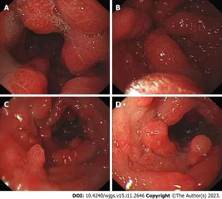
Figure 3 Gastroscopy of Patient 1. A and B: Thickened edematous mucosa with extensive congestion in the gastric body and antrum,with scattered,nodularlike mucosal uplift in the latter;C and D: Multiple mucosal protrusions of different shapes and sizes in the duodenal bulb and descending segment.

Figure 5 Biopsy from Patient 1 shows atrophic and dilated mucosal glands with infiltration of lymphocytes and eosinophils. A: Stomach;B:Intestine.Magnification: 4 ×.
Case 2:Both plain and three-stage enhanced CT of the abdomen showed gastric wall thickening in the lesser curvature and antrum.
Gastroscopy showed hyperemic and edematous mucosa (with a hyperplastic and nodular appearance) in the fundus,angle,antrum,duodenal bulb,and descending segment;this was prominent between the lesions (Figure 6).Colonoscopy showed similar results in colonic mucosa.However,the nodules were of different sizes.A mucosal intumescent lesion measuring 2.5 cm × 2.5 cm was observed 10 cm from the anal verge (Figure 7).EMR was performed,with biopsy samples obtained at multiple sites.
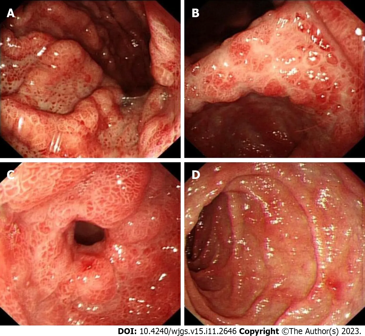
Figure 6 Endoscopic examination of Patient 2. A-D: Mucosae of the body (A),angle (B),antrum (C),and duodenal (D) descending segment were hyperemic and edematous,with a hyperplastic and nodular appearance.
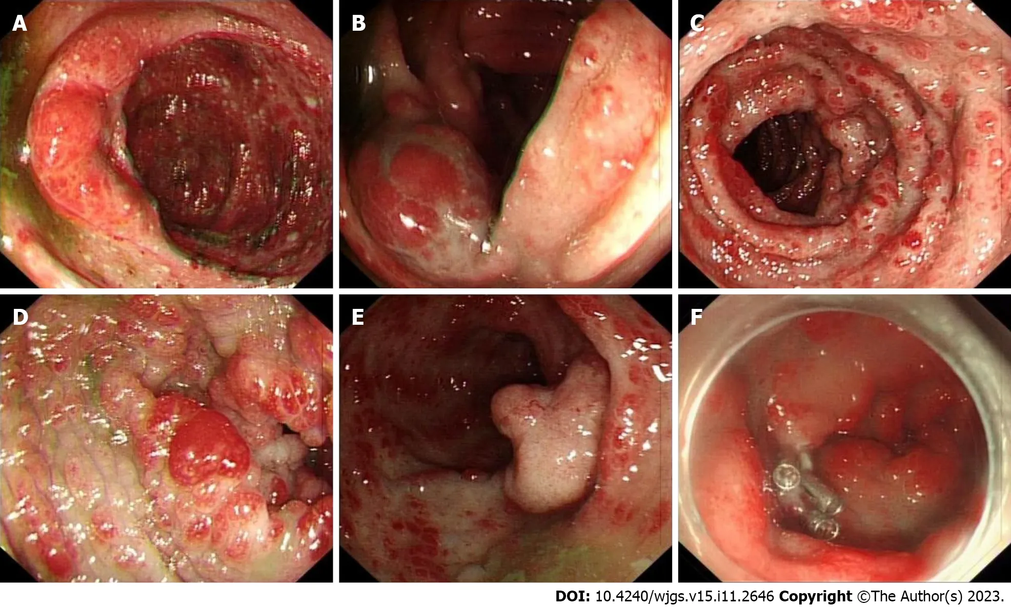
Figure 7 Colonoscopic examination of Patient 2. A-F: Mucosa of the entire colon was diffusely hyperemic and edematous,with nodules of different sizes.A mucosal intumescent lesion measuring 2.5 cm × 2.5 cm was observed 10 cm from the anal verge.
Gastroscopic pathology suggested that the lesions of the gastric body were consistent with hyperplastic polyps,acute and chronic mucosal inflammation.In the duodenum,distorted,branched,and hyperplastic glands were observed,indicating active chronic inflammation (Figure 8).Colonoscopy pathology showed active chronic enteritis and cryptitis in the terminal ileum,with round and blunt villi,and distorted glands,with infiltration of about 50 eosinophils per highpower field.Additionally,the pathology reports showed evidence of active chronic colitis,cryptitis,and crypt abscesses,with hyperplastic or atrophic,dilated,and distorted glands.This was accompanied by interstitial edema and eosinophil infiltration.The mucosal glands of the sigmoid colon and rectosigmoid junction were hyperplastic,with interstitial edema and crypt abscesses.CCS-related polyps could not be excluded (Figure 8).Examination of the rectal mass removed by EMR (Figure 7F) showed a villous tubular adenoma,consistent with high-grade intraepithelial neoplasia,with no apparent involvement of blood vessels or surgical margins.Immunohistochemical examination showed that about 60% of the cells were positive for Ki67,with high expression of MLH1 (+++),MSH2 (+++),MSH6 (+++),and PMS2 (+++),slight expression of P53 (+),and CK (+),and negative for CD31 and CD34 (Figure 8).
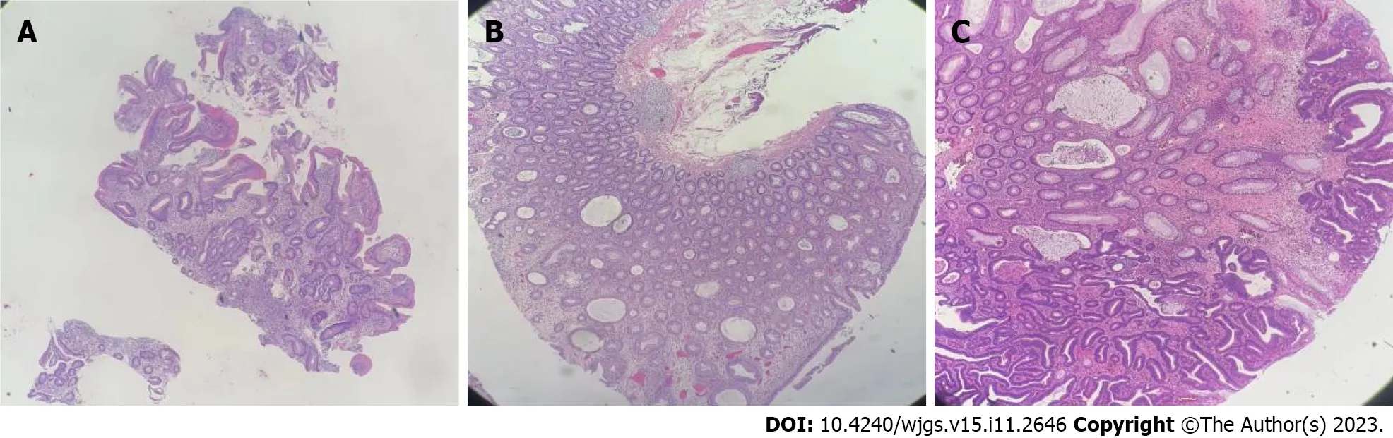
Figure 8 Histologic examination of biopsy samples from Patient 2. A: Gastroscopic pathology,suggesting active chronic inflammation with distorted,branched,and hyperplastic glands;B: Presence of active chronic colitis,cryptitis and crypt abscesses,with hyperplastic or atrophic,dilated,and distorted glands.Interstitial edema and eosinophil infiltration were also observed;C: The rectal mass was a villous tubular adenoma,indicative of high-grade intraepithelial neoplasia,with no apparent involvement of blood vessels or surgical margins.Magnification: 4 ×.
FINAL DIAGNOSIS
Case 1:Based on the aforementioned findings,Patient 1 was diagnosed with CCS and hypothyroidism.
Case 2:Based on the aforementioned findings,Patient 2 was diagnosed with CCS.
TREATMENT
Case 1:Treatment of Patient 1 included nutritional support,thyroid hormone supplementation (12.5 ug/d for the 1st3 d,25 ug/d thereafter),administration of prednisone tablets (30 mg/d),and symptomatic treatment during hospitalization.After discharge,she was continued on prednisone tablets (30 mg/d).
Case 2:The patient was initially treated with acid suppression (proton pump inhibitors) and nutritional support,followed by EMR for the removal of colonic polypoid lesions and the rectal mass.After endoscopic surgery,however,the patient and his family refused further hormonal or biological treatments and asked to be discharged.
OUTCOME AND FOLLOW-UP
Case 1:After 9 d of comprehensive treatment,her symptoms,including diarrhea and bloody stool,significantly improved.Hair and nail loss,however,did not significantly improve.Two months after discharge,this patient elected to reduce her dose of prednisone to 10 mg/d,and 3 mo later to 5 mg/d.Four months after the diagnosis of CCS,the patient suddenly developed nausea,vomiting,abdominal distension,and other manifestations of intestinal obstruction.Unfortunately,we did not obtain the results of thyroid function reevaluation for this patient after discharge.Because histopathologic examination of her colon showed tubular adenoma and moderate to severe epithelial dysplasia,the possibility of malignant transformation was considered.The patient died of multiple organ failure after 1 wk of treatment in a local hospital.
Case 2:One month later,the patient was followed up by telephone.At present,the patient is receiving symptomatic and supportive treatment and has been treated with an oral Chinese medicine for more than 20 d.His hypogeusia and appetite have improved,his new nails are soft,and his skin pigmentation has improved.However,his alopecia has not changed,and he continues to have yellow watery diarrhea,2-8 times per day.His body weight is 4.5 kg lower than his pre-discharge weight.
DISCUSSION
CCS is a rare,non-genetic syndrome characterized by ectodermal abnormalities and diffuse gastrointestinal polyps with protein loss.Since first described in 1955[2],more than 500 patients with CCS have been reported worldwide;most of these patients were Asian,with Japan accounting for more than 75%[3].The average age at onset is 59 years,with morethan 80% of these patients aged over 50 years at diagnosis[4].The male to female ratio ranges from 1.5 to 2:1[5].Patient prognosis is poor,with a 5-year mortality rate as high as 55%[1].The 2 patients described in this study included one woman and one man,both aged over 50 years.
At present,the etiology of the disease is unclear.Autoimmune factors have been reported to be involved in its possible etiology and pathogenesis.Many patients with CCS show positive plasma antinuclear antibody (ANA) series,elevated IgG4 levels,or IgG4 (+) plasma cells infiltrating into the polyps.Elevated levels of plasma IgE have also been reported in 1 patient with CCS[5].CCS was also found to be associated with hypothyroidism and other autoimmune diseases[6],such as membranous nephropathy[7],systemic lupus erythematosus,rheumatoid arthritis,and scleroderma.The involvement of autoimmune factors is supported by the overall good clinical response of CCS patients to immunosuppressive therapy.Both patients in the present study had normal ANA and plasma IgG4 levels,although Patient 1,who had CCS and hypothyroidism,had a significantly increased TSH level,accompanied by apathy and loss of appetite.To date,7 patients with CCS have been diagnosed with associated hypothyroidism,making this condition extremely rare (Table 2).Furthermore,prior to this article,there was only one reported case of CCS with concomitant hypothyroidism in China.All patients were aged over 59 years at diagnosis,with the oldest patient being 82 years.All 7 patients had alopecia,nail dystrophy,diarrhea,and multiple polyps.After clinical treatments,the symptoms of 6 patients were relieved to varying degrees,whereas the 7thpatient died of respiratory failure 1 year after treatment.This patient had hypothyroidism following surgery for Graves’ disease.The original text did not mention whether auto-antibodies were positive.However,because Graves’ disease is associated with autoimmune antibodies,we can be certain that the patient had autoimmune abnormalities.This further confirms the association of CCS with autoimmune factors[8].Qiaoetal[9]reported a CCS case in which the patient,however,had normal serum levels of antithyroglobulin and anti-thyroid peroxidase antibodies.Taken together,these findings indicate that the etiology of CCS and its relationship to autoimmunity are unclear,necessitating further research.
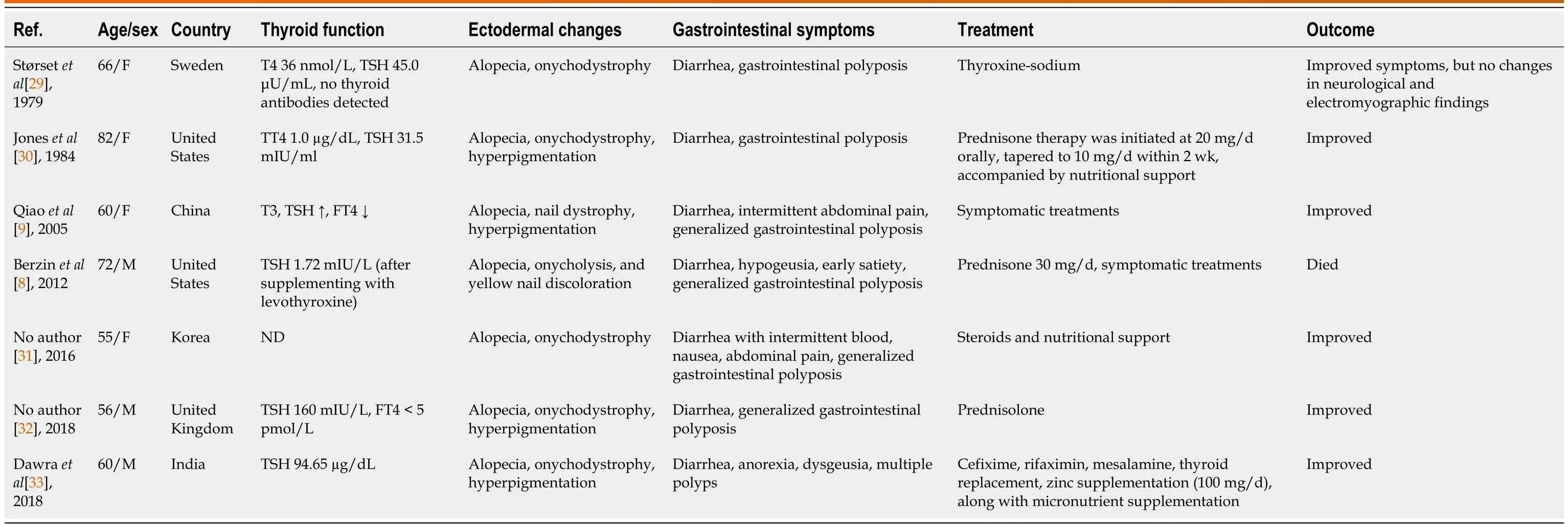
Table 2 Description of patients with Cronkhite-Canada syndrome combined with hypothyroidism
Infectious factors have also been associated with CCS.For example,Helicobacterpylori(H.pylori) infection has been associated with CCS,with symptoms being relieved after anti-H.pyloritreatment[10].Antibodies to Saccharomyces cerevisiae have also been detected in the plasma of CCS patients[11-13],and Clostridium difficile has been identified in the feces of patients with CCS[14].These findings suggest that infection with several types of pathogenic bacteria is associated with CCS.
Although CCS is regarded as a non-genetic disease,genetic factors may be associated with its development.Although both patients in the present study denied that anyone in their families had a similar history,genome-wide association study revealed that mutations in thePRKDCgene,normally encoding the catalytic subunit of DNA-dependent protein kinase,may play a role in the pathogenesis of CCS[15].Genetic analysis of a Chinese mother and child who were diagnosed with CCS revealed C.3921-3925delAAAAG (p.Ile1307fsX6) mutation in theAPCgene[5].
Other factors associated with CCS included mental and physical stress[6],allergy[12],and gut microbiota[16].Discontinuation of allergy-inducing agents,such as hair dyes and certain drugs,was found to reduce IgE concentrations and eosinophil infiltration,as well as to improve clinical symptoms in patients with CCS[12].Moreover,the remission and regression of polyps after prednisolone (PSL) treatment of patients with CCS was accompanied by changes in the abundance and diversity of gut microbiota[16].PSL can not only inhibit proinflammatory cytokines but can also mediate polyp regression by altering the composition of gut microbiota.Additional studies are needed to better understand the associations between proinflammatory cytokines and microecological dysbiosis,which might be involved in the pathogenesis of CCS.
CCS is characterized by ectodermal abnormalities,gastrointestinal symptoms,and protein loss.Ectodermal changes include alopecia,skin pigmentation,and nail dystrophy,including nail yellowing,atrophy,and loss.The main gastrointestinal symptoms are abdominal pain and diarrhea,frequently accompanied by nausea,acid regurgitation,anorexia,and abnormal taste.The occurrence of fractures has also been reported[17].Based on the initial symptom,CCS can be divided into five types: Diarrhea (type 1),hypogeusia (type 2),dry or strange sensation in the mouth (type 3),abdominal pain (type 4),and alopecia (type 5)[18].Patients 1 and 2 in the present study had types 1 and 2 CCS,respectively.Both patients had three main ectodermal changes,abdominal pain and nausea,with Patient 1 also having vomiting.Due to oral mucosal lesions,including inflammation and infection,and zinc and copper deficiency,patients may lose their taste[19].Alopecia can be caused by malnutrition[20],whereas nail dystrophy has been associated with the bad nutritional status and inflammatory response[19].Polyps in patients with CCS are distributed throughout the digestive tract,being common in the stomach and colon,less common in the small intestine and rectum,and almost nonexistent in the esophagus[10,21].Polyps are usually diffusely distributed and nodular,with different shapes and sizes.Histologically observed glandular hyperplasia and cystic dilatation are accompanied by the infiltration of inflammatory cells,especially eosinophils.Both patients in the present study had both gastric and intestinal polyps,with findings on gastrointestinal endoscopy being consistent with CCS.
The pathology of CCS is not specific,with four histological types: hyperplastic,adenomatous,juvenile,and inflammatory polyps.The evolution of polyps may follow the mucosal hyperplasia (C-C polyps)-adenoma-carcinoma pathway[22].About 12.5% of polyps are estimated to become cancerous,with this being a significant cause of death.Pathologic analysis of the polyps in Patient 1 showed tubular adenoma and moderate to severe epithelial dysplasia,complicated with an intestinal obstruction 4 mo after the diagnosis of CCS,suggesting malignant transformation of the adenoma.The rectal mass in Patient 2 was pathologically diagnosed as a high-grade intraepithelial neoplasia,necessitating immediate EMR.These findings emphasize the importance of close surveillance and prompt removal of polyps.
The average recovery times for diarrhea,taste abnormality,and ectodermal changes in patients with CCS are 51,84,and 9 d,respectively,and the mean times to resolution of gastric and colonic polyps are 248 and 238 d,respectively[23].Currently,there are no standard treatment guidelines,including duration of treatment,with most CCS patients receiving comprehensive empirical treatment based on glucocorticoids.Several studies have recommended treatment for 6-12 mo[24],suggesting that the steroid dose should be slowly tapered only after endoscopic confirmation of the regression of polyposis[25].Although 30-49 mg/d oral prednisone was reported to have the optimal effect during the active stage of CCS[26],patients may relapse when glucocorticoid dose is gradually reduced.Additional studies in larger patient cohorts are needed to determine whether to use glucocorticoids,their duration and dose,regimens for reduction,and the need for maintenance therapy with other medications.Nutritional support is often combined with other treatments,making it difficult to accurately determine the effectiveness of nutritional support in patients with CCS[10].Other treatments can include immunosuppressive agents,acid suppression,traditional Chinese medicines,salicylic acid preparations,TNFinhibitors[27],and endoscopic or surgical treatment.Achieving a sustained endoscopic response is the therapeutic goal and associated with a reduced risk of cancer[4,25].Kimetal[28] reported a successful case of fecal microbiota transplantation in the treatment of steroid-refractory CCS.The etiology and pathogenesis of this disease are not yet fully understood,and various other methods are still being explored.
CONCLUSION
In summary,CCS is a rare syndrome primarily affecting male patients with a relatively poor prognosis.Its etiology remains unclear,but current research suggests a strong association with autoimmunity.Based on the results of literature review in this article,it can be inferred that a clear diagnosis and treatment of hypothyroidism contribute to improving the prognosis.Clinical manifestations are diverse,including diarrhea,gastrointestinal polyps,skin hyperpigmentation,alopecia,and nail atrophy.Comprehensive treatment based on hormone therapy can lead to partial or complete remission of clinical symptoms.Polyps meeting the indications for endoscopic surgery should be actively treated surgically,which can prevent polyp malignancy and the occurrence of complications,such as intestinal obstruction and intussusception.Early diagnosis and treatment are crucial for inducing remission and improving disease prognosis.Long-term follow-up is necessary for subsequent treatment of this disease.In the future,this disease will still require further basic and clinical research,especially regarding its etiology and treatment approaches.
FOOTNOTES
Author contributions:Lv YQ collected and sorted out the cases,reviewed the literature,and wrote the manuscript;Wang ML reviewed the literature;Tang TY contributed to the content and editing of the manuscript;Li YQ reviewed and revised the manuscript;All authors have read and approved the final manuscript.
Supported byJilin Provincial Science and Technology Department Project,No.20200201343JC;and Science and Technology Development Program of Jilin Province,No.20210402013GH.
Informed consent statement:This study was approved by the Ethics Committee of the First Bethune Hospital of Jilin University to waive informed written consent about personal and medical data collection.
Conflict-of-interest statement:The authors declare that they have no conflict of interest to disclose.
CARE Checklist (2016) statement:The authors have read the CARE Checklist (2016),and the manuscript was prepared and revised according to the CARE Checklist (2016).
Open-Access:This article is an open-access article that was selected by an in-house editor and fully peer-reviewed by external reviewers.It is distributed in accordance with the Creative Commons Attribution NonCommercial (CC BY-NC 4.0) license,which permits others to distribute,remix,adapt,build upon this work non-commercially,and license their derivative works on different terms,provided the original work is properly cited and the use is non-commercial.See: https://creativecommons.org/Licenses/by-nc/4.0/
Country/Territory of origin:China
ORCID number:Yu-Qin Li 0000-0002-5505-3906.
S-Editor:Lin C
L-Editor:Filipodia
P-Editor:Lin C
 World Journal of Gastrointestinal Surgery2023年11期
- World Journal of Gastrointestinal Surgery的其它文章
- Systematic sequential therapy for ex vivo liver resection and autotransplantation: A case report and review of literature
- Gastric inflammatory myofibroblastic tumor,a rare mesenchymal neoplasm: A case report
- Isolated traumatic gallbladder injury: A case report
- Metachronous primary esophageal squamous cell carcinoma and duodenal adenocarcinoma: A case report and review of literature
- Organ sparing to cure stage IV rectal cancer: A case report and review of literature
- Effect of perioperative branched chain amino acids supplementation in liver cancer patients undergoing surgical intervention: A systematic review