All-trans retinoic acid regulates the expression of MMP-2 and TGF-β2 via RDH5 in retinal pigment epithelium cells
Yu-Mei Mao, Chang-Jun Lan, Qing-Qing Tan, Gui-Mei Zhou, Xiao-Ling Xiang,Jia Lin, Xuan Liao
1Department of Ophthalmology, Affiliated Hospital of North Sichuan Medical College, Nanchong 637000, Sichuan Province, China
2Medical School of Ophthalmology and Optometry, North Sichuan Medical College, Nanchong 637000, Sichuan Province, China
Abstract● AlM: To investigate the effect of all-trans retinoic acid (ATRA) on retinol dehydrogenase 5 (RDH5), matrix metalloproteinase-2 (MMP-2) and transforming growth factor-β2 (TGF-β2) transcription levels, and the effect of RDH5 on MMP-2 and TGF-β2 in retinal pigment epithelium(RPE) cells.
● KEYWORDS: retinol dehydrogenase 5; matrix metalloproteinase-2; transforming growth factor-β2; all-trans retinoic acid; ARPE-19
INTRODUCTION
Despite the dramatic increase in incidence, the mechanisms underlying myopia were not well understood[1].The current research supports the hypothesis that the visual control of eye size and refractive state may involve a cascade of retinal chemical signals that arise from the processing of visual input.These signals passed through the retinal pigment epithelium (RPE) and choroid and eventually affected the sclera, causing its remodeling and axial growth.However, the role of RPE in this process remains uncertain.Increasing evidences showed that all-trans retinoic acid (ATRA) promotes differentiation of RPE cellsin vitro[2],and is involved in the regulation of RPE barrier function[3].Ⅰn this epithelial differentiation process, some factors such as matrix metalloproteinase-2 (MMP-2) and transforming growth factor-β2 (TGF-β2) play an important role.Previous studies have found that ATRA may play a role in the occurrence and development of myopia by regulating the expression of MMP-2 and TGF-β2[4-5].
ATRA is the main active form of retinoic acid (RA) which is a potent regulator of corneal epithelial integrity and transparency, photoreceptor development and maturation, and the pigmentation of RPE cells[6].As a key enzyme, retinol dehydrogenase 5 (RDH5) not only participates in the oxidation of 11-cis retinol to 11-cis retinal in the visual cycle, but also regulates the metabolism of RA/ATRA.These substances may be involved in the signal cascade controlling development of the eyeball.The aim of the present study is to explore the possible mechanism by studying the effects of ATRA on the expressions of RDH5, MMP-2 and TGF-β2 in RPE cells, as well as the changes in the expressions of MMP-2 and TGF-β2 after RDH5 knockdown.
MATERIALS AND METHODS
Materials and ReagentsThe human retinal pigment epithelium cell line ARPE-19 was purchased from PROCELL(Wuhan, China).ATRA was purchased from Sigma-Aldrich(Poole, UK); Lipfectamine@2000, Opti-MEM Reduced Serum Medium, Annexin V-FITC/PI Apoptosis Kit were purchased from Invitrogen (USA); RDH5 siRNA and negative control siRNA were purchased from Public Protein/Plasmid Library(China).FastKing gDNA Dispelling RT SuperMix and SuperReal PreMix Color (SYBR Green) SuperReal were purchased from TIANGEN (Beijing, China).
Cell Culture and TreatmentARPE-19 cells were routinely resuscitated and passaged when the cell confluency reached 80%–90%.Cells at passages 3–6 were used for experiments and were divided into normal control (NC) group and ATRA intervention group.Dilute ATRA (molecular weight 300.4)powder with sterile DMSO solution to obtain an ATRA solution with a final concentration of 0.25 mol/L.ARPE-19 cells cultured in normal DMEM/F12 medium for 48h were taken, and then diluted with DMEM/F12 medium to different concentrations of ATRA (0–20 μmol/L); 0 was the control group, and subgroups of ATRA intervention group were divided into 0.5, 1, 5, 10, and 20 μmol/L.
Chromatographic Purification of siRNACells in each group were treated for another 24h for subsequent experiments.From the AUG start codon of the transcript, the “AA”duplex sequence was searched, and the 19-base sequence at the 3’end was used as a potential siRNA target site.The total length of the primers was 21 bases.According to the splicing positions of different targets, they were divided into 6 groups:RDH5 siRNA-435 group, RDH5 siRNA-799 group, RDH5 siRNA-441 group, negative control group, positive control group and fluorescently labeled negative control (FAMsiRNA) group.Different target siRNAs were diluted to a final concentration of 50 μmol/L in sterile non-enzymatic DMEM/F12 medium, and cells in each group were treated for 48h for subsequent experiments.
Flow CytometryThe ARPE-19 cell suspension obtained by digestion was counted, and 1×10⁵/mL was inoculated into a 24-well plate, with 1 mL of medium per well and cultured for 24h, cells were starved for 24h with serum-free DMEM/F12 medium.The cells in each well were processed according to cell grouping, and after 24h of culture, the cells in the 24-well plate were digested into a suspension for cell cycle detection.Cells were digested with trypsin and then collected from each well, washed twice with PBS solution, centrifuged at 2000 rpm for 5min, and resuspended with 500 μL binding buffer.5 μL Annexin V-FⅠTC and 5 μL propidium lodide were added and mixed in the dark and incubate at room temperature for 5–15min.Flow cytometry with excitation wavelength Ex=488 nm and the emission wavelength Em=530 nm.Three replicate wells were set for each group, and the average value was taken for statistics.
Quantitative Real-time Polymerase Chain ReactionAccording to the manufacturer’s protocol, total RNA was extracted from APRE-19 cells in different groups using trizol reagent, respectively.This was followed by reversetranscription of 1 μg of total RNA to cDNA using FastKing gDNA Dispelling RT SuperMix.Then, qPCR was performed on a PCR instrument with SYBR green using resultant cDNA as template.Procedure was as follow: pre-denaturation at 95℃for 15min; denaturation at 95℃ for 15s, annealing at 60℃ for 34s; extension at 60℃ for 60s, with 40 cycles.According to the formula 2-△△Ct, the relative expression of the target genes were calculated in each group.The primer sequences were designed and provided by Sangon Bioengineering Company(Shanghai, China), and listed in Table 1.
Statistical AnalysisThe software SPSS 26.0 and GraphPad Prism 8 were used for statistical analysis and graphing.Data were presented as the mean±standard deviation (SD).Student’st-test (SNK) and One-way ANOVA were used to determine the level of significance;P<0.05 was defined as statistically significant.Allin vitrocell experiments were repeated at least three times by a single well-trained researcher.
RESULTS
Effects of Different Doses of ATRA on Apoptosis of ARPE-19 CellsTo identify whether ATRA can affect ARPE-19 cells in cell viability, we examined the apoptosis in the ATRA treatment group and control group.According to the results of flow cytometry, it was found that the early apoptosis and total apoptosis levels of ARPE-19 cells were increased after treatment with ATRA above 5 μmol/L for 24h, and the differences were statistically significant compared with the control group (P=0.01,P=0.03); when the ATRA concentrationexceeded 10 μmol/L, the early apoptosis, late apoptosis and total apoptosis were significantly increased compared with the control group (P=0.002,P=0.02,P=0.003).After 24h of culture, the apoptosis level of ARPE-19 cells increased with the increase of ATRA concentration, showing a dose-dependent apoptosis effect (Figure 1).

Table 1 Primer sequences of qRT-PCR
mRNA Expressions of MMP-2, TGF-β2, and RDH5 Regulated by ATRAAccording to the results of quantitative real-time polymerase chain reaction (qRT-PCR), with the increase of ATRA concentration, the expression of MMP-2 and TGF-β2 mRNA increased in a dose-dependent manner; when the concentration was 5 μmol/L, the expression of MMP-2 and TGF-β2 mRNA increased most significantly, compared with the control group, the difference was statistically significant(P=0.03,P<0.001; Figure 2A, 2B).The expression of RDH5 mRNA in ARPE-19 cells was gradually decreased after ATRA treatment for 24h.When the concentration reached 5 μmol/L ATRA, RDH5 mRNA decreased significantly, and the difference was statistically significant compared with the control group (P<0.001; Figure 2C); it was shown that ATRA can inhibit the expression of RDH5 molecule in ARPE-19 cells at the mRNA level, while promoting the expression of MMP-2 and TGF-β2 signaling molecules in a concentration-dependent manner.
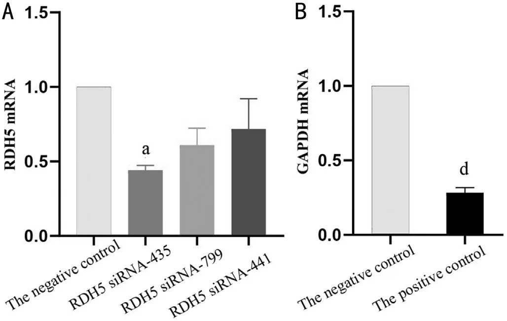
Primer Sequences and Purity Identification Results of Different siRNAsThe primer sequences for different siRNAs were shown in Table 2.The negative control was a random RNA sequence that does not down-regulate the expression of the gene; the fluorescently labeled negative control (FAM-siRNA)was only labeled with fluorescence to facilitate the observation of transfection under a fluorescence microscope, which was used to optimize transfection conditions and evaluate transfection efficiency; The positive control was an RNA sequence that can down-regulate the expression of GAPDH gene, which was used to detect the transfection efficiency.
Effect of Different siRNA Targets on RDH5 Gene Knockdown EfficiencyThe samples were collected after 48h of siRNA transfection in each group for quantitative detection.qRT-PCR results showed that the knockdown efficiency of RDH5 siRNA of different targets was different, and theknockdown efficiency of the RDH5 siRNA-435 target was the most obvious, which was higher than the negative control group, the expression of RDH5 mRNA decreased more than 50%, and the difference was statistically significant (P=0.02;Figure 3A).By collecting the same batch of transfected cells to quantitatively detect the transfection efficiency, the qRTPCR results were found to be similar at the negative control.In comparison, the expression of GAPDH mRNA in the positive control was reduced about 70%, and the difference was statistically significant (P<0.001; Figure 3B).

Table 2 Primer sequences of different target siRNAs and information for testing
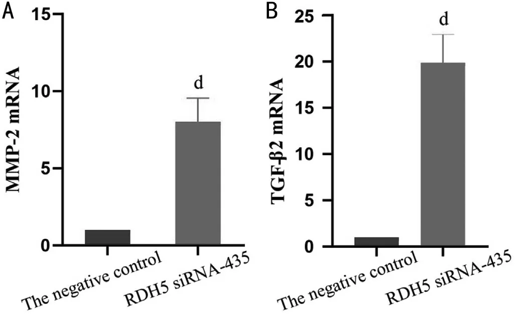
Effects of RDH5 on the Expression of MMP-2 and TGF-β2 mRNA in ARPE-19 cellsAfter 48h of transfection with the target RDH5 siRNA-435, the results of qRT-PCR were shown in Figure 4.Compared with the negative control, the expressions of MMP-2 and TGF-β2 mRNA were significantly increased, and the difference was statistically significant(P<0.001; Figure 4A, 4B), suggesting that knockdown of RDH5 can significantly promote MMP-2 and TGF-β2 mRNA expression in ARPE-19 cells.

Figure 1 Changes of apoptosis level of ARPE-19 cells induced by ATRA The upper right quadrant was the early apoptotic cell population, and the lower right quadrant was the late apoptotic cell population.ATRA: All-trans retinoic acid; ARPE: Adult retinal pigment epithelium.

Figure 2 Quantitative analysis of the effects of different concentrations of ATRA on the mRNA expressions of RDH5, MMP-2, and TGF-β2 Compared with the control group, aP<0.05, bP<0.01, cP<0.001.ATRA: All-trans retinoic acid; ARPE: Adult retinal pigment epithelium; RDH:Retinol dehydrogenase; MMP: Matrix metalloproteinase; TGF: Transforming growth factor.
DISCUSSION
The retinal local regulation theory believed that the stimulation of optical defocus or form deprivation can induce the retina to produce primary signaling factors (such as RA, dopamine,insulin-like growth factor,etc.) and act on RPE to produce secondary signaling factors (such as MMP-2, TGF-β2,hepatocyte growth factor and glycosaminoglycan,etc.), thereby regulating the development of the eyeball[7].RA plays a key role in this pathway[8].RA is a derivative of vitamin A, which can be classified as ATRA, 9-cis RA and 13-cis RA[9].Among them, ATRA is not only the main active form of RA, but also the most active metabolite of vitamin A[9].Studies have found that ATRA acts as a signaling molecule in the retina, promoting the secretion of MMP-2 and TGF-β2, which were associated with increasing axial length in a myopic animal model[10].

Figure 3 The effect of RDH5 siRNA with different targets on the knockdown efficiency of RDH5 gene Compared with the negative control, aP<0.05, dP<0.001.RDH: Retinol dehydrogenase.

Figure 4 The effect of target RDH5 siRNA-435 on MMP-2 and TGF-β2 mRNA expression Compared with the negative control, dP<0.001.RDH: Retinol dehydrogenase; MMP: Matrix metalloproteinase; TGF:Transforming growth factor.
As a key catalytic enzyme, RDH5 protein is involved in the oxidation of retinol to retinal in the visual cycle, and further affects the production of RA and ATRA.Previous studies have shown that RDH10 and RDHE2, members of the retinol dehydrogenase family, as important enzymes in the visual cycle, assist RDH5 in the process of oxidizing retinol to retinal[11].Strateet al[12]demonstrated that RDH10 can act as a feedback regulator that negatively feedback regulates the concentration gradient of RA.It is worth noting that the expression of RDH10 is suppressed in the presence of RA excess[13].Adamset al[14]reported that RA strongly down regulated the expression of RDHE2, and human RDHE2 appears to be regulated at the transcriptional level by RA through a negative feedback loop.In this study, it was found that the expression of RDH5 mRNA was significantly down-regulated after treating ARPE-19 cells with different concentrations of exogenous ATRA.Moreover, the present study also showed that when excessive exogenous ATRA was stimulated, it would have a certain toxic response to RPE cells and promote their apoptosis, which was consistent with the previous study[15].It suggested that ATRA regulated the transcription level of the RDH5 gene through negative feedback to achieve the dynamic balance of RA metabolism.In addition, the transcription factor binding sequence of human RDH5 gene was searched in the Eukaryotic Promoter Database, and it was found that ATRA can bind to RA response element in the regulatory region of RDH5 gene through RARB(encoding RARβ nuclear receptor) transcription factor, thereby regulating the expression of RDH5 gene (P=0.001).Research mentioned above provided more adequate evidence support.
The present study discovered that 10 μmol/L ATRA treated ARPE-19 cells for 24h resulted in the mRNA expression of TGF-β2 and MMP-2 increased, especially in TGF-β2, which was consistent with the results reported in the literatures[4-5].Further, when the expression of RDH5 mRNA was knocked down, both MMP-2 and TGF-β2 mRNA secreted in RPE cells were significantly up-regulated.As an endogenous key enzyme, RDH5 is involved in the key step in the synthesis of RA/ATRA, that is, the oxidation of retinol to retinal.This study demonstrated for the first time that RDH5 gene knockdown promotes secretion of MMP-2 and TGF-β2 mRNA transcription levels in RPE cells, speculating that this gene regulates the expression of MMP-2 and TGF-β2 by interaction with ATRA.This study also has some limitations.First, although only mRNA was measured in the study, given that transcript level is the basis of all changes, the current study is still of certain significance.Further studies are needed to better understand the effective correlation among MMP-2, TGF-β2 and RDH5in vivoexperiments.Second, this study showed that overdose-ATRA not only promoted apoptosis, but also inhibited the expression of RDH5.However, whether RDH5 is related to apoptosis needs further study.Third, MMP-2 and TGF-β2 are both the marker of epithelial mesenchymal transition (EMT) in RPE trans-differentiation, the relevance of EMT in myopia may be better understood with more research, to prove RA signal pathway involved in the pathogenesis of shortsightedness.
It is worth mentioning that RPE trans-differentiation can be inducedin vitroby RA[16], where increased expression of MMP-2 and TGF-β2 may indicate EMT[17-18].EMT was initially discovered as a key developmental mechanism, a cellular process in which epithelial cells form mesenchymal characteristics and express several cellular markers[19].TGF-β2 promotes the expression of MMP-2, a mesenchymal marker,the transformation of RPE cells into myofibroblasts and the production of the components of extracellular matrix, that is,controls the development of EMT[20].In addition, a synthetic retinoid derivative, fenretinide, has been shown to promote trans-differentiation of ARPE-19 cells towards a neuronal-like phenotype[21-22].Whether the EMT mechanism exists in RPE cells during myopia deserves further investigation.
Ⅰn summary, the present study indicates the effects of RDH5-mediated ATRA on MMP-2 and TGF-β2 in RPE cells.These results are expected to provide innovative information for molecular research and clinical treatment in myopia.Furtherin vitroandin vivoexperiments are needed to better understand its specific effects and the underlying mechanisms behind them.
ACKNOWLEDGEMENTS
Authors’contributions:Mao YM performed experimental assays and drafted the manuscript; Lan CJ and Tan QQ reviewed the manuscript; Zhou GM, Xiang XL, Lin J performed laboratory data and literature review; Liao X organized this study and performed manuscript revision.All authors read and approved the final manuscript.
Foundations:Supported by Project of Science & Technology Department of Sichuan Province (No.23NSFSC1940); City and College Cooperation (No.22SXFWDF0003).
Conflicts of Interest:Mao YM, None; Lan CJ, None; Tan QQ, None; Zhou GM, None; Xiang XL, None; Lin J, None;Liao X, None.
 International Journal of Ophthalmology2023年6期
- International Journal of Ophthalmology的其它文章
- Preliminary proteomic analysis of human tears in lacrimal adenoid cystic carcinoma and pleomorphic adenoma
- Assessment of the effects of intrastromal injection of adipose-derived stem cells in keratoconus patients
- Evaluation of optic nerve head vessels density changes after phacoemulsification cataract surgery using optical coherence tomography angiography
- Stability of neodymium:YAG laser posterior capsulotomy in eyes with capsular tension rings
- Comparison of the efficacy and safety of ultrasonic cycloplasty vs valve implantation and anti-VEGF for the treatment of fundus disease-related neovascular glaucoma
- Volumetric fluid analysis of fixed monthly anti-VEGF treatment in patients with neovascular age-related macular degeneration
