Comparison of biological behavior of lacrimal gland adenoid cystic carcinoma with high-grade transformation cells
Chuan-Li Zhang, Li-Min Zhu, Xun Liu, Mei-Xia Jiang, Ting-Ting Lin, Yan-Jin He
1Oculoplastic and Orbital Disease Department, Tianjin Medical University Eye Hospital, Eye Institute and School of Optometry, Tianjin Key Laboratory of Retinal Functions and Diseases, Tianjin Branch of National Clinical Research Center for Ocular Disease, Tianjin 300384, China
2Tianjin Eye Hospital Optometry Center, Tianjin 300384,China
Abstract
● KEYWORDS: lacrimal gland; lacrimal gland adenoid cystic carcinoma; high-grade transformation; primary cell culture; biological behavior; mRNA array
INTRODUCTION
Lacrimal gland adenoid cystic carcinoma (LACC) is the most common malignant epithelial tumor of the lacrimal gland[1-3], with poor prognosis. After surgical treatment, the 5-year survival rate is less than 50%, and the 10-year survival rate is less than 20%[1,4]. High-grade transformation (HGT)of carcinomas leads to loss of morphological characteristics and turns them into high-grade cancer, which is usually undifferentiated. In 2017, the WHO’s “Classification of Head and Neck Tumors” defined the concept of HGT for salivary adenoid cystic carcinoma[5]. Several studies have shown that LACC could also undergo HGT (LACC-HGT), and LACCHGT has a higher malignant degree and worse differentiation than non-HGT[6-7]. At present, no human-derived LACC-HGT cell lines could be found in domestic and foreign cell banks.In previous studies, non-HGT primary cells were successfully obtained by primary tissue culture[8]. In the present study,LACC-HGT primary cells were cultured and identified in the same manner. The differences in the biological behavior between LACC-HGT and non-HGT primary cells and the differentially expressed genes were observed and compared to study LACC pathogenesis and malignant transformation.
MATERIALS AND METHODS
Ethical ApprovalThe study samples were only used for relevant scientific research. A written consent for the study was obtained from the patients prior to tissue collection, and the tumor tissues used in this study were approved by The Ethics Committee of Tianjin Medical University Eye Hospital[No.2017KY(L)-23].
Experimental SpecimensTwo patients were diagnosed with LACC-HGT in Tianjin Medical University Eye Hospital. After the tumor tissues were removed, the specimens were stored in pre-cooled culture medium and transferred to the laboratory for primary cell culture as soon as possible. LACC-HGT-1 was derived from a 47-year-old woman who underwent left eye orbital tumor resection after progressive eyeball protrusion.LACC-HGT-2 was derived from a 49-year-old man who underwent right eye orbital tumor resection due to gradual aggravation protrusion over 2mo. Both cases were primary and had no history of chemotherapy or radiotherapy before surgery.In this study, LACC-HGT-1 primary cells were used as an example for subsequent experiments.
The LACC-HGT diagnostic criteria referred to the HGT histological and morphological diagnostic criteria proposed by Seethala and Stenman[5]. 1) In terms of growth pattern:significantly enlarged solid cell nests, larger than a low-fold field of vision; acne-like necrosis; calcification; phosphating or papillary growth. 2) For obvious cell atypia: nuclear enlargement; nuclear membrane thickness; chromatin vacuoles; obvious nucleoli; common nuclear fission; increased Ki-67 in immunohistochemical (IHC) staining. 3) IHC staining shows a decrease or disappearance of myoepithelium. 4) The interstitium shows proliferation of fibrous connective tissue. A diagnosis of LACC-HGT was made after meeting the reference criteria of four major items or 12 minor items.
Hematoxylin and Eosin StainingTissue sections were dewaxed with xylene (Solarbio, China), dehydrated with gradient ethanol (Solarbio, China), stained with hematoxylin(Solarbio, China) for 10 min, and rinsed with water. The tissue sections were then stained with 1% hydrochloric acid ethanol(Solarbio, China) and rinsed with water. Finally, the sections were stained with eosin, dehydrated with gradient ethanol,rendered transparent with xylene, and sealed with neutral gum.
Immunohistochemical StainingTissue sections were dewaxed with xylene, dehydrated with gradient ethanol, and washed with phosphate buffer saline (PBS; Gibco, USA).After dripping with 3% peroxide solution (Zhongshan Jinqiao Biotechnology Co., Ltd., China) for 10min, citrate buffer was used for antigen repair. Normal goat serum (Zhongshan Jinqiao Biotechnology Co., Ltd., China) was sealed at room temperature for 30min. After the blocking solution was removed, the primary antibody was added at 4℃ overnight.On the next day, the second antibody was incubated at room temperature. Fresh DAB solution (Zhongshan Jinqiao Biotechnology Co., Ltd., China) was added in a wet box and redyed with hematoxylin for 5min. After dehydrating and sealing, the results were observed under microscope.
LACC-HGT Tissue Primary Cell CultureTumor tissue samples were rinsed with culture medium 2-3 times. LACCHGT tissues were cut into 1 mm3pieces and then slowly transferred into a T25 cell culture flask (Nest, China), which contained RPMI-1640 (Gibco, USA), 10% fetal bovine serum(ExcellBio, China), and 1% penicillin and streptomycin(Gibco, USA). The cells were cultured in an incubator(Thermo, USA) at 37℃ with 5% CO2. After the cells crawled out of the tissue mass, the culture medium was changed every 2d. After the first digestion with trypsin, LACC-HGT primary cells were resuspended and re-cultured in a T75 cell culture flask (Nest, China). The culture medium was changed every 2d. When the cells fused to approximately 80%, the cells were digested and passed on 1:2, and then the 4th-10thgenerations cells were selected as the research object.
ImmunofluorescenceThe fifth-generation LACC-HGT primary cells, with a concentration of about 5×10⁵/mL, were inoculated into 24-well plate crawling tablets (Nest, China). On the next day, the cells were fixed with 4% paraformaldehyde(Solarbio, China). Subsequently, 0.5% Triton X-100 reagent was added to each well for 10min. An appropriate amount of goat serum sealant was added, and mouse anti-human monoclonal antibodies cytokeratin (CK; Zhongshan Jinqiao Biotechnology Co., Ltd., China) and CK7 (Zhongshan Jinqiao Biotechnology Co., Ltd., China) were added at 4℃overnight. Fluorescent secondary antibody (Zhongshan Jinqiao Biotechnology Co., Ltd., China) was added and incubated in the dark for 1h. After DAPI (Zhongshan Jinqiao Biotechnology Co., Ltd., China) staining was performed, the cells were observed under the microscope (Olympus, Tokyo, Japan), andpictures were taken. PBS was used as negative control instead of primary antibody.

Table 1 Primer sequence
CCK-8 ExperimentAfter the fifth-generation LACC-HGT and non-HGT primary cells were collected and counted,a 100 µL cell suspension was inoculated in 96-well plates,and the number of cells was approximately 5000. After incubation was conducted at 37℃ with 5% CO2for 3h, 10 µL of CCK-8 liquid (Solarbio, China) was added to each well. The absorbance was measured at 450 nm to compare the optical density (OD) values of the two kinds of primary cells.
Wound Healing ExperimentApproximately 2 mL fifthgeneration cell suspensions of non-HGT and LACC-HGT primary cells were inoculated into six-well plates, and the number of cells was approximately 4×10⁵/mL. On the next day, a 100 µL pipettor tip was vertically scratched. Roughly 2 mL of 1% low-concentration serum medium was replaced, and incubation was continued in the incubator. The relative cell migration distance to the scratch area was photographed at 48h after the scratch test.
Drug Sensitivity ExperimentApproximately 100 µL of fifth-generation LACC-HGT and non-HGT cell suspensions were inoculated in a 96-well plate, with the number of cells at approximately 5000. Different concentrations (0, 5, 10,20, 30, 40, and 50 µmol/L) of cisplatin (Solarbio, China) and paclitaxel (Solarbio, China) were co-cultured for 48h with the cells. Subsequently, roughly 10 µL of CCK-8 was added into each well, and the OD was measured at 450 nm. The difference in the OD of the two types of primary cells was compared,and the 50% inhibitory concentration (IC50) was calculated as follows: IC50=min+(max-min)/[1+10(logEC50-x)×absolute value of maximum slope].
Differentially Expressed Genes and Bioinformatic AnalysisThe differentially expressed genes between non-HGT and LACC-HGT primary cells were detected using mRNA array, and the threshold was set as fold change ≥2.Gene Ontology (GO) and Kyoto Encyclopedia of Genes and Genomes (KEGG) analyses were used to determine the roles and signal pathway of these differentially expressed genes.GO enrichment analysis included biological process (BP),molecular function (MF), and cellular components (CC).
Real-time Quantitative Polymerase Chain ReactionAll experimental procedures were performed strictly following the manufacturer’s instructions. The mRNA from non-HGT and LACC-HGT cells was extracted using an EZ-press RNA Purification kit (EZBioscience). The FastKing RT kit(Tiangen Biotech Co., Ltd., China) was used to obtain firststrand cDNA. SYBR Green quantification PCR analysis was performed using the SuperReal PreMix Plus kit (Tiangen Biotech Co., Ltd., China) under the following thermocycling conditions: 95℃ for 15min, followed by 40 cycles of 95℃ for 10s and 60℃ for 30s. The expression levels were quantified using the 2-ΔΔCtmethod and GAPDH as the internal control.The primer sequences are shown in Table 1.
Statistical AnalysisSPSS23.0 software was used for statistical analysis. The data between the two groups were expressed as the mean±standard deviation (SD). Student’st-test and variance analysis were conducted to compare the relationships between groups. The difference was statistically significant atP<0.05.
RESULTS
LACC-HGT Tissue DiagnosisThe LACC tissue blocks in the primary culture showed significant high-grade transformation. LACC-HGT and non-HGT tissues existed at the same time, with a clear boundary between them.Hematoxylin and eosin (HE) staining results showed that the solid cell nests of LACC-HGT carcinoma tissues were significantly larger than those of non-HGT tissues, and LACCHGT cells were more closely arranged. Moreover, LACCHGT tissues exhibited obvious nuclear pyknosis, nucleolysis,and large necrotic areas; the cells were irregular in shape (such as round, spindle, and square), the nuclei were enlarged, and cell atypia was more obvious. Immunohistochemical staining(IHC) results showed that LACC-HGT tissue epithelial marker CK and glandular epithelial marker CK7 were positive, myoepithelial marker P63 and smooth muscle actin(SMA) expression levels were negative, and the expression of proliferation index Ki-67 and P53 significantly increased compared with that in non-HGT tissues. In summary, HE and IHC revealed that the tissue blocks of this primary cell culture met the LACC-HGT diagnostic criteria (Figure 1).

Figure 1 HE and IHC staining of human lacrimal gland adenoid cystic carcinoma LACC-HGT tissue HE staining showed dissolution, necrosis, and obvious atypia of cancer cells (×200). IHC staining of LACC-HGT tissue showed positive staining for CK and CK7, negative staining for P63 and SMA, positive staining for Ki-67 and P53 (×200). HE staining of non-HGT tissue showed orderly and compact cancer cells (×200). IHC staining of non-HGT tissue showed positive staining for CK and CK7, positive staining for P63 and SMA, and weak positive staining for Ki-67 and P53 (×200).HE: Hematoxylin and eosin; IHC: Immunohistochemical; CK: Cytokeratin; SMA: Smooth muscle actin; LACC-HGT: Lacrimal gland adenoid cystic carcinoma with high-grade transformation.

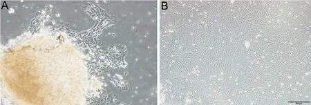
Figure 2 Human LACC-HGT primary cells culture A: Primary LACC-HGT cells crawled out of the tissue block (×100) on the third day of growth;B: On the 10th day of primary culture, tissue mass had fallen off and cell growth was in good condition (×40). LACC-HGT: Lacrimal gland adenoid cystic carcinoma with high-grade transformation.
LACC-HGT Tissue Primary Cell Culture and PurificationAfter the first day of primary culture, the tissue block could be firmly attached to the culture flask wall. On the third day,the cells could be seen crawling out from the side of the tissue block under an inverted microscope, forming a growth halo(Figure 2A). Subsequently, the number of cells crawling out of the tissue gradually increased. The cells grew slowly and gradually merged, and then they grew well. On the 10thday of primary culture, the tissue pieces fell off, and the cells were observed to grow well. The cells were fusiform or polygonal,tightly arranged, and connected into pieces, forming a monolayer layer with a clear outline (Figure 2B). The cells grew slowly during early culture. The initial expansion of the T25 flask to 80%-90% fusion was completed in approximately 2wk, and the cells were digested and passed on when they were 80%-90% full. In the initial cell culture, fusiform fibroblasts with small nuclei, abundant cytoplasm, and small nucleolar ratio were observed. Given the different sensitivities of LACC-HGT cells and fibroblasts to trypsin, more uniform and stable LACC-HGT cells could be obtained in the fourth generation after purification. In addition, after purification,the cells were polygonal or spindle shaped; closely arranged;contained a large nucleus, less cytoplasm, high nucleoplasmic ratio, and obvious cell atypia.
LACC-HGT Primary Cell IdentificationThe immunofluorescence results ofLACC-HGT primary cells confirmed that the LACC-HGT cells epithelial marker CK and glandular epithelial marker CK7 were positively expressed in the cytoplasm (Figure 3). This result showed that the cultured LACC-HGT primary cells retained the phenotypic characteristics of LACC epithelial tissue.
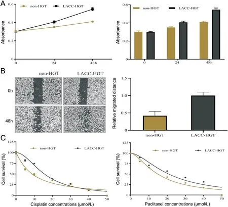
Comparison of Biological Behavior Between LACC-HGT Primary Cells and Non-HGT Primary CellsThe CCK-8 results showed that the OD value of LACC-HGT primary cells was significantly higher than that of non-HGT primary cells. The proliferation fold of LACC-HGT cells was 1.343±0.003, and that of non-HGT cells was 1.198±0.009 after 24h (P<0.001). Meanwhile, the proliferation fold of LACCHGT cells was 1.830±0.016, and that of non-HGT cells was 1.374±0.007 after 48h (P<0.001; Figure 4A).

Figure 3 Immunofluorescence staining of primary LACC-HGT cells CK and CK7 staining of LACC-HGT primary cells were positive (×100). LACCHGT: Lacrimal gland adenoid cystic carcinoma with high-grade transformation; CK: Cytokeratin.

Figure 4 Comparison of biological behavior between human LACC-HGT primary cells and non-HGT primary cells A: The proliferation ability of LACC-HGT primary cells was stronger than that of non-HGT primary cells; B: The migration ability of LACC-HGT primary cells increased compared with that of non-HGT primary cells; C: LACC-HGT primary cells had higher IC50 of cisplatin and paclitaxel than non-HGT primary cells.IC50: The 50% inhibitory concentration; LACC-HGT: Lacrimal gland adenoid cystic carcinoma with high-grade transformation.
The wound healing experiment results showed that the wound healing area of LACC-HGT primary cells was significantly higher than that of non-HGT primary cells after 48h (P<0.01;Figure 4B).
After various concentrations of cisplatin treatment were implemented, the IC50 values of LACC-HGT and non-HGT cells were 15.65±0.61 and 8.89±0.41, respectively (P<0.001).After paclitaxel treatment, the IC50 values of LACC-HGT and non-HGT cells were 20.16±1.91 and 15.82±0.67, respectively(P<0.05). The sensitivity of LACC-HGT primary cells to cisplatin and paclitaxel was significantly lower than that of non-HGT primary cells, and the difference was statistically significant (Figure 4C).
Differentially Expressed Genes and Bioinformatic AnalysisCompared with non-HGT primary cells, the results of mRNA array showed 9566 differentially expressed genes in LACCHGT primary cells, of which 5162 were upregulated and 4404 were downregulated (Figure 5A). GO analysis results indicated that the differentially expressed genes were mainly located in“plasma membrane”, “basement membrane”, and “cytoplasm”.The differentially expressed genes were also closely associated with “extracellular matrix organization”, “cell adhesion”,“signal transduction”, “extracellular matrix structural constituent”, and “actin binding” (Figure 5B). KEGG pathway results indicated that the differentially expressed genes were mainly involved in “protein digestion and absorption”,“pathways in cancer”, “focal adhesion”, and “ECM-receptor interaction” pathway (Figure 5C).
RT-qPCR Analysis of Differentially Expressed GenesAccording to mRNA array fold change, the top five differentially expressed genes were N-acetylneuraminate pyruvate lyase(NPL), MARVEL domain containing 3 (MARVELD3),periostin (POSTN), syntabulin (SYBU), and allograft inflammatory factor1 (AIF1). The RT-qPCR result showed that the expression of NPL, MARVELD3, SYBU, and AIF1 was higher in LACC-HGT cells than in non-HGT cells, whereas that of POSTN was lower (Figure 6).
DISCUSSION
Adenoid cystic carcinoma is a highly malignant tumor originating from different parts of the epithelium, such as salivary gland,lacrimal gland, submandibular gland, and breast. It is prone to local spread and distant metastasis, and the prognosis of patients is poor[9-10]. In 2017, the WHO’s “Classification of Head and Neck Tumors” explicitly added the concept of highgrade transformation to salivary gland tumors, which should be paid close attention because high-grade transformation causes poorer prognosis in patients. In addition to salivary adenoid cystic carcinoma, high-grade transformation could be found in various adenoid cystic carcinomas, such as maxillary sinus adenoid cystic carcinoma high-grade transformation[11]and LACC-HGT[6]. The prognosis of patients with adenoid cystic carcinoma high-grade transformation is significantly reduced.LACC is one of the most common lacrimal gland epithelial malignant tumors, and treatment mainly depends on local surgery, adjunctive radiation, and chemotherapy. Although comprehensive treatment could improve the treatment effect to a certain extent, some patients with LACC still face metastasis,recurrence, and drug resistance[12]. Studies have shown that LACC could also undergo high-grade transformation. And LACC-HGT showed similar characteristics to salivary gland adenoid cystic carcinoma high-grade transformation, such as poorly differentiated or undifferentiated carcinoma, frequent mitoses and extensive necrosis. Patients with LACC-HGT have higher histological grade than those with non-HGT,resulting in poorer prognosis among the former[6]. In view of this phenomenon, the present study aimed to isolate tumor cells that may be different from the two types of tissues to determine the possible mechanisms from the aspect of cell biological behavior.
Tissue mass culture is a relatively common primary cell culture method in solid tumors, and it is convenient and easy to operate; various primary cells have been successfully cultured through this method[13-14]. In previous studies, non-HGT cells were successfully obtained by primary culture[8]. On this basis, LACC-HGT cells were cultured in the present study to understand the differences in biological behavior between the two types of cells.
CK and CK7 are cytokeratins, which are cytoskeleton proteins specific to epithelial cells and often used as epithelial tumor markers. LACC is the only malignant tumor of epithelial origin among orbital malignancies. The immunofluorescence results showed that LACC-HGT primary cells were consistent with LACC tissue samples. Therefore, LACC-HGT primary cells were relatively stable and could be used for biological behavior study.
The biological behavior between LACC-HGT cells and non-HGT cells was significantly different. In this study, CCK-8 and wound healing experiment results showed that the proliferation and migration ability of LACC-HGT primary cells were significantly higher than those of non-HGT cells,corresponding to a shorter symptom onset time, higher recurrence rate, and shorter survival rate in patients with clinical LACC-HGT than in patients with non-HGT. Cisplatin and paclitaxel are commonly used chemotherapy drugs in patients with LACC. Cisplatin, as one of the platinum chemotherapy drugs, could induce DNA damage and promote the death of tumor cells. It is a commonly used chemotherapy drug for various malignant tumors[15], such as breast cancer[16]and esophageal squamous cell carcinoma[17]. Paclitaxel, as a mitotic inhibitor, inhibits the division and proliferation of tumor cells by blocking the cell cycle, and it has become a commonly used chemotherapy drug for clinical malignancies,such as lung cancer[18], nasopharyngeal cancer[19], and esophageal squamous cell cancer[20]. The IC50 of LACCHGT cells was significantly higher than that of non-HGT cells for cisplatin and paclitaxel. Thus, the sensitivity of LACC-HGT cells to cisplatin and paclitaxel was lower than that of non-HGT primary cells. In addition, LACC-HGT cells were more sensitive to cisplatin than to paclitaxel, providing certain guiding significance for the chemotherapy of LACC-HGT in clinical practice.

Figure 5 Differentially expressed genes and bioinformatics assay between non-HGT and LACC-HGT cells A: Heatmap of differentially expressed genes between non-HGT and LACC-HGT cells. B: GO analyses of differentially expressed genes between non-HGT and LACC-HGT cells. The BP bar chart represents extracellular matrix organization, cell adhesion, cell-cell adhesion, signal transduction, positive regulation of cell migration, response to mechanical stimulus, response to virus, positive regulation of phosphatidylinositol 3-kinase signaling, axon guidance, and angiogenesis in descending order. The MF bar chart represents extracellular matrix structural constituent, extracellular matrix structural constituent conferring tensile strength, actin binding, heparin binding, proteoglycan binding, SH3 domain binding, cytoskeletal protein binding, calcium ion binding, muscle alpha-actinin binding, Rho guanyl-nucleotide exchange factor activity in descending order. The CC bar chart represents plasma membrane, basement membrane, cytoplasm, integral component of plasma membrane, extracellular matrix, cell surface, Z disc, intracellular membrane-bounded organelle, postsynaptic density, and adherence junction in descending order. C: KEGG pathway analyses of differentially expressed genes between non-HGT and LACC-HGT cells. GO: Gene ontology; KEGG: Kyoto Encycl opedia of Genes and Genomes; BP: Biological process; MF: Molecular function; CC: Cellular components; LACC-HGT: Lacrimal gland adenoid cystic carcinoma with high-grade transformation.

Figure 6 Relative expression of NPL, MARVELD3, POSTN, SYBU, and AIF1 in non-HGT and LACC-HGT cells aP<0.05, bP<0.001. NPL:N-acetylneuraminate pyruvate lyase; MARVELD3: MARVEL domain containing 3; POSTN: Periostin; SYBU: Syntabulin; AIF1: Allograft inflammatory factor 1; LACC-HGT: Lacrimal gland adenoid cystic carcinoma with high-grade transformation.
On the basis of mRNA array, differentially expressed genes were detected and analyzed and bioinformatic assay was performed. These differentially expressed genes were closely associated with “extracellular matrix organization”, “cell adhesion”, and “signal transduction” and mainly involved in “ECM-receptor interaction”, “PI3K-Akt signaling pathway”, “MAPK signaling pathway”, and “Ras signaling pathway”. These signaling pathways are typical tumorrelated signaling pathways that play important roles in the occurrence and development of multiple carcinomas, such as hepatocellular carcinoma[21], ovarian cancer[22], pancreatic cancer[23], and breast cancer[24]. These differentially expressed genes and pathways may play important roles in LACCHGT pathogenesis. According to literature review, NPL and SYBU were rarely reported in cancer. MARVELD3, POSTN,and AIF1 play an oncogenic or tumor suppressor gene in different malignant tumors. MARVELD3, a tight junction membrane protein, was higher in endometrial carcinoma[25].POSTN is a secreted cell adhesion glycoprotein that plays an important role in proliferation, adhesion, migration, and epithelial-mesenchymal transition in invasive ductal breast carcinoma, renal cell carcinoma, and laryngeal squamous cell carcinoma[26-28]. AIF1 was highly expressed in esophageal carcinoma and clear-cell renal cell carcinoma, and it is a promising predictor of prognosis[29-30]. AIF1 promoted the migration of hepatoma cells and tumor growth by inducing a M2-like phenotype of macrophages, thus playing a pivotal role in the interaction between macrophages and hepatoma cells[31].These genes have not been reported to be associated with LACC-HGT. Therefore, future gene verification and related biological functions may provide more information to elucidate the mechanism of LACC-HGT. Additional investigation is also needed to compare the difference and identify the possible mechanism between non-HGT and LACC-HGT.
In summary, we successfully conducted primary cell culture,and compared the biological behaviors and differential genes of the two kinds of primary cells. LACC-HGT cells showed significantly higher proliferation, migration capacity, and antitumor drug resistance than non-HGT primary cells. Thus, this study provided a cell model for the further study of LACCHGT. Clinicians should correctly diagnose LACC-HGT and adopt reasonable strategies for personalized treatment as soon as possible, which is of vital importance for the clinical treatment and prognosis of patients with LACC-HGT. A limitation of this study is the small number of experimental sample size. In addition, although differential genes were detected by mRNA array, the role and mechanism of these differential genes for the high-grade transformation of LACC still require further experimental investigation. Therefore, in the future, a larger sample number and the function of these differential genes between non-HGT and LACC-HGT cells need to be further verified to provide more information to elucidate the possible pathogenesis in LACC-HGT.
ACKNOWLEDGEMENTS
Foundations:Supported by the Tianjin Key Medical Discipline (Specialty) Construction Project (No.TJYXZDXK-037A); Tianjin Medical University Eye Hospital.
Conflicts of Interest: Zhang CL,None;Zhu LM,None;Liu X,None;Jiang MX,None;Lin TT,None;He YJ,None.
 International Journal of Ophthalmology2023年2期
- International Journal of Ophthalmology的其它文章
- Comment on: Amniotic membrane for covering high myopic macular hole associated with retinal detachment following failed primary surgery
- Trend of glaucoma internal filtration surgeries in a tertiary hospital in China
- Effects of slanted bilateral lateral recession vs conventional bilateral lateral recession on convergence insufficiency intermittent exotropia: a prospective study
- Optical coherence tomography enhanced depth imaging of chorioretinal folds in patients with orbital tumors
- Trends in operating room-based glaucoma procedures at a single eye center from 2016-2020
- Small incision lenticule extraction and femtosecondassisted laser in situ keratomileusis in patients with deep corneal opacity: case series
