Development and validation of a predictive model for patients with post-extubation dysphagia
Jia-ying Tang, Xiu-qin Feng, Xiao-xia Huang, Yu-ping Zhang, Zhi-ting Guo, Lan Chen, Hao-tian Chen, Xiao-xiao Ying
Nursing Department, the Second Affiliated Hospital of Zhejiang University School of Medicine, Hangzhou 310009, China
KEYWORDS: Post-extubation dysphagia; Nomogram; Predictive model
INTRODUCTION
As a result of the development of critical medical technology, the number of hospitalized patients on mechanical ventilation has climbed every year, with an average yearly increase of 27.48%,[1]thereby becoming the primary consumer of healthcare resources. Following endotracheal intubation, some patients develop dysphagia (post-extubation dysphagia [PED]), with choking when drinking water, no visible swallowing movements, and impaired speech as the predominant clinical signs.[2]According to reports, 41%-93% of patients with endotracheal intubation may experience varying degrees of dysphagia after extubation, and 17%-36% patients have occult aspiration (no cough refl ex during aspiration).[1,3,4]
PED is related to many factors, including the patient’s own illness, deep tissue damage caused by long-term throat compression, and local neurosensory anomalies.[5]The presence of PED can have some negative effects, including an increased risk of aspiration pneumonia, tracheal reintubation, and malnutrition, all of which contribute to prolonged hospitalization and higher medical costs.[6,7]Furthermore, PED frequently persists, thereby negatively affecting the patient’s quality of life and longterm prognosis even after hospital discharge. According to previous research, up to 80% of critically ill patients still have dysphagia when they are transferred out of the intensive care unit (ICU), and more than 60% of patients have symptoms that do not resolve until discharge from the hospital.[1]The inability to eat by mouth and the longterm use of nutritional tubes can affect the patient’s psychological state and result in a care and financial burden on family members. According to a previous study,[8]the United States spends more than $500 million each year on medical bills for hospital care and follow-up rehabilitation of PED patients, contributing greatly to the cost burden on the public healthcare system.
Based on these findings, clinical practitioners are paying increasing attention to the swallowing ability of tracheally intubated patients. Nevertheless, the etiology of PED is unclear, and the risk factors have not been identified. Although advanced age,[9]use of sedatives,[10]and other factors[11]have been identified as the main causes of PED, the extent to which these factors affect the occurrence of dysphagia is unknown. The existing risk predictive models have not been widely used clinically due to large quantities of missing data based on selection bias and the lack of external validation.
Early identification of high-risk individuals and taking appropriate measures can prevent the occurrence and deterioration of PED, thereby improving the prognosis of patients. The aim of this study was to develop and internally validate a predictive model for PED, providing an assessment tool that allows clinicians to forecast which individuals are at high risk and to guide treatment and rehabilitation.
METHODS
Inclusion and exclusion criteria
The study involved ICU patients who met the following inclusion criteria: age ≥18 years, treated with tracheal intubation and mechanical ventilation, awake and able to complete command actions, and having no dysphagia (Kubota water swallowing test < Grade 2) before tracheal intubation. Informed consent was obtained.
Patients were excluded if they had other pre-existing conditions that had affected their swallowing function prior to intubation, such as neck surgery, Parkinson’s disease, cerebrovascular accident, neuromuscular disease, oropharyngeal or esophageal tumor, or inability to drink or eat by mouth due to illness or treatment.
Data source and study population
We obtained approval from the Institutional Review Board. A retrospective selection of patients who had been admitted to the emergency ICU, central ICU, and cardiovascular surgery ICU from June to December 2021 was conducted, and these patients served as the derivation cohort. Patients recruited from the same departments of the same hospital who received endotracheal intubation therapy from March 2022 to June 2022 served as the external validation cohort for the predictive model.
Risk factors
Risk factors were considered based on the following: (1) risk factors for PED identified in previous studies; (2) bedside evaluation completed within 4-6 h after extubation; and (3) variables observed by clinicians or nurses before the onset of dysphagia. Based on these criteria, a total of 35 risk factors were considered, including demographic characteristics (age, gender, body mass index [BMI], smoking history); admission diagnosis and pre-admission complications; surgical data (e.g., operation time, aortic occlusion time, esophageal ultrasound); intubation conditions (e.g., time, model, depth, air pressure, urgent tracheal intubation, repeated intubation); treatment (nasogastric tube, nasogastric tube insertion length, use of sedatives, use of analgesic drugs, pause of sedatives, pause of analgesic drugs); level of consciousness (somnolence); and various evaluations after extubation (e.g., activities of daily living [ADL] score, Acute Physiology and Chronic Health Evaluation II [APACHE II] score, Confusion Assessment Method for the Intensive Care Unit [CAM-ICU], New York Heart Association (NYHA) classification,[12]integrity of oral mucosa, voice disorder, sore throat).
Among these, age, BMI, operation time, aortic occlusion time, endotracheal intubation time, APACHE II score, cardiac function evaluation, pause of sedatives, and sore throat were included as continuous or ordinal variables. Dichotomous variables included gender, admission diagnosis and pre-admission complications, use of sedatives or analgesic drugs, esophageal ultrasound, urgent tracheal intubation or repeated intubation, nasogastric tube, CAM-ICU, integrity of oral mucosa, and voice disorder. Detailed information on the independent variable codes is available in supplementary Table 1.
Primary outcome: dysphagia after endotracheal intubation
Swallowing function was measured using the standardized swallowing assessment (SSA), which can be performed efficiently by bedside clinicians or nurses.[13]SSA has high sensitivity and specificity in PED screening, and it is easy for ICU nurses to master.[14]In all ICUs of our hospital, the SSA score is a routine record that is completed within 4-6 h after extubation as a part of the nursing standards.
Data measurement and collection
All the patients received a preliminary clinical assessment including risk factors. Prospective methods were used in the validation group. Three clinical nurses who served as team members in the project were trained and passed the exam to ensure proficiency in SSA, Voice Disability Assessment (VDA), and throat pain score. The nurses used the SSA, VDA, and throat pain score to assess the patient’s swallowing ability, voice interruption, and throat pain within 4-6 h after extubation. Patients with special conditions were evaluated jointly by intensive care physicians.
We obtained the demographic characteristics and general information, including disease information and treatment information before extubation, from the patients’ medical records, and no imputation techniques were used to address missing data. The remaining data, such as ADL score, consciousness level, and NYHA classification, were collected through on-site evaluation. Once a patient received the diagnosis of dysphagia, the clinicians or nurses recorded the specific symptoms and results from medical records. The patient’s recovery and prognosis were also documented. Before the evaluation of the validation group, the patients or their families were informed of the research purpose and evaluation procedures, and we obtained their written consent. Due to the retrospective study design, the need for informed consent was waived for the derivation cohort. Details of the research process are shown in supplementary Figure 1.
Development and validation of a PED predictive model
A PED predictive model was developed using a combination of variable screening and least absolute shrinkage and selection operator (LASSO) regression, as shown in supplementary Figure 2. Univariate regression analyses were performed on each potential predictor, and we selected variables withP<0.20. We further used LASSO regression to select the most useful candidate predictors. We checked the multicollinearity of the independent variables using the variance inflation factor (VIF) method. By combining the statistically significant variables from univariate regression analysis and LASSO regression, multivariate logistic regression analysis was performed to calculate the odds ratio (OR; 95% confidence interval [95%CI]) andP-value for each variable to predict a potential diagnosis.
The receiver operating characteristic (ROC) curve was performed to assess the ability of the model to discriminate between patients diagnosed with PED and those not diagnosed with PED. The predictive value of the model was evaluated by area under the ROC curve (AUC), and the optimal cutoff threshold values were determined using the maximum Youden’s index (sensitivity+specificity-1). The closer the AUC is to 1, the better the predictive model is. Using these optimal cut-off values, we calculated the sensitivity, specificity, positive predictive value (PPV), and negative predictive value (NPV) of the model. Then, we prospectively validated this model for the risk of PED in external validation cohort. The patients were included if they underwent a swallowing evaluation with the SSA. Model adjustmentfit tests were assessed using Hosmer-Lemeshow goodness. If the test result was statistically significant (P<0.05), there was a certain difference between the predicted value and the observed value, and the model was poor.
Statistical analysis
Statistical software SPSS 23.0 and R4.1.0 were used for data analysis. The measurement data conforming to the normal distribution are described by mean±standard deviation; the measurement data of the skewed distribution are expressed by the median (interquartile range); and the count data are described by frequency and percentage. Baseline characteristics of the groups were summarized using Student’st-test, Chi-square test, and Wilcoxon ranksum test. The significance level of the above statistical analyses was set atα=0.05 andP<0.05 (two-tailed) for statistical significance.
RESULTS
Baseline characteristics of patients
A total of 305 patients were included in the study, including 235 patients in the derivation cohort (53 PED vs. 182 non-PED) and 70 patients in the validation cohort (17 PED vs. 53 non-PED). There was no statistically significant difference in the frequency of PED between the derivation cohort (22.56%) and the validation cohort (24.29%). The characteristics of the patients are available in supplementary Table 2.
Identification of predictive factors in the derivation cohort
In the derivation cohort, 35 variables were included in the univariate regression analysis, among which 17 variables hadP<0.20 (supplementary Table 3). These 17 variables included age, BMI, heart disease, diabetes, esophageal ultrasound, urgent tracheal intubation, repeated intubation, intubation model, intubation depth, pause of sedatives, pause of analgesic drugs, APACHE II score, ADL score, level of consciousness, nasogastric tube, sore throat, and voice disorder.
These 17 variables were further screened through LASSO regression. The presence/absence of PED was used as the dependent variable for internal cross-validation selection. The process of LASSO regression is shown in supplementary Figure 3. Whenλwas 0.0228, the model performed best. Finally, we found 13 non-zero coefficient characteristics. The characteristics included age, heart disease, intubation model, intubation depth, repeated intubation, pause of sedatives, pause of analgesic drugs, APACHE II score, ADL score, level of consciousness, nasogastric tube, sore throat, and voice disorder.
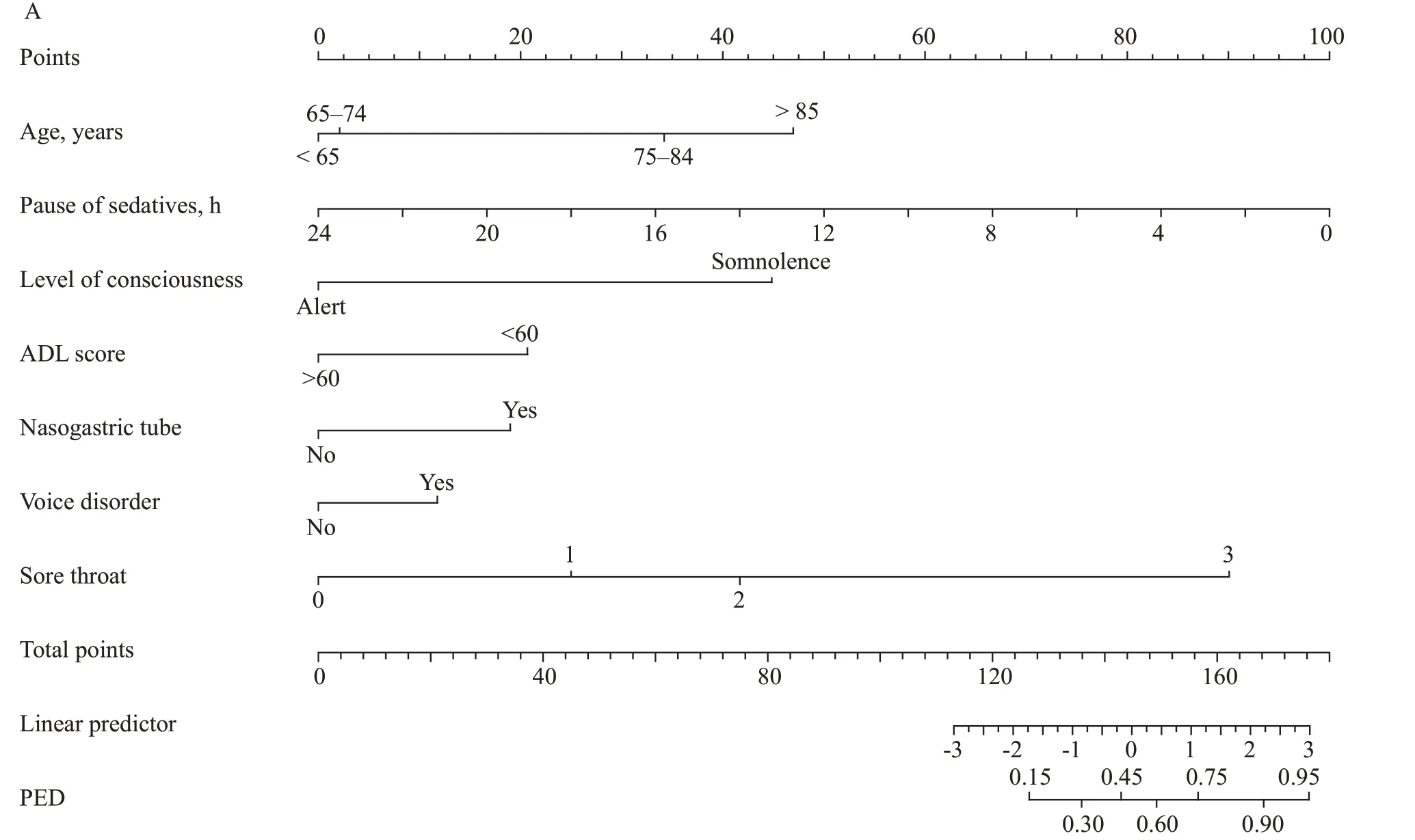
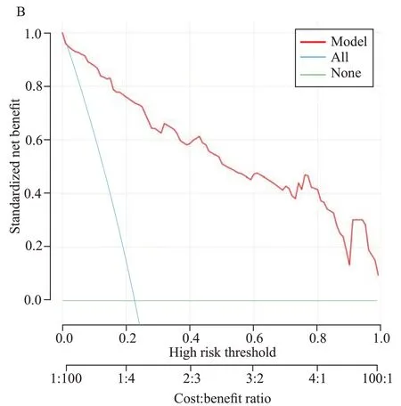
To further eliminate the redundant features, the abovementioned 13 PED-predicting features were subjected to backward stepwise logistic regression. Seven risk factors, including age, pause of sedatives, level of consciousness, ADL score, nasogastric tube, sore throat, and voice disorder, were found to be independent predictors of PED (Table 1), and they are visualized as a nomogram in Figure 1A. The VIF of all dependent variables was close to 1, with a range of 1.059-1.363. Figure 1B presents the decision-curve analysis of the nomogram. The decision curve of this predictive model was more efficient than that of other predictive strategies.
Development of the PED predictive model
The PED predictive model and the scoring criteria of the model were developed based on the seven risk factors selected by backward stepwise regression.
The results indicated that the Youden index was higher when the total score was between 0.1 and 0.2. Therefore, a score of 0.158 was used as the cut-offvalue of the model. At this point, the sensitivity and specificity of the predictive model were both high. The details of the sensitivity and specificity of the PED predictive model under different cut-offvalues are shown in supplementary Table 4. The AUC and 95%CIwere 0.945 (0.907-0.969). The accuracy, specificity, and sensitivity were 84.68%, 81.87%, and 94.34%, respectively. The PPV was 60.24%, and the NPV was 98.03%, withR2=0.461 and adjustedR2=0.445. The calibration curve showed good performance (χ2=4.5329,df=8,P=0.8061). The ROC curve and the scoring standard of the predictive model are shown in supplementary Figure 4.

Table 1. The independent predictors of PED based on multivariate logistic regression analysis in the derivation cohort
The model equation was as follows: logit (P) = -2.287 + age (>85 years) × 4.421 + age (65-74 years) × 0.209 + pause of sedatives × -0.393 + level of consciousness (somnolence) × 4.236 + ADL score (>60) × -1.952 + nasogastric tube (yes) × 1.791 + voice disorder (yes) × 1.085 + sore throat (Grade I) × 2.380 + sore throat (Grade II) × 3.955 + sore throat (Grade III) × 16.830.
Evaluation of the PED predictive model
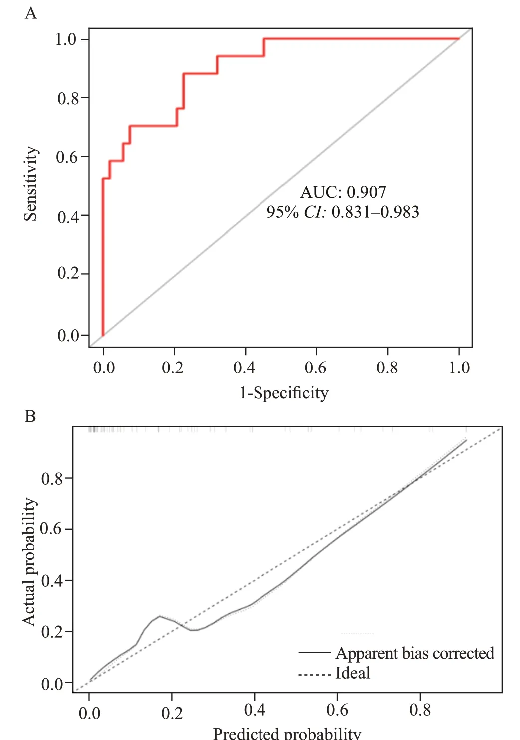
The performance of the proposed model was evaluated by prospectively recruiting 70 patients with tracheal intubation and extubation in the ICUs of the same hospital. The AUC corresponding to the validation set model was 0.907 (95%CI0.831-0.983; Figure 2A). The accuracy, sensitivity, and specificity of the model were 84.29%, 70.59%, and 88.68%, respectively. The calibration curve showed good performance, indicating that the model had good discrimination and calibration (Figure 2B).

Figure 1. Development of the nomogram and decision curve for the predictive model in the derivation cohort. A: nomogram visualization of the PED predictive model; B: decision curve of the PED predictive model in the derivation cohort. PED: post-extubation dysphagia.

Figure 2. ROC validation of the PED predictive model and the calibration curves of the model in the verification group. A: AUC that corresponds to the validation set model; B: calibration curves in the validation cohort. ROC: receiver operating characteristic; AUC: area under the ROC curve; PED: post-extubation dysphagia.
DISCUSSION
In recent years, research has shown the need to evaluate a patient’s swallowing function after extubation and to develop a predictive model. After patients are extubated, medical staffcan use a predictive model to identify patients at high risk of PED as early as possible and provide a basis for making plans to improve the prognosis.
Rumbach et al[15]used burn patients as research subjects to develop an early warning model for PED and found that the sensitivity and specificity were 100% and 83.74%, respectively. Due to the particularity of burn patients, thismodel cannot be directly applied to ICU patients. The predictive model developed by Guo et al[16]for PED in ICU endotracheal intubation patients showed that the sensitivity was 70.3% and the NPV was 88.2%, with a PPV of 57.1%. The predictive abilities were considered low, and the overall predictive accuracy was limited. Based on these findings, we attempted to develop a PED risk predictive nomogram for patients with endotracheal intubation and applied simplefactor logistic regression analysis combined with a LASSO algorithm in the analysis of risk factors as a means to avoid linear relationships between factors and the phenomenon of model overfitting as well as to improve the predictive effect of the model.
Our study showed that advanced age was an independent risk factor for dysphagia after endotracheal intubation and extubation, which is consistent with the results obtained by Oliveira et al.[17]Physiological weakness in elderly individuals can affect their swallowing function. A previous study showed that compared with younger patients, the incidence of swallowing dysfunction was higher in elderly patients, which may be related to physiological changes. In addition, the direct injury to the throat caused by endotracheal intubation and the disuse of oropharyngeal muscles caused by intubation greatly affect the ability of elderly individuals to masticate and control swallowing muscles. We also found that patients who had low self-care ability (ADL score <60) were more likely to have dysphagia, and the incidence of PED in patients with ADL score >60 was 6.82 times lower (P<0.05) than that in patients with ADL score <60, which is consistent with previous research.[18]This may be related to the patients’ weak state, low level of consciousness, and lack of coordination.

The risk of PED was higher in patients with somnolence and a shorter duration of sedative drug pause. Somnolence is a risk factor for PED; it is often accompanied by dyskinesia of the tongue and pharynx, paresthesia, and impaired protective reflexes, thereby prolonging the oral preparation period and the time for food to pass through the pharynx, eventually resulting in swallowing dysfunction and even aspiration. The use of sedatives, especially midazolamtype sedative drugs, inhibits the swallowing reflex to a certain extent and prolongs oral preparation for swallowing operations.[19]During this period, the peristaltic movement of the pharynx is weakened, which causes uncoordinated swallowing movements that seriously affect their speed and accuracy. Thorn et al[20]found that propofol-type sedative drugs can cause damage to throat muscle endurance and strength and affect esophageal motility, resulting in difficulties with swallowing. The uncoordinated activities of the oral cavity and the esophagus cause swallowing disorders and greatly increase the risk of aspiration. In addition, the use of sedatives can lead to decreased consciousness or even disturbance of consciousness, thereby resulting in unnecessary tracheal intubation and nasogastric tube indwelling time.[21]
The nasogastric tube was an independent risk factor for PED in our study. Analysis of the reasons shows that the nasogastric tube can cause discomfort and foreign body sensation, delayed elevation of the larynx, and esophageal sphincter insufficiency, which in turn leads to a lower swallowing speed.[22]Long-term indwelling nasogastric tubes can also cause gastroesophageal reflux, which can lead to mucosal damage or ulcers in the throat,[23]further exacerbating patients’ poor swallowing function. At the same time, tube-feeding nutrition causes the throat reflex to become passive, thereby producing swallowing difficulties. To reduce these problems, intermittent tube feeding has been employed in the rehabilitation of stroke patients with dysphagia as a nutritional support and dysphagia strategy,[24,25]but its application in PED patients needs further study.
Patients with voice disorder and sore throat have a higher risk of PED. In our study, 50.1% of the patients had a certain degree of hoarseness and disturbance, and 50.4% complained of throat pain. This is consistent with the results obtained by Medeiros et al.[26]The reason may be associated with direct damage to the tissue structure or mucous membrane of the throat during orotracheal intubation.
CONCLUSION
A predictive model that incorporates age, pause of sedatives, level of consciousness, ADL score, nasogastric tube, sore throat, and voice disorder may have the potential to predict PED in patients in the ICU.
Funding:This study was supported by the Health Science and Technology Project of Zhejiang Province(2022KY173) and the Health Science and Technology Project of Zhejiang Province (2023KY761).
Ethical approval:The local institutional review board reviewed and approved the study (2022-0795).
Conflicts of interest:The authors declared there are no conflicts of interest.
Contributors:JYT conceived the study, designed the trial, and obtained research funding. XQF was responsible for supervising the implementation of the project. XXH was responsible for administrative and technical support. ZTG and HTC provided statistical analysis and interpretation of data. LC, YPZ, and XXY reviewed, collected, and managed the data. All the authors participated in drafting the manuscript and read and approved the final manuscript.
All the supplementary files in this paper are available at http://wjem.com.cn.
 World journal of emergency medicine2023年1期
- World journal of emergency medicine的其它文章
- Patient care during interfacility transport: a narrative review of managing diverse disease states
- Endothelial cell metabolism in sepsis
- Nutritional status and prognostic factors for mortality in patients admitted to emergency department observation units: a national multi-center study in China
- Prolonged dual antiplatelet therapy after drug-eluting stent implantation improves long-term prognosis for acute coronary syndrome: five-year results from a large cohort study
- Efficacy and safety of remimazolam-based sedation for intensive care unit patients undergoing upper gastrointestinal endoscopy: a cohort study
- Glutamine supplementation attenuates intestinal apoptosis by inducing heat shock protein 70 in heatstroke rats
