Information standardization for typical toxic and harmful algae in China’s coastal waters—a case study of Karenia mikimotoi*
Ting ZHAO , Huidi CAO , Yanfen JIA , Xiaotian HAN ,**, Tian YAN , ,Rencheng YU ,
1 Changjiang River Estuary Ecosystem Research Station, Institute of Oceanology, Chinese Academy of Sciences, Qingdao 266071, China
2 Center for Ocean Mega-Science, Chinese Academy of Sciences, Qingdao 266100, China
3 Eco-Environmental Monitoring and Research Center, Pearl River Valley and South China Sea Ecology and Environment Administration, Ministry of Ecology and Environment, PRC, Guangzhou 510610, China
4 Qingdao Municipal Bureau of Ecology and Environment, Qingdao 266003, China
5 Key Laboratory of Marine Ecology & Environmental Sciences, Institute of Oceanology, Chinese Academy of Sciences, Qingdao 266071, China
Abstract To assist the researches of toxic and harmful algae and provide government workers with judgment basis for decision-making related events, we established a biological information management system for toxic and harmful algae in China’s off shore to assist relevant research. In this study, Karenia mikimotoi was studied as a typical toxic and harmful algae species, and the basic biological information and biogeographic distribution information of K. mikimotoi were systematically studied and collected. In the part of basic biological information, the name, toxin, and molecular characteristic sequence of K. mikimotoi were sorted out by literature searching and website browsing. Through experimental means, the relevant information of morphological identif ication, pigment composition, and lipid composition were obtained.In the part of biogeographic distribution information, this study sorted out the information of K. mikimotoi,analyzed the characteristics of its occurrence, and completed the standardized construction of biogeographic distribution information. Through the collation of basic biological information and biogeographic distribution information of K. mikimotoi, the standardization of related information was completed, which provided template and method reference for information collection of other toxic and harmful algae species,which was benef icial to the database analysis and design.
Keyword: harmful algae; basic biological information; biogeographic distribution information; information standardization
1 INTRODUCTION
Harmful algal blooms not only harm marine life but also pose a threat to higher trophic organisms (including humans) through the food chain. At the same time, they also limit the development of the marine economy and are an important stress factor for the health of marine ecosystems and the safety of f ishery resources.
Regarding current red tide events caused by toxic and harmful algae, many scholars have performed detailed studies (Zhou et al., 2001). For example,Gillibrand et al. (2016) conducted a systematic study on factors including the environment and vertical movement and migration ofKareniamikimotoiin the waters off the coast of Scotland from July to September 2006. Aoki et al. (2017) also investigated the causes of theK.mikimotoitide that occurred in Iwanli Bay,eastern Japan, in 2014. However, these studies were aimed at a specif ic species in a certain period of time or in a certain region, including the location,area and economic loss of red tides. Knowledge of the long-term evolution law of red tides caused by diff erent species is limited compared with that of their temporal and spatial distribution characteristics.From the perspective of environmental protection management, there is an urgent need for studies on the biogeographic distributions of diversif ied toxic and harmful algae, which require the introduction of an information management system as a comprehensive carrier of biological information to be managed.
An information management system (IMS) is an eff ective information carrier. It is the product of accounting, statistics, organizational theory,mathematical modeling, and other disciplines.Information management systems have been applied to marine biological information (Yamamoto et al.,2012). For example, for NCBI, Algae-Base, Bigelow,and other websites, most content includes data such as biological information and molecular information for algal species, but little information about the spatial and temporal distributions of alga-induced red tides is displayed, or it remains unavailable.The aims of this study are to digitize, unify, and standardize multidisciplinary information on diff erent toxic and harmful algae and carry out standardized storage and management to rapidly disseminate the multidimensional information on toxic and harmful algae to researchers and decision managers.
In this paper,K.mikimotoiwas selected as a typical toxic and harmful algal species.K.mikimotoibelongs to the order Dinof lagellata, widely distributed in temperate and tropical shallow waters, and is a typical ichthyotoxic red tide alga. In recent years,K.mikimotoihas caused several red tides in China’s waters, the polluted area has gradually expanded, and the number of events has gradually increased. The toxins secreted by this microalga can seriously harm or kill f ish and invertebrates, cause economic losses in f isheries, damage marine ecosystems, and even threaten human health through the food chain. For this paper,K.mikimotoiwas selected as the research object, and its collected information was standardized to provide a reference for the collection and collation of biological information for other toxic and harmful algae in China’s off shore waters.
2 MATERIAL AND METHOD
2.1 Information collection
Before information standardization, information collection should be carried out. The purpose of information collection is to sort and classify available information, but it is also crucial to the construction of subsequent biological information management systems. In general, at the beginning of information collection, toxic and harmful algae distributed in China’s off shore waters were determined as the target subjects. After establishing the research target subjects, the next step is to determine the detailed specif ic information needed about these toxic and harmful algal species. The basic process of information collection is shown in Fig.1.
The basic biological information ofK.mikimotoi, such as taxonomic status, toxins,morphological identif ication, pigment composition,lipid composition, and molecular characteristic sequences, were summarized and sorted. The biogeographic distribution information on off shore red tides ofK.mikimotoiin China was sorted, the characteristics of red tide occurrence were analyzed,and standardization was completed. The obtained information, pictures, and other data were analyzed on the basis of their information characteristics and stored according to diff erent data attributes to provide a basis for further database design.
2.2 Information acquisition
2.2.1 Document acquisition
From the perspective of basic biological information and biogeographic distribution information, the data items to be entered into the database during initial development (such as algal species name, density,life history, toxin, collection and culture, molecular biology information) are selected and collected,the standardization of data is performed, and the information standardization of the data required by the system is described.
2.2.2 Experimental acquisition
2.2.2.1 Microalgal culture
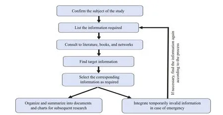
Fig.1 Basic ideas for collecting information
TheK.mikimotoisamples used in this work were provided by the Key Laboratory of Marine Ecology and Environmental Science, Institute of Oceanology,Chinese Academy of Sciences. All glassware was immersed in diluted HCl (10%) and washed several times with deionized water. Natural seawater from the East China Sea was f iltered through a 0.45- μ m mixed-f iber membrane. An algal inoculum of 400 mL was transferred into culture vessels (5-L conical f lasks containing 2 L of culture medium), the cultures were acclimatized with the medium of f/2 (Guillard and Ryther, 1962). The cultures were then acclimatized under an average irradiance of 150 μmol photons/(m2·s) with a light꞉dark regime of 12 h꞉12 h with three replicates.
2.2.2.2 Morphological observation
Optical microscope observations:K.mikimotoiwas f ixed with Lugo reagent, and an appropriate amount of algal liquid was taken for observation under an optical microscope (Nikon 1000).
Scanning electron microscope observation: a scanning electron microscope (SEM, TESCAN VEGA 3) was used to study the micromorphology ofK.mikimotoi.
2.2.2.3 Pigment extraction and analysis
High-performance liquid chromatography (HPLC,Waters e2695) was used for pigment composition analysis (Lai et al., 2012).K.mikimotoiwere f iltered through a GF/F f ilter membrane with a diameter of 47 mm. The obtained sample membrane (parallel samples P1 and P2) was wrapped with aluminum foil and stored in an ultra-low temperature refrigerator until analysis. The f ilter membranes were taken out from the low-temperature refrigerator and cut them into small pieces. Then transfer them into a brown sample bottle with a volume of 1.5 mL, and add 1 350-μL HPLC grade methanol and 150-μL internal standard mother liquor (8ʹ-apo-beta-carotenal).Squeeze and crush the f ilter membranes to completely immerse them in the added methanol extract. Place the brown sample bottle in an ice water bath, after ultrasonic treatment for 5 min, suck the supernatant and f ilter through 0.2-μmol/L pore diameter needle type organic phase f ilter head, f inally the f iltrate was injected into a clean 1.5-mL sample bottle.After these steps, accurately suck 700-μL f iltrate to another sample bottle, 140-μL ultrapure water was immediately add for analysis. The analysis method followed that of Zapata et al. (2000).
2.2.2.4 Lipid extraction and analysis
The experimental algal species were f iltered by GF/F glass f iber membrane with a diameter of 47 mm. After f iltration, the f ilter membranes were cut into small pieces and then 2.4 mL of solution(methyl tert-butyl ether (MTBE)꞉MeOH=5꞉1) and a steel ball were added, the mixture was ground at 35 Hz for 4 min, and the process was repeated 4 times in an ice water bath for 5 min. The sample was centrifuged at 4 °C and 10 000 r/min for 15 min, and 1.75 mL of the supernatant was placed into an eppendorf (EP)tube for vacuum drying. Next, 200 μL of solution(dichloromethane (DCM)꞉MeOH=1꞉1) was mixed,vortexed for 30 s, and ultrasonicated in an ice water bath for 10 min. Then, the sample was centrifuged at 4 °C and 13 000 r/min for 15 min, and 75 μL of supernatant was collected in an injection bottle.
For the detection method, refer to Dunn et al.(2011) and Want et al. (2010). Lipid components were detected using a SCIEX ExionLC™ AC system coupled to the SCIEX X500R Q-TOFMS (AB SCIEX, USA). The chromatographic separation was conducted on a Phenomen Kinetex C18 (2.1 mm×100 mm, 1.7 μm). The mobile phase A was 40% Milli-Q water and 60% acetonitrile solution, containing 10-mmol/L ammonium formate,and the mobile phase B was 10% acetonitrile and 90% isopropanol solution. 50 mL of 10-mmol/L ammonium formate aqueous solution was added to every 1-L phase B. The separation was achieved with the following gradient elution condition: 40% B(0–12 min), linear increase to 100% B (12–13.5 min),100% B (13.5–13.7 min), and f inally decrease to 40%B (13.7–18 min). The injection volume was 1 μL(positive ion) and 2 μL (anion) and the f low rate was 0.3 mL/min for both positive and negative ion modes.The column temperature was 45 °C.
2.2.3 Data storage and classif ication
When sorting the obtained information, it was classif ied according to its collectors and stored centrally. First, an original folder named after the algal species was established. Then, two subfolders of basic biological information and biogeographic information were set up, and several specif ic information subfolders were created in the two folders to classify and summarize the collected information in the form of tables or pictures.
2.2.4 Analysis of information characteristics
To keep the data types entered into the database consistent, the data characteristics of the collected information were analyzed, the type of data was determined, and the data format was standardized,providing a reference for def ining the data attributes in the database later.
3 RESULT AND DISCUSSION
3.1 Basic biological information
3.1.1 Names of algal species
In the early stage, there were many diff erent names for the typical species studied, resulting in many aliases (former names), such asGymnodiniumsp., while the more recognized name is “Kareniamikimotoi”, in which “mikimotoi” comes from the name of a Japanese farm owner (Lü et al., 2019) who funded research on the red tide ofK.mikimotoiin his farming area. All relevant names were sorted in the study.
3.1.2 Classif ication status
Hansen et al. (2000) separated this species from the genusGymnodiniumaccording to relevant data and established the genusKarenia. Bergholtz et al.(2006) synthesized the morphological characteristics,ribosomal DNA sequence (LSU rDNA) and pigment composition ofK.armiger, and found the rDNA sequences ofK.armigerand the genusTakayamaare highly homologous. The authors considered the two taxa to belong to the same species. It is suggested to combine the three into a new family,Kareniaceae. By browsing the AlgaeBase website,we can f ind thatKareniamikimotoibelongs to the Eukaryota empire, Chromista kingdom, Harosa(supergroup stramenopiles-alveolates-rhizaria(SAR)) subkingdom, Harvard Infrakingdom, Miozoa phylum, Myzozoa subphylum, Dinophyceae class,Gymnodiniales order, Kareniaceae family, and Karenia genus, which are divided in great detail. The obtained classif ication status information will be used to establish the toxic and harmful alga directory on the information platform.
3.1.3 Life history
Kareniamikimotoican adapt to various light,temperature, salinity, and nutrient conditions (Li et al., 2019). It can assimilate diff erent forms of phosphates f lexibly, and could take nutrients for growth autotrophically (by photosynthesis) and heterotrophically (by phagotrophy) simultaneously.K.mikimotoipropagates asexually mainly by division.No temporary cysts were found through molecular and morphological observation (Hansen et al., 2000).In culture experiments, it can be observed that some strains can get small cells through cell division. So far, there is no suffi cient evidence to prove thatK.mikimotoican reproduce sexually, and its main mode of reproduction is asexual reproduction through cell division (Lü et al., 2019).
3.1.4 Toxin information
Chen et al. (2015) and Li et al. (2017) conducted systematic studies of several toxins inK.mikimotoiand selected the three most common toxins:hemolytic toxins, cytotoxins, and f ish toxins. Among them, hemolytic toxins (HTs) are the main cause of f ish death during the red tides ofK.mikimotoi. These toxins will cause pathological phenomena such as adjacent gill leaf adhesion, gill blood vessel rupture,blood cell exudation, and so on (Liu et al., 2015).Cytotoxins mainly consist of two toxins, gymnocin A and gymnocin B, which have similar characteristics to hemolytic toxins, but the diff erence is that hemolytic toxins mostly act on the cell membrane, while cytotoxins inhibit cell growth (Guo, 2014). However,many studies have also shown that hemolytic toxins and cytotoxins are not the main causes of death(Chang and Gall, 2013; Lin et al., 2016; Li et al.,2017). More in-depth research is needed on the toxins and toxicity mechanism ofK.mikimotoi.
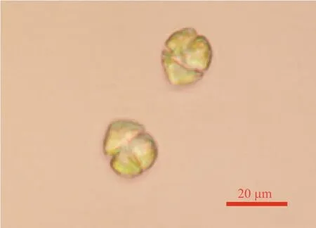
Fig.2 Optical microscope observation
3.1.5 Morphological identif ication
The optical microscopy images show that the algal body ofK.mikimotoiis single-celled and greenish brown. At rest, only the dorsoventral surface can be seen, which is slightly f lat (Fig.2).
According to morphological identif ication via scanning electron microscopy,K.mikimotoicells are nearly round and slightly f lat in dorsal and ventral view. The side, top, and bottom surfaces are hemispherical to widely conical, the upper cone has a groove at the top that extends upward from the upper end of the longitudinal groove to the top and reaches the back, and the bottom end of the lower cone has a depression (Fig.3). The results of this morphological identif ication are consistent with records in the literature. It should be noted that during scanning electron microscope photography, it was found that the algal body was folded, which may be related to the growth period of the algae, f ixation of the algal body and other factors. Their length was about 20–30 μm, and their width was about 16–30 μm. Basic on the Technical Specif ication for Red Tide Monitoring(HY/T 069-2055, 2005 (China’s National Standard)),when the density were higher than 106cells/L, it can be judged as the harmful algal blooms.
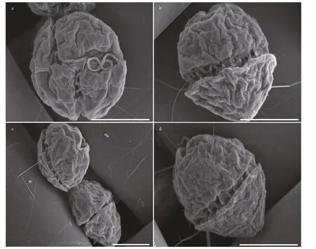
Fig.3 Morphological identif ication of K. mikimotoi with SEM
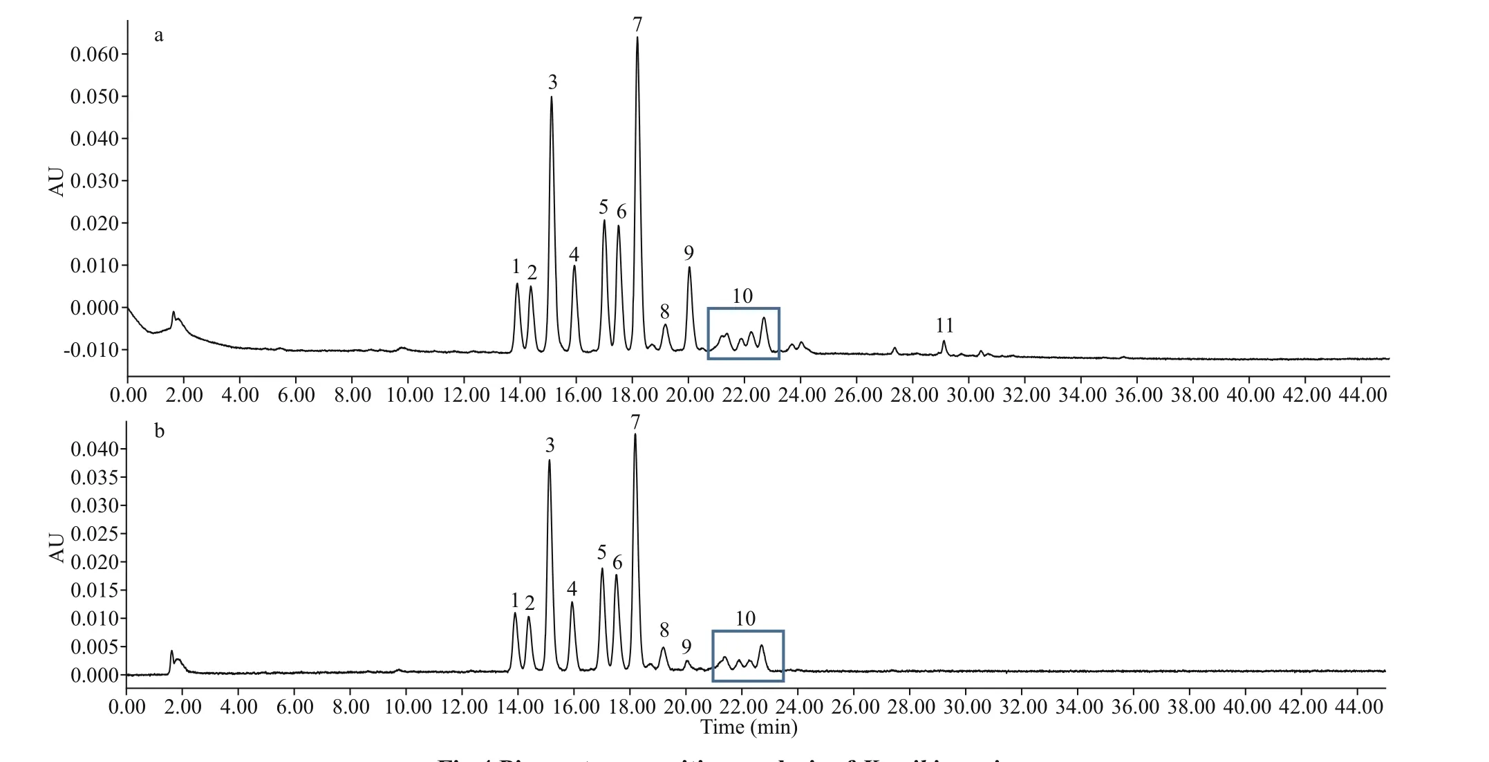
Fig.4 Pigment composition analysis of K. mikimotoi
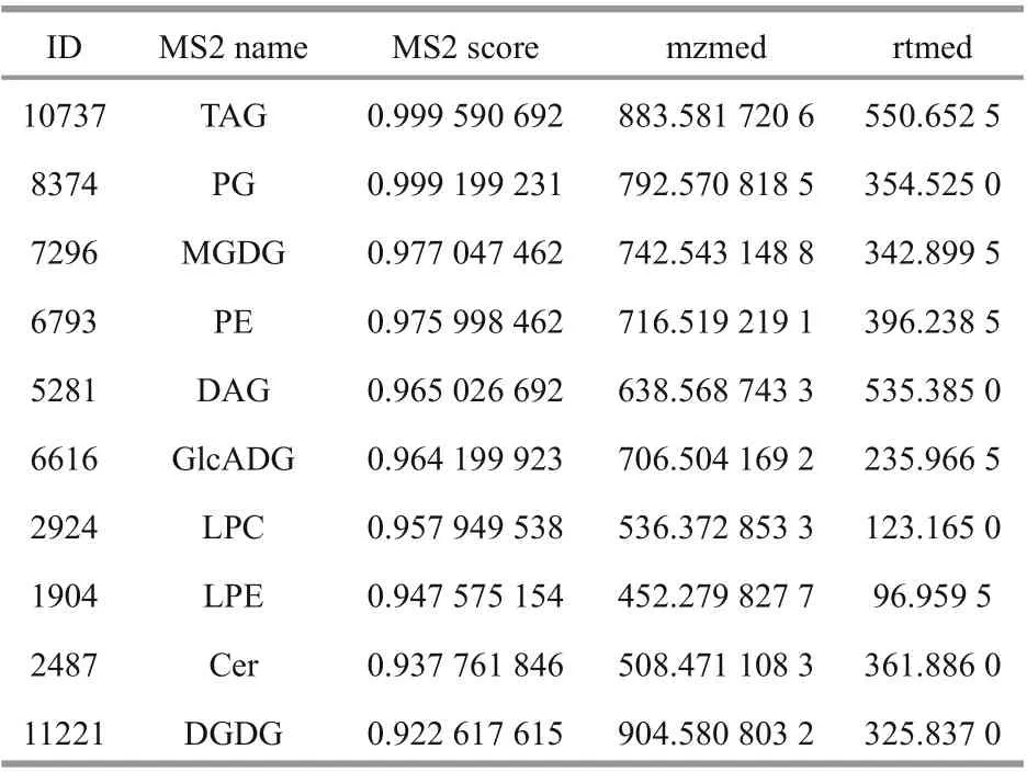
Table 1 The detected lipids
3.1.6 Pigment composition analysis
The pigment compositions of P1 and P2 of the two control samples were detected by HPLC (Fig.4). The results show that the pigments detected by the two samples were basically the same. The marker pigment ofK.mikimotoiis fucoxanthin (FUCO), which includes 19ʹ-butyryl FUCO and 19ʹ-hexoyloxyfuco(hex-FUCO) (Suzuki and Ishimaru, 1992). In addition,K.mikimotoialso contains pigments such as didinoxanthin and 4-keto 19ʹ hexyl oxyfucophyll.Gyroxanthin diester pigment was also found in sample P1, which is a characteristic pigment ofK.mikimotoi. Li et al. (2009) used this pigment and gyroxanthin to characterizeK.mikimotoiin the largescale red tide of dinof lagellates in the East China Sea,but this characteristic pigment peak was not obvious or absent in the two samples, which may be due to the low content of gyroxanthin diester pigment in this strain ofK.mikimotoi. However, no obvious chlorophyll peak was detected during the experiment.Sample processing may take a long time, resulting in the degradation of chlorophyll.
3.1.7 Lipid composition
The detected lipids mainly included triglyceride(TAG), phosphatidylglyceride (PG), monogalactose diglyceride (MGDG), phosphatidylethanolamine(PE), diglyceride (DAG), glucuronic acid glycolipid(GlcADG), diglyceride (DGDG), etc. (shown in Table 1). As a typical red tide alga producing f ish toxins,K.mikimotoiis a haemolytic, cytotoxic species,and can produce substances with strong hemolytic activity, which are mainly glycolipids and unsaturated fatty acids (Yasumoto et al., 1990). MGDG and DGDG were haemolytic (Parrish et al., 1998). In addition,relevant substances such as lysophosphatidylcholine(LPC), lysophosphatidylethanolamine (LPE),and ceramide (Cer) were also found in this study.Lysophospholipids are single-chain fatty acyl phospholipid derivatives produced by the breaking of ester bonds at position 1 or 2 of phospholipids under the action of natural degradation or phospholipase(Li et al., 2018). Their components include measured LPC, LPE, and other substances.

Table 2 Characteristic molecular sequences
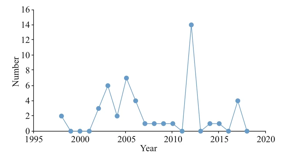
Fig.5 The year and number of occurrences of K. mikimotoi along the coast of China
3.1.8 Molecular characteristic sequence
Molecular biology combined with morphological characteristics has gradually become a more common research method. Based on ultrastructural and molecular biological data, Hansen et al. (2000)distinguished the two species ofK.mikimotoifromGymnodiniumaureolumand independently established a new genus forK.mikimotoi. Although the similarity of 18S rDNA sequences of diff erent algal species in the same genus is very high, their sequences show great diff erences, especially in the ITS1 and ITS2 regions, which are more suitable for molecular identif ication among species. Kimura et al. (2015) constructed an RNA library by molecular means. Through analysis, it was found that some genes were similar to some toxin genes in other algal species, which provided a basis for studying the toxicological mechanism ofK.mikimotoi. In addition, Zhang et al. (2009) also used loop-mediated isothermal amplif ication (LAMP) technology to identify the molecular sequence ofK.mikimotoifrom the South China Sea and achieved ideal results.According to this series of studies, information related to the identif ication of algal species at the molecular level has increasingly become an important tool and focus in algal research.
According to the relevant molecular sequences of algal species provided by the NCBI, the four regions of 18S rDNA, 28S rDNA, ITS1, and ITS2 and the sequences with obvious characteristics of the COX1 mitochondrial gene were selected (Table 2).
3.1.9 Sorting of basic biological information
After collection of the required information, the information was integrated in the form of tables(Table 3). Other unused information was stored for later use.
3.2 Biogeographic distribution information
3.2.1 Analysis of red tides ofK.mikimotoi
According to the bulletin on the state of China’s marine ecological environment, the bulletin on China’s marine disasters, and a literature query, the distribution information ofK.mikimotoired tides in China’s off shore areas is from south to north. From Zhejiang to Fujian waters are the mainly region ofK.mikimotoibloom (Liu et al., 2014; Ding and Zhang,2018; Lü et al., 2019). Figure 5 shows many red tide events ofK.mikimotoiin China’s off shore areas.Red tides occurred almost every year from 1998 to the present (Fig.5). Meanwhile, the reproduction ofK. mikimotoiwere distributed in the four sea areas of China, and the Donghai Sea has the most outbreaks(Fig.6).
3.2.2 Biogeographic distribution information sorting
At the initial stage of the study, the names of typical toxic and harmful algae that induce red tides,the locations of red tides (longitude and latitude,province, sea area), scale, duration, a description of red tide characteristics, and the temperature and salinity of the sea area were sorted (Table 4). The reason for performing information collection is to determine the cause of red tides. Second, we sorted relevant information about the place of occurrence,including the sea area of occurrence and the longitude and latitude of the site. In addition, it was also necessary to record relevant information for each red tide, such as scale, duration, water temperature, and salinity of the sea area. Finally, it was also necessary to describe each red tide event, which will provide an intuitive representation for users.
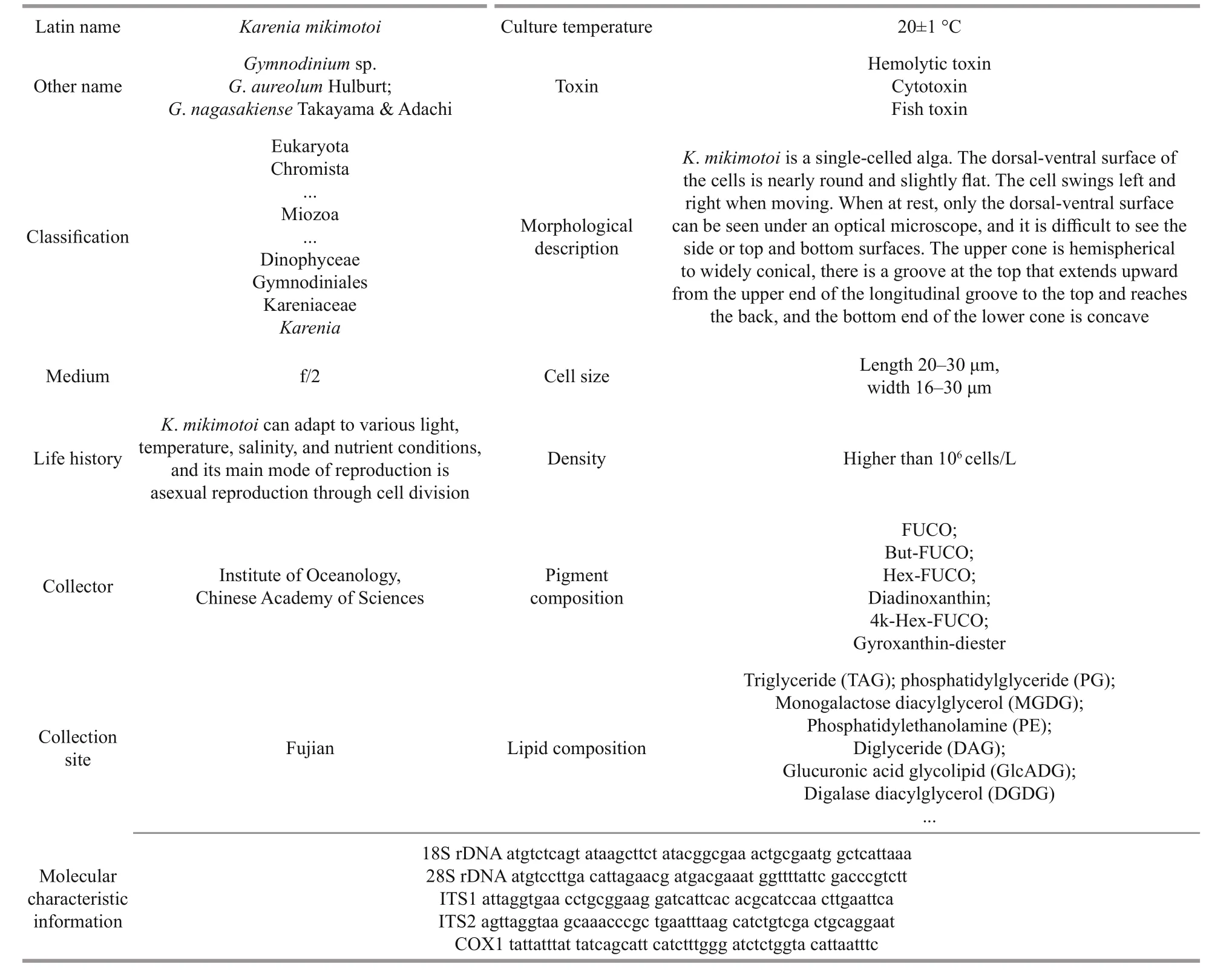
Table 3 Basic biological information of K. mikimotoi
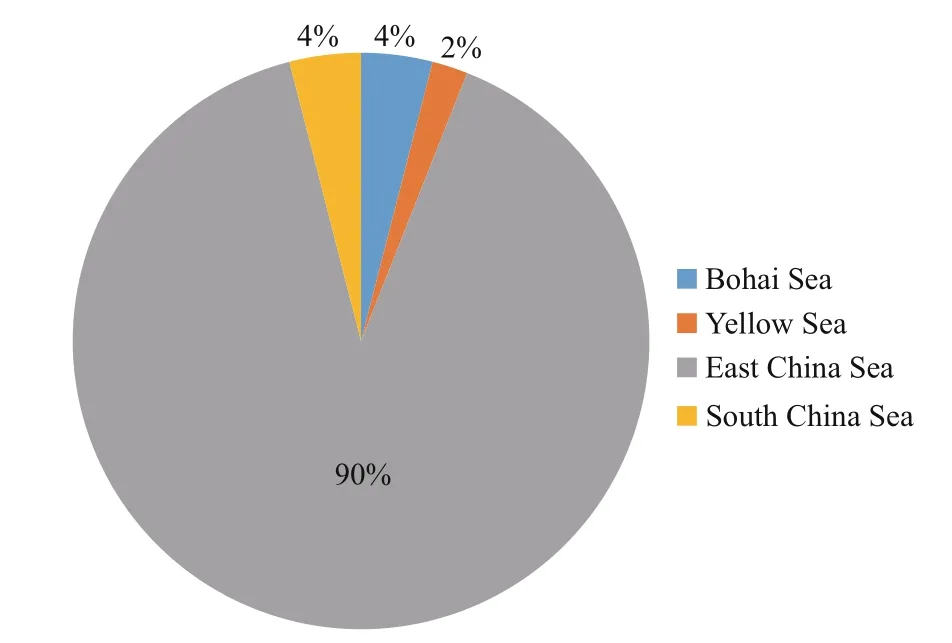
Fig.6 Karenia mikimotoi red tide distribution in various sea areas of China
3.3 Data classif ication
Due to the wide range of information obtained,the information should be eff ectively classif ied and summarized during data storage. This paper classif ies and arranges the collected information forK.mikimotoiinto two parts: basic biological and biogeographic distribution information (Fig.7).

Fig.7 Information classif ication of K. mikimotoi

Table 4 Karenia mikimotoi biogeographical distribution information
For basic biological information, 18 f ield names,such as Chinese name, Latin name, classif ication status, size, morphological description, suitable growth temperature, pigment composition, and lipid composition information, were obtained according to the collected information (Table 5). Many characters were required for classif ication status, morphological description, pigment composition, lipid composition,molecular feature sequence information, and references. These f ields were set to contain text for storage. Morphological images were stored in JPG format. Other information required fewer characters and was stored in character form.
For biogeographic distribution information,according to the collected information, the names of typical toxic and harmful algae inducing red tides,the occurrence locations of the red tides (longitude and latitude, province, and sea area), scale, duration,a description of red tide characteristics, and the temperature, salinity and references of the sea area were sorted, and the information characteristics were analyzed (Table 5). Except for the description of red tide characteristics and references, which required a certain amount of text, the data were stored as characters; other f ields could be represented by a small number of characters or text, so they were set to the character type.
4 CONCLUSION
This work collected and arranged the basic biological information ofK.mikimotoiand obtained the data through auxiliary means (i.e., experiments). The biogeographic distribution information was collected comprehensively as a goal of this study. The harmful algal distribution map obtained through the secondaryanalysis of data will also be used as a reference for designing a biogeographic distribution function on the information platform in the future. According to basic biological information and biogeographic distribution information, the relevant information on the typical toxic and harmful algaK.mikimotoiwas sorted, and information standardization was provided a template and method reference for the study of other toxic and harmful algae.
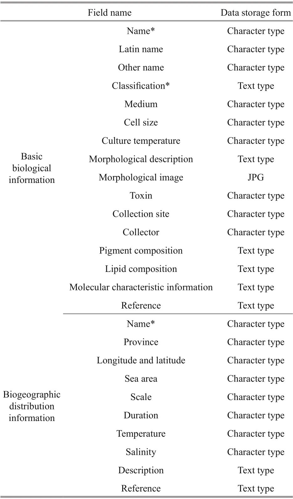
Table 5 Characteristic analysis of basic biological information
5 DATA AVAILABILITY STATEMENT
The datasets generated and/or analyzed during the current study are available from the corresponding author on reasonable request.
 Journal of Oceanology and Limnology2022年6期
- Journal of Oceanology and Limnology的其它文章
- Overview of harmful algal blooms (red tides) in Hong Kong during 1975–2021
- Biochemical composition of the brown tide causative species Aureococcus anophageff erens cultivated in diff erent nitrogen sources*
- Identif ication of paralytic shellf ish toxin-producing microalgae using machine learning and deep learning methods*
- Screening for lipophilic marine toxins and their potential producers in coastal waters of Weihai in autumn, 2020*
- First observation of domoic acid and its isomers in shellf ish samples from Shandong Province, China*
- Spatial variation of lipophilic marine algal toxins and its relationship with physicochemical parameters in spring in Laizhou Bay, China*
