基于两性离子配体构筑的Eu(Ⅲ)配位聚合物的晶体结构和对呋喃西林的检测
王凯民 李立凤 石明凤 叶艳青*, 王玉娜 郭津榕 唐怀军 马钰璐
(1云南民族大学化学与环境学院,昆明 650504)(2昆明医科大学药学院暨云南省天然药物药理重点实验室,昆明 650500)
0 Introduction
Coordination polymers(CPs),as a new multifunctional material,have attracted extensive attention of chemists not only because of their unique network structure but also rapid chromic behavior,including gas separation[1],chemical catalysis[2],magnetism[3],optical properties[4],biological application[5],and so on[6-10].Compared with the conventional neutral ligands,the zwitterionic types of organic ligands have obvious advantages owing to their good solubility,improved adsorption selectivity of guest molecules,and strong coordination ability.However,to date,only limited CPs based on zwitterionic organic ligands have been demonstrated[11-16].Therefore,the CPs based on this class of ligands still need further exploration.
Furacilin is a synthetic antibiotic that can treat livestock diseases.It was widely used in animal husbandry and aquaculture a few years ago.Later,it was found that the residues of furacilin and the metabolites in animal-derived foods can be transmitted to humans through the food chain.Long-term intake of furacilin will cause various diseases and have side effects such as carcinogenesis and teratogenesis on the human body.The United States,Australia,Canada,Japan,Singapore,and the European Union have expressly prohibited the use of such drugs in the food industry,and strictly implemented the detection of nitrofuran residues in aquatic products.Furacilin is listed as a banned drug in China[17-19].Therefore,the development of new materials for the detection of furacilin is critical in terms of environmental considerations.Although various sophisticated methods[20-22]such as GC-MS,LC-MS,and LC-UV have been developed for furacilin detection,these advanced analysis and testing techniques are limited by their inconvenient to carry,timeconsuming and tedious operation,high cost,and not real-time monitoring drawbacks[23-25].Compared with the traditional detection methods,fluorescence detection based on complexes has the advantages of high sensibility,easy synthesis,low cost,and simple operation[26].
In this work,a novel Eu(Ⅲ) CP,{[Eu(L)2(H2O)4]Cl3·2H2O}n(1)(L=1,1′-((2,3,5,6-tetramethyl-1,4-phenylene)bis(methylene))bis(pyridin-1-ium-4-carboxylate)),was synthesized under hydrothermal conditions by a zwitterionic organic ligand.X-ray diffraction analysis shows that 1 has a cationic skeleton on the main network and the Cl-ions used to balance the charges just exist in its pores.Its structure,photoluminescence,and detection properties were systematically studied.Firstly,the solidstate luminescence properties of 1 were investigated,realizing the zwitterionic ligand is an excellent antenna chromophore for sensitizing Eu3+ions.In addition,this water-stable 1 was utilized as a chemosensor to detect various common antibiotics to find that this one can exhibit high selectivity,sensitivity,and recyclability in the detection of furacilin molecules in aqueous phases.
1 Experimental
1.1 Materials and methods
All reagents used are commercially available analytical pure and can be used directly without purification.Powder X-ray diffraction(PXRD)was determined by a Bruker D8 Advance X-ray diffractometer(CuKα,λ=0.154 056 nm),in a 2θrange of 5°-50°,with a voltage of 35 kV and a current of 50 mA.Thermogravimetric analysis(TGA)was performed on a Perkin Elmer thermogravimetric analyzer from room temperature to 800℃ with a heating rate of 10℃·min-1under an N2atmosphere.Elemental analyses(C,H,and N)were performed by an Elementar Vario EL Ⅲ analyzer.The infrared spectrum was measured by the KBr tablet pressing method on FTS-400 Fourier transform spectrometer,with a wavelength range of 4 000-400 cm-1.The UV spectrum was scanned by a Beijing Puxi TU-1901 double beam UV-Vis spectrophotometer.Solid and liquid fluorescence spectra were measured at room temperature using a HITACHI F-4500 fluorescence spectrophotometer.
1.2 Synthesis of CP 1
1,1′-(1,4-Phenylenebis(methylene))bis(4-carboxypyridin-1-ium)chloride ligand((H2L)Cl2,0.02 mmol),EuCl3·6H2O solid(0.04 mmol),deionized water(2 mL),and methanol(1 mL)were added into a 10 mL beaker and mixed well.A NaOH aqueous solution(0.01 mol·L-1)was added drop by drop to adjust the pH value of the mixture to about 6.The mixture was stirred at room temperature for 30 min,then placed in a hydrothermal reactor and heated to 120℃for 2 d.The mixture was cooled naturally to room temperature to obtain light yellow block crystals.The yield was 32.1%(based on(H2L)Cl2). Elemental analysis Calcd. for C48H60Cl3EuN4O14(%):C,49.05;H,5.15;N,4.77.Found(%):C,48.76;H,5.20;N,4.74.IR(KBr,cm-1):3 423(br),1 608(s),1 564(s),1 446(w),1 384(s),1 237(m),1 120(m),1 076(m),1 042(m).
1.3 Crystal structure determination
Light colorless single crystals with a suitable quality of 1 were obtained under hydrothermal conditions.Crystallographic data for 1 were recorded on a Bruker APEX-Ⅱ diffractometer with graphite monochromated MoKαradiation(λ=0.071 073 nm)at 293(2)K usingωrotation scans.The crystal data was solved by direct methods using the SHELXS program and refined by full-matrix least-squares onF2using SHELXS in Olex2-1.3 software[27-29].Non-hydrogen atoms were refined anisotropically.All hydrogen atoms were generated theoretically and were placed at geometrically estimated positions.The guest solvent molecules were disordered and not located,so the SQUEEZE method was added to the CIF file to remove the contributions of disordered guest molecules.The number of lattice water molecules and the final chemical formula of 1 were determined by combining the single-crystal structures and TGA results.The crystallographic data and structure refinement details of 1 are summarized in Table 1.The main bond lengths and bond angles for 1 are shown in Table 2 and the hydrogen bond parameters for 1 are listed in Table 3.

Table 1 Crystal data and structure refinement of 1

Table 2 Selected bond lengths(nm)and bond angles(°)for 1

Table 3 Hydrogen bond parameters for 1

Continued Table 3
CCDC:2150460.
2 Results and discussion
2.1 Crystal structure of CP 1
Single-crystal X-ray diffraction measurement displays that CP 1 crystallizes in the monoclinic space groupP2/c.As shown in Fig.1c,the asymmetric unit consists of a crystallographically independent Eu(Ⅲ)ion,two fully deprotonated linker L,four coordinated water molecules,and three free Cl-ions.The distorted guest solvent molecules in 1 were subtracted by the SQUEEZE method in the PLATON program.Each Eu(Ⅲ)center is eight-coordinated to four O atoms(O1,O3,O1a,and O3a)of four L ligands and four O atoms(O2,O4,O2a,and O4a)of four water molecules,forming the primary building unit[EuO8](Fig.1a).The two carboxylic acid groups of the zwitterionic ligand(H2L)Cl2in 1 are completely deprotonated,forming two carboxylate groups which adopt the monodentate bridging mode(μ1-η1)to coordinate to Eu(Ⅲ) centers.Due to the presence of freely rotatable methylene(—CH2—),the two pyridine rings rotate to the same side of the benzene ring,making each L ligand presents a concave shape.It should be noted that the dihedral angles produced between the benzene ring and the two pyridine rings are slightly different,which are 82.87°and 86.77°respectively.Besides,the two pyridine rings are not in the same plane,but are distributed at a dihedral angle of 10.40°(Fig.1b).Adjacent[EuO8]building units are connected by two L ligands in opposite orientations,getting a hexagonal cage containing two L ligands and two Eu(Ⅲ)centers.The[EuO8]vertexsharing channels expand into a 1D bead chain extending along theb-axis with an effective pore size of 1.098 nm×1.579 nm(Fig.1d).Then,through theπ…πstacking interactions between neighboring benzene rings(centroid-centroid:0.373 82 nm),the mutually parallel 1D bead chains are unfolded into a 2D layer on the(10-1)plane(Fig.1e).Adjacent layers are further connected by the C—H…Cl hydrogen-bonding interactions between phenylmethyl groups and the free Clions to give a 3D supramolecular network(Fig.1f and 1g).It should be pointed out that the L molecules used to form 1 are pyridinium zwitterionic ligands with dispersed positive and negative charge centers.The positive charges of the L molecule are mainly concentrated on the N atoms of the pyridine rings,and the negative charges are mainly centralized on the O atoms of the carboxylate groups.Therefore,1 continues this characteristic of L,and its final structure also has separate charge centers.Interestingly,the negative charges of the carboxylate groups are neutralized by the Eu(Ⅲ)ions,so the main skeleton of 1 is positively charged.Its positive charges are distributed on the pyridine N atoms,as well as the Cl-ions used to balance the charges just exist in the pores of the 3D supramolecular structure in the free form.
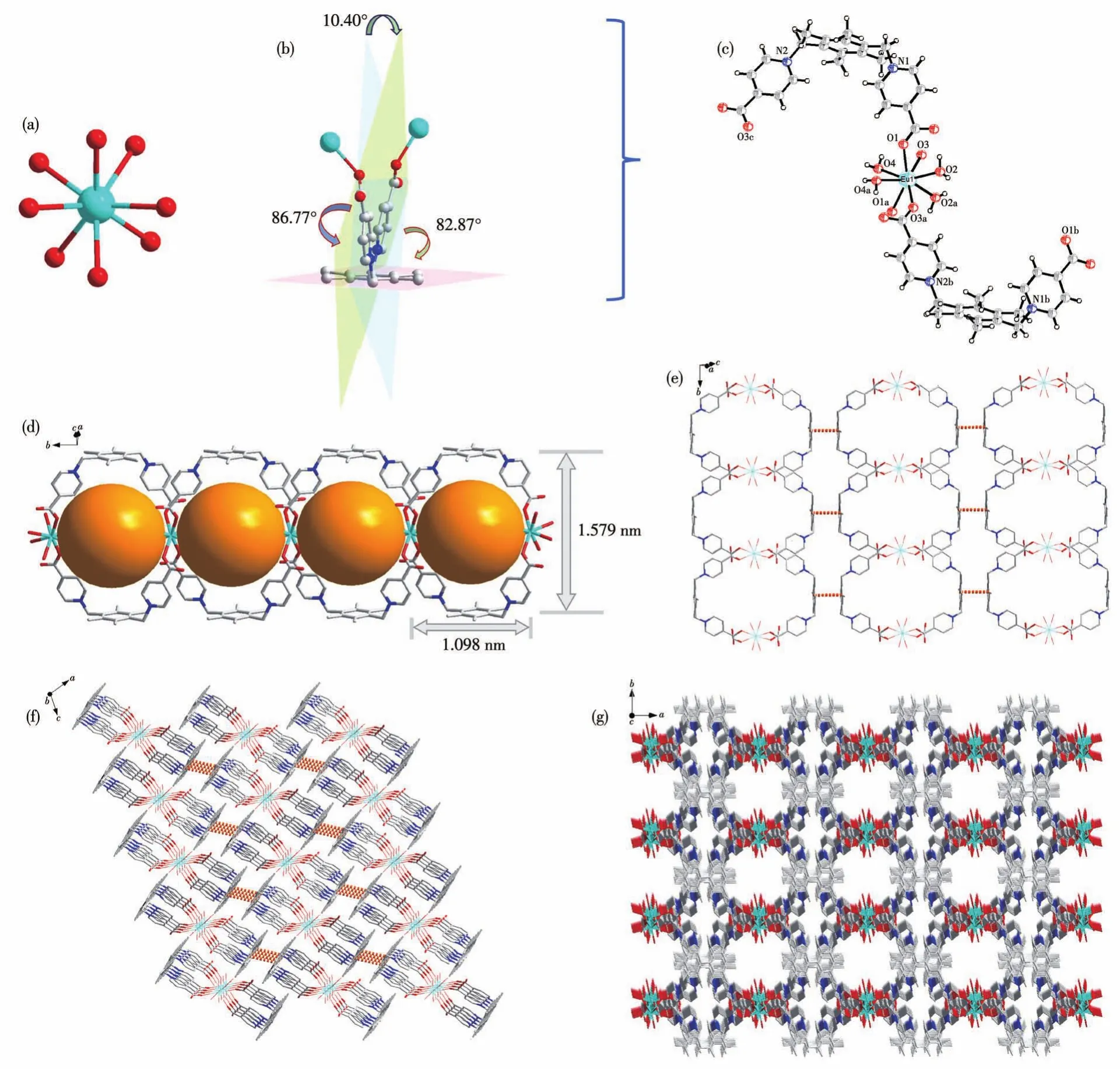
Fig.1 Structure of CP 1:(a)[EuO8]primary building unit of 1;(b)coordination mode of L;(c)coordination environment;(d)1D chain structure;(e)π…π stacking interactions;(f,g)overviewing of 3D packing plot
2.2 PXRD and TGA
TGA and PXRD were used for thermal and chemical stability analysis.The phase purity of CP 1 was confirmed by the PXRD pattern.The peaks of the synthesized sample matched well with the simulated results generated from single-crystal diffraction data,suggesting that the crystal of 1 was pure(Fig.2).To testify to the pH and water stability of 1,the corresponding measurements were performed at ambient temperature,in which the ground samples of 1 were treated with different pH-value solutions(pH=7 and 10)or purified water,and immersed for 3 d,respectively.The results display that the main diffraction peaks of 1 treated under different pH-value solutions or purified water coincide with those of the initially synthesized sample,indicating that 1 had excellent stability in water(Fig.2).The stability of 1 in water is a necessary condition for it to be used as an aqueous phase detection material.
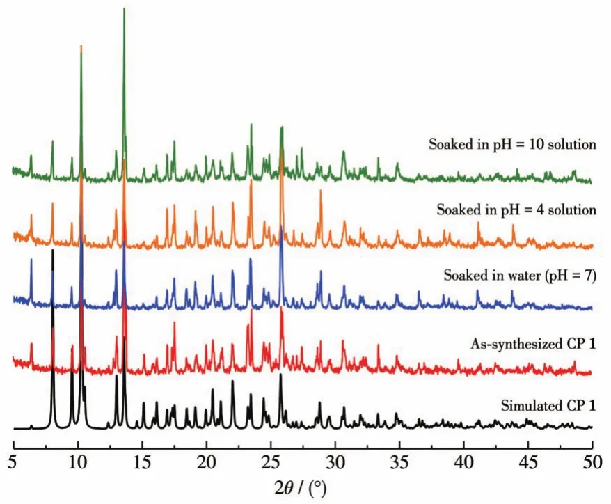
Fig.2 PXRD patterns of CP 1
The thermal stability of CP 1 was studied under an N2atmosphere from 20 to 800℃(Fig.3).The TG curve showed a weight loss of 3.25% from 80 to 170℃,which is attributed to the loss of the two uncoordinated free water.With further being heated,1 lost weight about 4.76% from about 180 to 310℃,which could be mainly ascribed to the loss of the three coordinated water.After 380℃,the crystal structure began to collapse due to the decomposition of organic ligands,indicating that 1 has good stability.
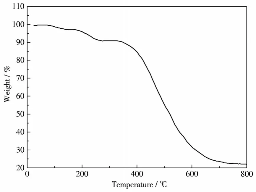
Fig.3 TG curve of CP 1
2.3 Photoluminescence properties
The solid luminescence properties of ligand(H2L)Cl2and CP 1 have been investigated at room temperature as shown in Fig.4.The free ligand(H2L)Cl2showed a wide emission peak at 472 nm(λex=416 nm),which is assigned to the typicalπ*-πtransition of ligands[15-16].The sample of 1 exhibited emission peaks of typical Eu(Ⅲ) ions at 590,616,650,and 695 nm(λex=397 nm),which belong to5D0→7F1,5D0→7F2,5D0→7F3,and5D0→7F4transitions of Eu3+ions,respectively.Notably,there was no emission band of the ligand observed in the emission spectra of the compounds,indicating the efficient energy transfer from the ligand to the Eu3+ion.These results indicate that ligand(H2L)Cl2is an excellent antenna chromophore for sensitizing Eu3+ions,which means that the energy transfers from the ligand to the Eu3+center effectively[16].
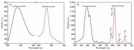
Fig.4 Solid-state luminescent spectra of free ligand(H2L)Cl2(a)and CP 1(b)
2.4 Selective detection of furacilin
Because of the excellent water stability and strong fluorescence of CP 1,the application in detecting eight daily common antibiotics in water systems was explored.First,the sensing ability of 1 toward different antibiotics was investigated by adding 100 μmol·L-1aqueous solutions of gentamicin(GEN),kanamycin(KAN),penicillin(PEN),cefixime(CFX),cefradine(CED),furacilin(FAC),roxithromycin(ROX),and azithromycin(AZM)to suspensions of 1(Fig.5a).Sensing experiments showed that among all antibiotics studied in this work,FAC showed a significant quenching effect on the luminescence intensity of 1,while other antibiotics showed a smaller quenching effect(Fig.5b).Hence,the preliminary sensing experiments revealed that 1 was capable of sensing FAC most selectively.

Fig.5 (a)Emission spectra of 1 adding 100 μmol·L-1aqueous solutions of different antibiotics;(b)Comparisons of the luminescence intensity of 1-suspension in different common antibiotics;(c)Luminescence spectra of 1-suspension titrated by adding different concentrations of FAC;(d)SV plot of 1 quenched by FAC in water
To further understand the ability of CP 1 to detect FAC,we continued to study the fluorescence titration obtained by adding different concentrations of FAC to 1-suspension(in water)to explore the relationship between FAC concentration and the fluorescence intensity.As shown in Fig.5c,when FAC was gradually added to 1-suspension,the fluorescence intensity decreased with the increase of FAC concentration.By measuring the fluorescence intensity at 616 nm,we found that the decrease in fluorescence intensity was particularly obvious.When FAC concentration reached 80 μmol·L-1,the fluorescence of 1-suspension was almost extinguished by 90%.Hence,we used the calculatedI0/Ias the ordinate and the FAC concentration(μmol·L-1)as abscissa,to draw the result,as shown in Fig.5d.We found that the fluorescence quenching efficiency of FAC in a concentration range from 5 to 44 μmol·L-1conformed to the Stern-Volmer(SV)linear fitting formula:I0/I=1+KSVcFAC,whereI0is the initial fluorescence intensity of 1-suspension without FAC,Iis the fluorescence intensity of 1-suspension after adding FAC,andcFACis the concentration of FAC added.After calculation,the quenching constantKSVwas 5.198×10⁴L·mol-1in the low FAC concentration range(R2=0.996 9),which corresponded to the highest known value of CP-based sensors previously reported[21-26](Table 4).At the same time,the limit of detection(LOD)was calculated by 3σ/K,whereσis the standard error andKis the slope obtained by linear fitting of concentration-dependent luminescence intensity at low concentration.The LOD value of 1 for FAC was found to be 0.13 μmol·L-1.This demonstrates the potential of 1 as a highly sensitive luminescent probe for FAC.

Table 4 Comparison of KSVand LOD values for CPs reported for aqueous phase sensing of furacilin
To further detect the application of CP 1 sensor in a biological system,we carried out the sensing experiment of 1 in 2-(4-(2-hydroxyethyl)piperazin-1-yl)ethanesulfonic acid(HEPES)simulated physiological environment(20 μmol·L-1HEPES aqueous buffer solution,pH=7)[30-32].We found that the fluorescence intensity in this simulated physiological environment was the same as that in pure aqueous solution;At the same time,we also selected FAC with a concentration of 50 μmol·L-1as the representative to investigate the fluorescence intensity of 1 in water and the simulated physiological environment.The intensity did not change significantly(Fig.6).This indicates that the fluorescence sensing of 1 to FAC may be of great use in physiology.

Fig.6 Emission spectra of 1 after immersing in HEPES aqueous buffer solution or pure water
In addition,considering that the reproducibility of the chemical sensor is an important parameter of practicability,the repeatability of CP 1 fluorescence detection was also studied.It is found that 1 can be reused at least five times after multiple dispersion,crystallization,and centrifugal washing in water.By monitoring the emission spectrum of 1-suspension in the presence of 100 μmol·L-1FAC,it was found that the quenching efficiency of each cycle was substantially unchanged(Fig.7).Additionally,it is important to investigate the luminescence quenching mechanism and selective detection ability of 1.The PXRD pattern of 1 recovered after five detection cycles showed almost the same as the initial sample,indicating the high stability of the material(Fig.8a).Then,by testing the FT-IR spectrum after five cycles,we found that the main spectrum(3 424(br),1 609(s),1 564(s),1 446(w),1 385(s),1 237(m),1 121(m),1 077(m),1 043(m))of 1 was also consistent with that of the original synthesized sample.In addition,an ICP experiment was also performed to monitor the release of Eu(Ⅲ)ions from 1 after five cycles of sensing experiments.The mass concentration of Eu (Ⅲ) ion in the filtrate was 0.17 μg·mL-1,which was close to that of the blank sample(0.14 μg·mL-1).This ICP test result showed that there was no release of Cd(Ⅱ)ions from the samples of 1 in the sensing experiments.Combining the XRD,FT-IR,and ICP results after five cycles,we find that the luminescence quenching is not induced by the framework collapse and 1 can be reused as a probe.
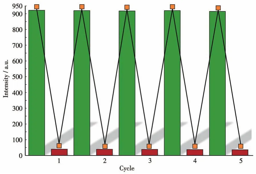
Fig.7 Quenching cycle test of 1 after adding 100 μmol·L-1FAC in aquatic system

Fig.8 (a)PXRD patterns of 1 after detection of FAC for five cycles;(b)Spectral overlap between the UV-Vis spectra of the antibiotics in aqueous solution and the excitation spectrum of 1
Then,the inner filter effects(IFE)can be used to explain the quenching effects.As shown in Fig.8b,there was a significant spectral overlap between the absorption band of FAC and the excitation spectrum of 1;however,there was no effective overlap for other antibiotics,indicating that there exists the competitive absorption of the excitation energy between 1 and the antibiotics analytes.The above results show that 1 can be used as a fluorescent probe for FAC and has high selectivity,high sensitivity,and reusability.
3 Conclusions
A novel Eu(Ⅲ)coordination polymer(1)was synthesized under hydrothermal conditions by a zwitterionic organic ligand.X-ray diffraction analysis shows 1 has a cationic skeleton on the main network.Firstly,the solid-state luminescence properties of 1 were investigated,realizing the zwitterionic ligand is an excellent antenna chromophore for sensitizing Eu3+ions.In addition,this water-stable 1 was utilized as a chemosensor to detect various common antibiotics to find that this complex can exhibit high selectivity,sensitivity,and recyclability in the detection of furacilin molecules in aqueous phases.Our laboratory is also carrying out follow-up research work.

