The latest trends in Asian rhinoplasty
Haibo Xiang,Wanwen Dang,Yang An ,Yonghuan Zhen ,Dong Li
Department of Plastic Surgery,Peking University Third Hospital,Beijing 100191,China
Keywords:Asian rhinoplasty Synthetic/autologous graft 3D technique
ABSTRACT Rhinoplasty in Asia is becoming increasingly widespread.When taking into consideration the anatomical differences between the features of Caucasian and Asian patients,it is unsurprising that knowledge gained from research on Caucasian rhinoplasty is not always applicable to Asian rhinoplasty.Furthermore,reviews aimed at describing the recent developments in Asian rhinoplasty are rare.This review aims to provide a comprehensive summary of the latest trends in rhinoplasty for Asians by classifying it into three parts:methods,materials,and three-dimensional techniques,hopefully providing surgeons with references to aid in conducting rhinoplasty on Asians.
1.Introduction
Rhinoplasty is now performed extensively worldwide,with the total volume of surgery inferior only to that of blepharoplasty.1Compared with the classic Caucasian nose,the Asian nose is typically characterized by a low,wide,flat nasal dorsum,an under-projected nasal tip,a retracted columella,a wide nasal base,and a flaring alar with a large amount of subcutaneous fatty tissues.1-4Such anatomical differences determine the needs of Asian patients,and the rhinoplasty surgery strategy for Asians is quite different from that of Caucasians,which is of particular interest to be studied.Therefore,this review aims to uncover how Asian rhinoplasty has developed and how it will progress in the future by looking at its latest developments.First,we discuss the methods,which are mainly composed of autologous material management and nasal tip and base management.The management of the costal cartilage,which is the most prevalent cartilage used in Asian rhinoplasty,along with the management of the septal cartilage and perpendicular plate of the ethmoid (PPE),which is aimed at solving the problem of inadequate septal cartilage in Asians,are discussed in detail.Six alloplasts are discussed in the “Materials” section.We believe that the alloplasts discussed in this section can represent the current status of Asian synthetic grafts because they comprise traditional and innovative materials and nonsurgical and surgical grafts.Finally,we briefly introduce the three-dimensional (3D) technique because it is making increasing contributions to preoperative evaluation and has a promising future in creating implants.
2.Methods
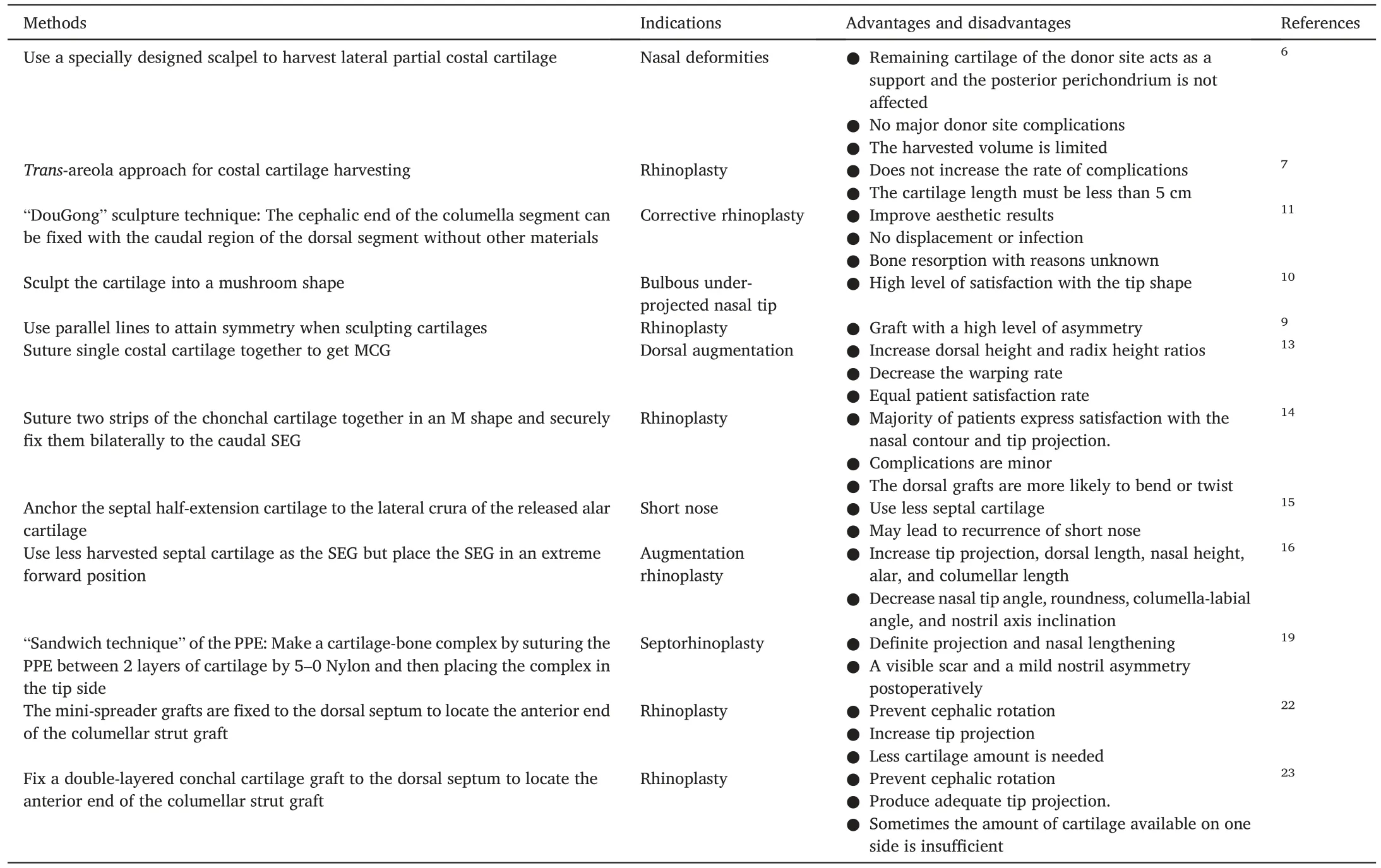
Usually,rhinoplasty in Asians includes several cardinal steps:cartilage harvesting(septal,auricular,or costal cartilage),nasal base stabilization,management of the nasal tip lobule,and dorsal augmentation.3Surgeons typically have their own specific method that they use in order to go through these steps and attain the required result.From this,many new methods have been developed,and these are making great improvements in these cardinal steps.These improvements have led to better rhinoplasty results.This review emphasizes the introduction of autologous material management and nasal base and tip management.The methods discussed are summarized in Table 1.

Table 1.Recently developed methods in the management of autologous material and the nasal base and tip in Asia.
2.1.Autologous graft management
Autologous materials,especially costal cartilage,have recently become the mainstay of Asian rhinoplasty.In this section,we discuss the management of four types of autologous grafts:costal cartilage,auricular cartilage,septal cartilage,and the PPE.
Costal cartilage management has gained considerable attention because this cartilage can provide adequate high-quality cartilage grafts.Moreover,it becomes extremely useful when the septal cartilage has been harvested during the first rhinoplasty,and the conchal cartilage is unavailable to support the anticipated structure.5From our experience,costal cartilage has become the most commonly used autologous tissue in Asian rhinoplasty because it has an adequate supply of tissue and is biologically stable.
While there are clear advantages to using costal cartilage,it still needs to be harvested before use.When compared to the septal and auricular cartilages,harvesting costal cartilage is relatively difficult and requires more attention.Recently,Jiang et al.6used a specially designed scalpel to harvest the lateral part of the costal cartilage.This suggests that partial lateral harvesting of the costal cartilage might be considered when aiming to reduce harm and complications.Costal cartilage harvesting has led to the development of a new technique that is used in women.A trans-areola approach for costal cartilage harvesting for females was applied in Asia to conceal the scar caused by the traditional approach.7The one limitation of this method is that the costal cartilage harvested must be less than 5 cm,which is the average length of the fifth costal cartilage.
After harvesting the costal cartilage,meticulous carving is the next step.In general,we use the central part of the cartilage for carving.To match the contour of the nasal dorsum,a slight concavity on the undersurface is often needed.8Avoiding wrapping and keeping the asymmetry of the costal cartilage are at the core of this step.A method to attain symmetry was introduced recently.9As to the special sculpture technique,mushroom-shaped costal cartilage has been shown to correct the bulbous under the projected nasal tip.10Another special sculpture technique is the “DouGong” technique.11This technique can fix the joint between the nasal dorsum without the columellar strut using a screw and K-wire.
What we have discussed above concerns one-block costal cartilage grafts.However,sometimes,an ideal tip shape cannot be obtained with only a tip onlay graft since it frequently results in an over-corrected tip that is much rounder than desired and is the exact opposite of an underprojected tip.12Multilayered costal cartilage might serve as a solution to this problem.By suturing it from convex to concave on one end and then concave to convex on the other end,the dorsal height and radix height ratios increase,and the warping rate decreases.13
The auricular cartilage is easy to obtain and has good flexibility and elasticity.It can be molded into a suitable shape for rhinoplasty.However,its high postoperative resorption rate and insufficient support,along with its deformation with scar contracture,have always been a concern for auricular cartilage.Therefore,it is usually used as supplementary material for inadequate septal cartilage volumes.Recently,some Chinese scholars have transformed the auricular cartilage into an M-shaped graft and transplanted it to the end of the nasal septum.Such a graft can act as a frame of the nose tip and can solely control the different base shape indices,such as projection and rotation,alar-to-columellar proportion,and nasolabial angle position and shape.14In addition,the shape and texture of the nose tip using such a graft are closer to the natural state.
Septal cartilage is the most common choice when performing rhinoplasty.However,septal cartilage is often inadequate in Asians.Therefore,taking advantage of the septal cartilage economically needs to be discussed.Lee et al.15made improvements in the traditional method of anchoring the septal half-extension cartilage to the dome of the alar cartilage.Another innovation was made in the traditional(Toriumi-style)tongue-and-groove method to take advantage of the additional support derived from the direct abutment and loading transmission through the caudal septum,16thus reducing the amount of cartilage used.
As mentioned previously,all traditional autologous grafts have disadvantages.The septal cartilage in Asians lacks volume.The conchal cartilage is weak and cannot support this structure.Harvesting the costal cartilage risks inducing donor site complications,and calcified costal cartilage is inevitable.In such a situation,the PPE,with its suitable hardness between that of the cartilage and bone and easily accessible surgical area,comes to mind.We have also confirmed,using morphological measurement on a 3D computed tomography (CT) reconstruction,17that the amount of graft provided by the PPE in people of Chinese Han nationality is sufficient to meet the requirements of clinical surgery.This supports the practicability of using the PPE in rhinoplasty.

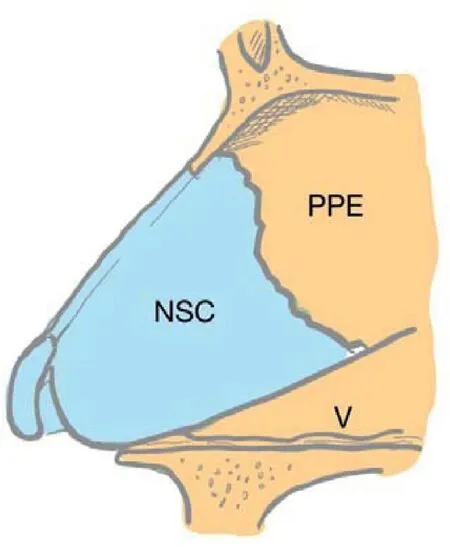
Fig.1.Location of the PPE.PPE,perpendicular plate of the ethmoid;NSC,nasal septal cartilage;V,vomer.
The PPE is a thin,flattened lamina.It is located in the posterosuperior part of the vomer and septal cartilage(Fig.1),adjacent to the sphenoid sinus and skull base.18It is usually used in combination with septal cartilage.The harvested PPE is trimmed into a slatted nasal septum reinforcement graft by a rongeur and fixed to the dorsal and caudal segments of the L-shaped nasal septal cartilage stent.This is done to enhance the mechanical support of the L-shaped stent and the tip of the nose,thus extending the support structure and elongating the nasal length.Another reported technique of using the PPE is the “sandwich technique,”which can provide optimal projection and lengthening.19
The most valuable utilization of the PPE is for short nose deformities.Short nose deformities involve a shortened nasal length,accompanied by excessive head rotation of the nasal tip and excessive exposure of the nostrils.Patients with short nose deformities lack sufficient septal cartilage volume and require strong,stable nasal tip support.Therefore,the PPE is suitable for these patients.We have also proved the effectiveness and merits of its use in correcting a short nose deformity in Chinese patients.20Despite the advantages discussed above,the shortcomings of the PPE are also notable.Incorrect harvesting of the PPE may lead to complications such as cerebrospinal fluid leakage,damage to the olfactory area,and bleeding.Meanwhile,it is difficult for beginners to harvest the PPE,which restricts its popularization.Harvesting the PPE under the guidance of a nasal endoscope can make the cut more precise and reduce the occurrence of complications,and harvesting the inferior portion of the PPE can avoid serious complications while providing a graft of the proper size,thickness,and quality.21
2.2.Nasal base and tip management
Nasal base and tip managements are the challenging aspects of rhinoplasty.They play a significant role in determining the nasal tip projection,length,and height.
The open approach is usually preferred in primary Asian rhinoplasty because it can meet the demand to visualize anatomical details when performing surgery.Traditionally,we stabilize the nasal base using a conventional columellar strut.The columellar strut is sutured in a pocket between the medial crura.Unfortunately,the columellar strut may result in cephalic rotation of the dome,which should be avoided in Asian patients with short noses and visible nostrils.In a recent study,a minispreader graft was used to connect the dorsal septum to the anterior end of the columellar strut,22which is called a rein-shaped columellar strut.This can prevent cephalic rotation caused by the traditional columellar strut.Such a method to prevent cephalic rotation was also reported by Mo et al.23

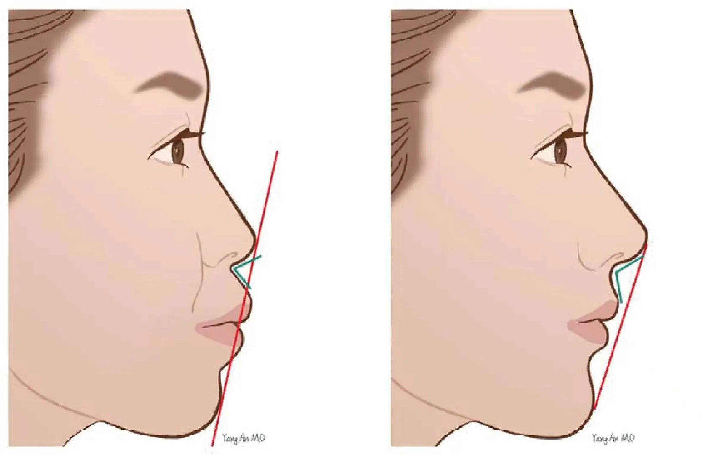
Fig.2.Features of patients with midfacial depression (left) compared to normal people (right):(1) decreased nasolabial angle (green line);(2) protruding mouth visually (red line);(3) flat nose visually;(4) deeper nasolabial fold.
Nasal base depression is a common facial feature among Asian patients.Compared with the Caucasian race,the nasolabial angle of Asians is about 5°smaller,which is related to the lack of strong bony support for the medial crura of the alar cartilage and the soft tissues underneath it.24The depression can make the nose look flat visually,thus resulting in an unsatisfactory nose appearance (Fig.2).Therefore,correcting the depression is an important step to ensure the outcome of rhinoplasty.In addition,recent advances have proven that the correction of the midface depression improves gingival exposure.25The most commonly used methods to treat the depression are surgical filling and injection filling.The intranasal approach is the first choice for surgical filling in Asia,while the upper gingivobuccal sulcus incision has been widely accepted because it helps to provide an open surgical field and contributes to the implantations of a variety of large grafts such as silicone,preshaped porous polyethylene,and autologous cartilage.26,27Autologous diced cartilage and expanded polytetrafluoroethylene (ePTFE) are used most commonly for surgical filling,while silicone and Medpor are less frequently used because of their high rate of complications.Injection filling can be adopted for patients who require a smaller incision or a shorter recovery period.Hyaluronic acid (HA) and autologous fat have been widely used as fillers for the correction of midfacial depression.28However,autologous fat is easily absorbed and displaced due to perioral movement.Moreover,owing to the operation being performed under blind vision,several significant risks should be noted:(1) the filler may enter the ophthalmic artery and embolize the middle retinal artery,thus causing temporary or permanent blindness;(2) it may embolize blood vessels and cause skin necrosis,which is consistent with the anatomy of the blood vessels in the area;(3)it may cause a clot to dislodge and cause embolization of a distant organ.
A septal extension graft(SEG)is often the first choice and the basis for Asian rhinoplasty when we need to manage tip support,projection,rotation,and nasal length.The central part of the quadrangular cartilage is usually harvested after dissecting the septum in the subperichondrial and subperiosteal facets.The SEG unilaterally overlaps the caudal septum by at least 5 mm,29and we should ensure that it is positioned at the midline.Subsequently,the lower lateral cartilage is repositioned by suturing it to the SEG.It can be placed on the anterior nasal spine,integrated with an extended columellar strut,overlapped with the caudal septum (unilaterally or bilaterally),or secured in an end-to-end fashion to the caudal septum.29The first choice for an SEG is septal cartilage,but in Asians,it is often inadequate,and hence,the costal cartilage may serve as an alternative.In Section 2.1,we introduce two recently reported methods to use the septal cartilage economically.An SEG can elongate the nasal length,enhance tip support,and control tip projection and rotation.When the superior margin of the SEG is longer,it results in counterrotation of the nasal tip.
Besides an SEG,some cartilages are also used to obtain an ideal postoperative result,which means a natural nasal tip with appropriate softness and mobility.There are often two options for achieving this goal:conchal cartilage and costal cartilage.There are three common techniques for using conchal cartilage in shaping nasal tips,30and two of them are suitable for patients with moderate or severe short noses.Conchal cartilage is recommended because it is easy to harvest,its elasticity is close to that of the alar cartilage,and it leads to an almost invisible scar.Costal cartilage is another common choice for shaping the nasal tips in Asians.Compared with auricular cartilage,it has higher stiffness and can provide better support for the nasal tip while partly sacrificing the natural appearance.No matter which option is chosen,what we are doing is a trade-off between stability and mobility.Auricular cartilage can provide better mobility and stiffness,simulating the alar cartilage,but less stability.In contrast,costal cartilages can provide higher stability because of their higher stiffness while risking nasal tip rigidity.
3.Materials
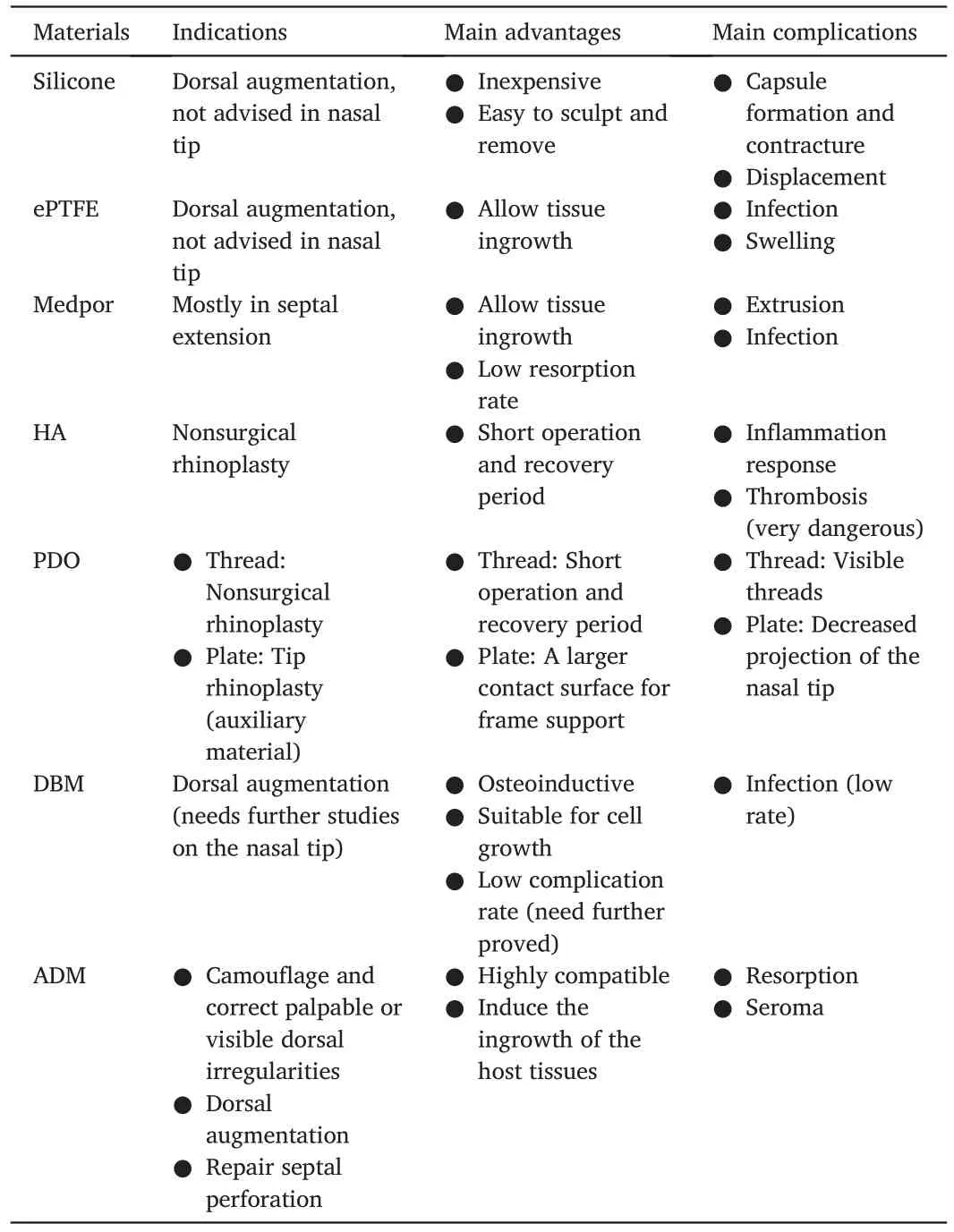
The graft materials used in Asian rhinoplasty are classified into three types:autogenous grafts,homologous grafts,and synthetic grafts (alloplasts).1,29Autogenous grafts have advantages over other grafts in the following aspects:high tissue availability,reliable long-term survivalrate,and most importantly,the lowest infection rate compared to that with other materials.29,31Donor site morbidity is also rare;they also have some limitations,as follows:risk of pneumothorax when harvesting rib cartilage,and a small amount of septal cartilage in Asians that cannot support revision surgery.As for homologous grafts,cadaveric costal cartilage is used due to its extensive availability.32Another extraordinary choice in this field is the acellular human dermis,which has the advantage of being relatively biocompatible.Synthetic grafts usually comprise silicone,ePTFE,porous high-density polyethylene,polydioxanone(PDO)plate,and HA.33It remains controversial which type of material is best when performing rhinoplasty.Recent advances in Asian rhinoplasty materials emphasize homologous and synthetic grafts such as PDO,HA,and acellular human dermis.From our perspective,this is partly because of the short history of these materials compared with materials with a long history,such as cartilage and silicone.Having discussed autologous material in Section 2.1,in this section,we will discuss some homologous and synthetic grafts used in Asian rhinoplasty.The indications,main advantages,and complications are summarized in Table 2.

Table 2.Indications,main advantages,and complications of homologous and synthetic grafts used in Asian rhinoplasty.
3.1.Silicone
Silicone is a pliable and elastic solid that has gained wide popularity in Asian rhinoplasty for many years,mainly because of its relatively low price,ease of sculpture,and ease of removal due to the fibrous capsule formed around the implant.Usually,two types of prefabricated silicone are used:L-shaped and I-shaped implants.The use of L-shaped implants carries a higher risk of extrusion,and I-shaped implants are therefore becoming increasingly preferred recently.34
Although silicone is widely used in Asia,it has been considered an inferior choice in recent years.It is proved that the lack of pores in silicone prevents tissue ingrowth,which makes micromotion inevitable when using silicone,thus leading to chronic inflammation around the tissue and thick fibrous capsule formation.35Furthermore,silicone may exert pressure on the nasal tip,putting it at risk of pressure necrosis.2Compared to other alloplasts,it has a relatively high rate of complications,especially asymmetric contracture of the capsule.A recent trend is to use an I-shaped implant at the dorsum in combination with autologous materials for tip augmentation to prevent such complications.
3.2.ePTFE (Gore-Tex)
ePTFE is the second-most commonly used alloplast in Asia.It is composed of carbon and fluorine molecules with pores and allows soft tissue ingrowth.36It is relatively biocompatible but prone to infection partly because of the ingrowth of fibroblasts,capillaries,and collagen,though animal tests imply that it has low rates of infection.37
In Asian rhinoplasty,the question of whether to use ePTFE or silicone has long been debated.To summarize,when considering the choice of these two materials,we should evaluate them from the following perspectives:(1)price:ePTFE is considerably more expensive than silicone;(2)Aesthetic outcome:in general,silicone has better long-term aesthetic satisfaction because it is easily sculpted into an ideal shape,and its hardness can better act as support in augmentation rhinoplasty compared to ePTFE;(3) complications:ePTFE implants have a lower chance of migrating and forming capsules but are more likely to result in infection;(4)whether to use conchal cartilage as a shield:it has been suggested that silicone implants should be used in combination with a cartilage shield in the nasal tip,while implanting ePTFE shows a 0% extrusion rate,indicating that the shield is not necessary for ePTFE implantation.38
3.3.Porous high-density polyethylene (Medpor)
Medpor is a nonantigenic,nontoxic,and nondegradable material that has been widely used clinically since the 1980s in Asia.It is manufactured by sintering small particles of polyethylene,forming an interconnected framework of pores ranging in size from 125 μm to 250 μm,39which enables it to allow the ingrowth of vascularized tissue.To some degree,it is a versatile material because it can exhibit better performance than silicone and ePTFE on some occasions.
Medpor is the most extensively used SEG in Asian rhinoplasty.40It is also reported to be used in the dorsum,columella,alar,tip lobule,and paranasal regions.41,42The most common complications associated with Medpor are extrusion and infection.Almost every report about Medpor in Asia stated these two complications to various degrees.40,43,44Recently,the first long-term complication study related to Medpor reported that the devastating destruction of nasal support lines may be related to Medpor implantation.45However,to date,no decisive evidence has proven the higher extrusion and infection rates of Medpor.Further long-term studies should be conducted to address this question.
3.4.Hyaluronic acid
HA is widely used in Asian rhinoplasty mainly because of its stable chemical properties,short operation time,quick recovery period for patients,reliable biocompatibility,malleability and durability,and low immunogenicity.
HA is typically used in injection procedures.Recent surveys have reached a consensus on the standard injection procedure,which comprises the following steps:(1) nasolabial angle:stab a sharp needle through the junction of the philtrum and columella and inject HA into the anterior nasal spine to enlarge the nasolabial angle;(2)nasal columella:injected through the midpoint of the nasal columella to elongate the columella and elevate the nasal tip.(3) nasal tip:injected between the middle crura of the nasal alar cartilage to enhance the contour and elevate the tip.(4) nasal dorsum:injected into the periosteum and perichondrium layer of the dorsum first,followed by the subcutaneous layer.(5)injected into the inner side of the brow,sliding subcutaneously to the bilateral junction region of the nasal root with the injection of HA during withdrawal.46,47
As an alloplast,the adverse outcomes of HA have always been a concern in Asian patients.Briefly,they comprise vascular thrombosis,inflammatory response (infectious or noninfectious),nodules and granuloma,and ache after injection.To avoid vascular thrombosis,surgeons must first have a comprehensive understanding of the vascular anatomy and its variants.Pulling back on the syringe before injecting also plays a significant role in avoiding vascular thrombosis.The inflammatory response,nodules,and granulomas are often caused by an immune response.Promoting the purity of HA is significant for preventing the immune response.A recent study to evaluate the safety of HA showed that the most frequently occurring complications were swelling,erythema,and bruising,and no severe adverse outcomes occurred,which supports the safety and efficacy of HA.48
3.5.Polydioxanone
PDO is a colorless crystalline resorbable polymer characterized by a high resorption rate because it can be degraded by hydrolysis.The biocompatibility and degradability of PDO have been tested in vivo and in vitro.49It is not recommended as mainstream in Asian rhinoplasty because we believe that it is more prone to complications.It is mainly used in two ways:PDO threads and PDO plates.
A recently widely accepted nonsurgical technique in Asian rhinoplasty involves PDO threads and fillers(gel-like substances).The surgical procedure consists of two steps:first,inject the filler into the supraperiosteal layer of the dorsal area,sometimes also into the nose tip and columella;second,insert a guided cannula into the subcutaneous plane,covering from the tip to the radix of the dorsum,and then place the PDO suture inside the cannula.50The same procedure is performed in the caudal part of the nose or the nasolabial angle area.50,51PDO threads act as a scaffold and provide physical augmentation,thus retaining the shape for a long period.In addition,it induces the production of type I and III collagen.
The PDO plate is an adjuvant material used in Asian patients.It can be used in Asian rhinoplasty in five ways,49but its function is to stabilize the nasal cartilage framework structure irrespective of the method used.
3.6.Demineralized bone matrix
Demineralized bone matrix (DBM) is obtained by degreasing,dehydrating,decalcifying,freeze-drying,and performing other treatments of allogeneic bones.The osteoinductive activity of DBM has been extensively studied in experiments and has been confirmed by a large number of clinical practices.52Compared with traditional alloplasts,DBM has three main merits:(1)It contains bone morphogenic proteins(BMPs)and a variety of growth factors,which makes it osteoinductive and osteoconductive.52After implantation in the body,it can induce the formation of new bone in the host,and it will eventually act the same as autologous bone.(2) DBM is a 3D porous sponge-like structure with high porosity and small pore diameter suitable for cell and blood vessel invasion,which provide favorable conditions for the formation of cartilage cells and matrix.53(3) According to recent domestic reports on DBM,it has a higher satisfaction rate compared with autologous cartilage,and the overall incidence of postoperative complications is low;however,no rejection has been reported.
Although DBM has not been widely reported in foreign rhinoplasty,Chinese reports on DBM application have been increasing recently.We believe that DBM is a safe alloplast with a certain degree of elasticity and toughness,and it has promising future applications in dorsal augmentation.However,we have reservations about its application in tip rhinoplasty since it remains unclear whether decalcified bone can provide sufficient support.In addition,long-term follow-up studies of DBM are lacking;thus,its long-term results,especially its resorption rate,need to be further verified in the future.
3.7.Acellular dermal matrix
The acellular dermal matrix(ADM),also an acellular human dermis,is processed from donated human skin using specialized techniques.This technique involves a proprietary freeze-drying process to remove dermal cells and the epidermis,which are immunogenic components of the skin,thus preventing immunologic reactions.As it is derived from human skin,it is highly compatible and not prone to incite inflammation.In addition,it provides a matrix consisting of a large amount of collagen and elastin fibers,which can induce the ingrowth of host tissues,including fibroblast infiltration and neovascularization,and contributes to maintaining a stable structure.Although there is no evidence that ADM can induce an immune response or inflammatory cell infiltration,a case of infection caused by ADM use has been reported,and the underlying reason remains unknown.54
The indications for using ADM include a deviated septum,nasal valve obstruction,external nasal deformity,tip rhinoplasty,and dorsal augmentation.55It is widely used for dorsal augmentation and has yielded satisfactory outcomes.Dorsal augmentation is usually performed through the open approach,which comprises three steps:(1)placing the ADM above the nasal dorsum to check its suitability for length and thickness;(2) inserting the ADM with the anticipated length and thickness into the nasal dorsum in a suitable position;(3) evaluating the height and shape of the nasal dorsum after covering the skin flap.If the height and shape are satisfactory,the skin is closed.56
Recently,novel uses of ADM have emerged.ADM is encouraged to be used in combination with other graft materials such as cartilage or silicone,which is the latest trend of innovations in ADM.Such a combination is safe and effective and can ensure the symmetry of rhinoplasty.In addition,when silicone is used in combination with ADM,the results show decreased rates of hypertrophic scarring and capsular contracture compared to those when using silicone alone.This may be explained by the decreased myofibroblast activity.57Cross-linked ADM is another recently reported type of ADM,which is irradiated with an electron beam.This irradiation can damage the DNA of microorganisms and induce adequate cross-linking of the collagen matrix.Compared with traditional ADM,this novel cross-linked ADM shows delayed degradation and can maintain its 3D matrix function during cell infiltration and neovascularization.Moreover,it delays the remodeling process via increased interfibrillar and intrafibrillar bonds.58
4.3D technique
Over the past few years,the use of the 3D technique,characterized by its high reproducibility and intuitiveness in rhinoplasty,has increased rapidly and indicates a promising trend.Recently,our team used the 3D imaging technique to prove that the cleft-side ala was anatomically ameliorated after muscle repositioning combined with diced costal cartilage grafting.59The 3D technique mainly includes 3D printing and 3D imaging techniques,and both of them have their own merits in assisting rhinoplasty performance.
4.1.3D imaging technique
3D imaging techniques can be classified into computer-aided 3D reconstruction and direct 3D imaging.The former refers to reconstructing 3D imaging using multi-directional two-dimensional images obtained by X-ray,ultrasound,CT,and magnetic resonance(MR)through specific algorithms;the latter refers to non-contact one-time stereo imaging technology.
Tools used in reconstructing 3D imaging,such as X-ray,ultrasound,CT,and MR,have different peculiarities.X-rays are relatively simple to perform,but their application is limited because of radioactivity,the cumbersome manual fixation process,and the inability to reconstruct soft tissue structures.Ultrasound is cheap and can extract the image of tissue with specific echoes,which is widely used in clinics.However,it squeezes the soft tissue and interferes with accurate reconstruction.CT,which is currently the best and most commonly used technique in relevant research,can not only obtain a comprehensive view from any angle by rotation but also extract the local imaging of specific tissue structures.Its main limitation is the loss of imaging information caused by sectional imaging and radioactivity.The resolution of 3D MR is significantly better than that of CT,and there is no radioactivity.However,it is not suitable for patients with metal implants in the oral and maxillofacial regions.It also has the disadvantages of extreme noise,a confined environment,and a slow imaging speed.
Currently,3D imaging techniques are mainly used for preoperative evaluation,simulation,and postoperative follow-up of rhinoplasty.For surgeons,the 3D imaging technique allows them to obtain relevant anatomical information intuitively,helps them design more suitable surgical plans for each individual,and increases the efficiency of communication between patients and surgeons.For patients,it provides a comprehensive understanding of the surgical plan and related risks,thereby establishing reasonable surgical expectations.A recent Chinese study showed that the 3D imaging technique did not affect the outcome of the surgery but indeed increased patient satisfaction with doctorpatient communication.60In addition,3D imaging techniques can help determine the anatomical features of people in a certain area.A study conducted in Korea using 3D imaging revealed that the nose on the face and nasal aperture of the skull were significantly correlated in terms of overall face and skull morphology in Korean subjects.61
4.2.3D printing technique
The 3D printing technique can create personalized organ models and implantation materials through computer-aided design,which can be applied in both preoperative simulation and implantation during operation.The three most frequently used 3D printing methods are inkjetbased,extrusion-based,and laser-based.The two former methods have an advantage in terms of printing speed and price,whereas the advantage of the latter method lies in the high-quality reconstruction of fine and complex structures.
Hitherto,the 3D printing model has been widely used in preoperative evaluation and simulation,although it is not able to simulate elasticity,hardness,and other characteristics.Polycaprolactone (PCL) is the preferred choice for implantable 3D printing materials.It has been proven to have better physical properties in in vitro tensile and flexural strength tests,a slower degradation rate,excellent biocompatibility,and surgical manipulability.62Because of its excellent mechanical properties,surgeons often use it in nasal tip rhinoplasty.Implanting 3D printed PCL as auxiliary tip support in combination with an SEG in Asian primary rhinoplasty can lead to more ideal tip projection and avoid secondary surgery caused by falling or bending of the nasal tip.63In another study that carried out long-term follow-up over at least two years,the result showed that the nasal tip projection remained stable even after absorption of PCL.64Moreover,it serves as a biological scaffold that promotes tissue growth within its tiny holes and contributes to forming neocartilaginous tissue.64,65

Fig.3.Schematic figure of the trend in Asian rhinoplasty.(1) Surgical method development:Emphasis mainly lies on autologous material management and nasal base and tip management.(2) Material development:Autologous cartilage such as costal and septal cartilage,along with bionic synthetic materials such as ADM,will be developed further.(3) 3D technique development:It will become more widely used in preoperative evaluation and,hopefully,in implantation.ADM,acellular dermal matrix;3D,threedimensional.
3D bioprinting uses bioink consisting of three major elements:porous scaffolds (usually polymers and decellularized matrix),growth factors,and cells such as stem cells and autologous or allogeneic chondrocytes as printing materials.It has attracted considerable attention because of its advantages in simulating human tissue,strong mechanical stability,and low incidence of calcification and inflammation.Augmentative implantation using a cartilage-derived hydrogel acting as a scaffold loaded with human adipose-derived stem cells(ADSCs)has gained success in mouse studies.Its unique advantage is to facilitate cartilage regeneration.66A bioactive 3D histotypic construct engineered with ADSCs and chondrocytes as loaded cells,together with decellularized porcine nasal cartilage graft as the scaffold,showed a large amount of production of cartilage,glycosaminoglycans,and collagen in vitro,indicating its potential in augmentation and reconstructive rhinoplasty.67To conclude,the different combinations of the three major elements determine the characteristics of printed implants and whether they are suitable for rhinoplasty;thus,the research focus has been mainly on configuring a scaffold that is more conducive to cell adhesion,proliferation,and differentiation.In addition,clinical studies and long-term follow-up of prostheses created by 3D bioprinting are rare and need to be performed.
5.Conclusion
Asian rhinoplasty is becoming increasingly prevalent,and the surgical methods and materials used are updated accordingly.
Regarding surgical methods,proper techniques to harvest and carve the cartilage are conducive to less suffering and better surgical outcomes.Nasal base and tip management strategies are discussed in succession to obtain satisfactory nasal height,tip projection,and rotation.Homologous and synthetic grafts,along with 3D printing materials,are discussed in the “Materials” section.Information regarding their merits and meticulous preoperative evaluation is required to make a proper choice.Inadequate septal cartilage has always been a barrier in Asian rhinoplasty,but it can be overcome by the economical use of septal cartilage and the PPE,as discussed in Section 2.1.Finally,we introduce the 3D technique,which is used extensively in preoperative evaluation,simulation,and postoperative follow-up of rhinoplasty.In the next stage of rhinoplasty,it can be used in implantation.
From what we have discussed above,we may look into the prospects for Asian rhinoplasty(Fig.3):(1)Traditional surgical methods continue to develop mainly in autologous material management and nasal base and tip management.(2) Autologous and artificial bionic materials should be further developed and favored because of their biocompatibility.(3)More personalized preoperative evaluation and surgical plans have emerged owing to the application of the 3D technique.
Ethics approval and consent to participate
Not applicable.
Competing interests
The authors declare no competing interests.
Authors’ contributions
Xiang H:Writing-Original draft.Dang W:Visualization.Zhen Y:Visualization.An Y:Writing-Review and Editing,Supervision.Li D:Supervision.
Acknowledgments
This work was supported by the Key Clinical Projects of Peking University Third Hospital(grant no.BYSYFY2021005).
 Chinese Journal of Plastic and Reconstructive Surgery2022年2期
- Chinese Journal of Plastic and Reconstructive Surgery的其它文章
- Foreword from Professor Yu-Ray Chen
- Analyzing the correlation among the five indications of the regenerative effectiveness of expanded skin:A retrospective study of 277 expansion cases
- Major and minor risk factors for postoperative abdominoplasty complications:A case series
- Anesthetic injection pain and hematoma occurrence during upper blepharoplasty:Comparison between thin needles and thick needles
- Assessment and management of immature facial scars by non-surgical methods
- Minimally invasive method to treat a rare wrist injury with simultaneous fractures of the scaphoid and hook of hamate:A case report and literature review
