Bowen’s disease with multiple lesions of the penis and scrotum:A case report
Lixun Zhng ,Wei Hn ,Guoling Shen
a Plastic Surgery Department,The First Affiliated Hospital of Soochow University,Suzhou 215001,Jiangsu,China
b Group Regenerative Biology and Medicine,Institute of Regenerative Biology and Medicine,Helmholtz Zentrum München,Neuherberg D-85764,Germany
c Department of Burns,The First Affiliated Hospital of Soochow University,Suzhou 215001,Jiangsu,China
Keywords:Bowen’s disease Squamous cell cancer in situ Multiple lesions
ABSTRACT Bowen’s disease is a rare squamous cell carcinoma in situ of the skin,the etiology and pathogenesis of which remain unclear.A 57-year-old man presented with the penis and scrotum erythema,with indistinct boundaries with the surrounding tissue.Pathology and histopathology of the biopsy specimen revealed Bowen’s disease.There are many clinical treatments for Bowen’s disease,including surgical excision,liquid nitrogen freezing,electrocautery,laser therapy,topical application of 5-fluorouracil ointment,and photodynamic therapy.Bowen’s disease mostly involves solitary lesions.This case involved multiple lesions,and its scope was extensive;therefore,surgical resection was performed.
1.Introduction
Bowen’s disease is a rare type of intraepidermal squamous cell carcinoma.It is also known as squamous cell carcinoma in situ of the skin and usually occurs in middle-aged and elderly populations.It can persist for several years to decades after its appearance and occur in any part of the skin or mucous as solitary or multiple lesions.
2.Case presentation
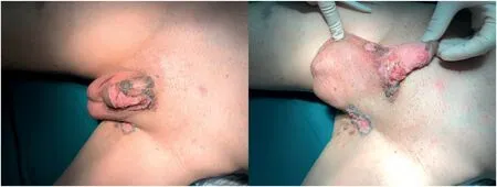
A 57-year-old male patient presented with erythema of 1-year duration of the skin of the penis and scrotum,which had been progressively increasing for two weeks.He was initially diagnosed with tinea corporis at a visit to another hospital and was treated topically with unknown creams.However,the patient’s symptoms did not improve.After the accelerated spread of erythema and the appearance of black deposits under the erythema,he underwent a biopsy in our Dermatology Department.The pathology was suggestive of Bowen’s disease.No evidence of invasive carcinoma was found.The patient’s family had no similar history.On physical examination,a 6 cm × 3 cm erythema extending to the coronal sulcus,a 1 cm×1 cm skin ulcer at the root of the penis,and a 2 cm×3 cm erythema extending from the left scrotum to the mid-inguinal region with a tough texture and indistinct boundaries with the surrounding tissues were observed;no enlarged lymph nodes were found in the groin (Fig.1).
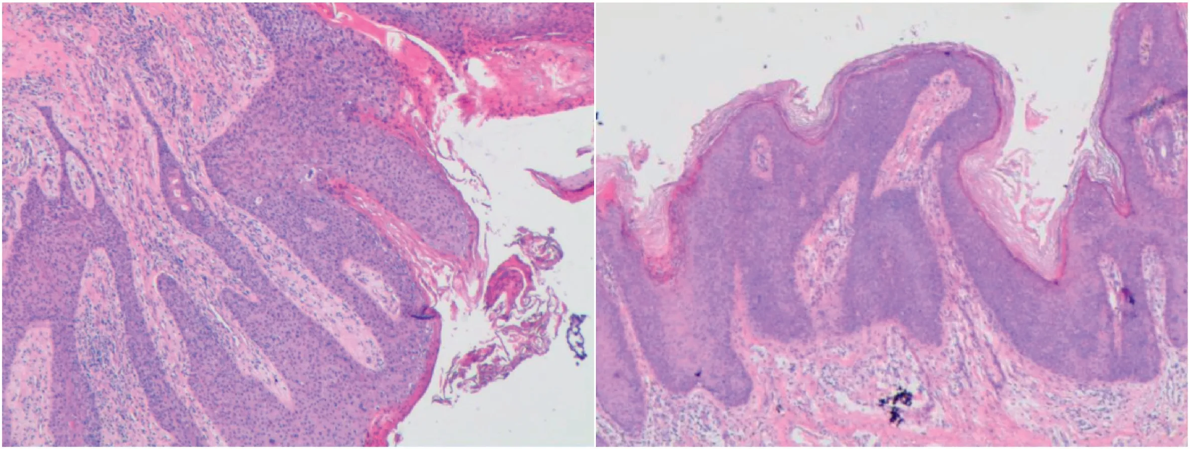
The lesion was excised under general anesthesia after preoperative evaluation of the patient.Due to the large extent of the lesion,the wound that could not be sutured directly after surgical excision was covered by split-thickness skin grafts harvested from the left lateral thigh.Histology showed atypical hyperplasia of the whole epithelium,hyperkeratosis of the epidermis,and disturbed polarity of the epidermal cells.Inflammatory cell infiltration was also observed in the dermis.Based on these pathological findings,the patient was diagnosed with Bowen’s disease(Fig.2).
Histological studies revealed atypical hyperplasia of the entire epithelium,hyperkeratosis of the epidermis,and disturbed epidermal cell polarity.Inflammatory cell infiltration was also observed in the dermis.
3.Discussion
Bowen’s disease,which was first described by Bowen JT in 1912,1is a carcinoma in situ of the skin with the potential to spread laterally,invading both the skin and mucous membranes.2Bowen’s disease lesions present as slow-growing,well-defined papules or patches with irregular margins,whose surfaces are covered with scales or crusts.The disease is common in middle-aged and elderly people and can be found on the skin or mucous membranes of any part of the body,usually singly;however,in a few individuals,multiple lesions may occur.3Pathologically,the distinctive features include full-thickness epidermal atypia with disordered architecture,abnormal mitoses,disorganized cell arrangement,and an intact basement membrane.If a small portion of the basal layer breaks down,it may result in genuinely invasive growth of squamous carcinoma.It has rarely been described in the penis or scrotum.

Fig.1.Gross appearance of the lesion.

Fig.2.Histological images of bowenoid papulosis.
The etiology and pathogenesis of Bowen’s disease remain uncertain.Substantial research suggests that it may be related to the following factors:exposure to arsenic agents,excessive ultraviolet radiation,human papillomavirus(HPV)infection,external stimuli,chromatophore quality,and genetic factors.Oncogenic HPVs (mainly HPV 16,HPV 18,HPV 31,and HPV 33)are associated with many cases of Bowen’s disease,with HPV 16 accounting for approximately 50%of reported cases.4Based on previously published reports,some patients with Bowen’s disease in the penile and scrotal regions have human immunodeficiency virus(HIV)infection;this may be related to the immune deficiency caused by HIV infection.5-7HPV studies were unavailable at our hospital.There was no history of arsenic exposure or long-term sun exposure,and the rapid development of lesions may be related to topical drug stimulation.
Several treatment modalities are available,including surgical excision,liquid nitrogen freezing,electrocautery,laser therapy,topical application of 5-fluorouracil(5-FU)ointment,and photodynamic therapy(PDT).The preferred treatment method remains surgical excision.8In this case,multiple lesions of Bowen’s disease of the penis and scrotum were surgically excised with a resulting large wound,and the site could not be directly sutured.Therefore,free skin grafting was performed.Jansen MH et al.9demonstrated that in the treatment of Bowen’s disease,surgical resection had a lower probability of recurrence compared to 5-FU application and PDT,while there was no significant difference between 5-FU and PDT after five years.ADA-PDT has fewer adverse reactions and does not affect esthetics;therefore,it has been widely used for precancerous lesions of the skin and superficial skin tumors with good efficacy in recent years.In addition,local cryotherapy and 5-FU treatment are effective but have a high recurrence rate;therefore,their application is limited.In cases in which the patient can tolerate surgery,we still recommend complete surgical excision for lesions that are extensive and scattered.Therefore,we selected a treatment plan involving complete surgical excision and free skin grafting in this case.The skin healed well,and there was no recurrence at the six-month follow-up.
The probability of Bowen’s disease developing into invasive squamous carcinoma is approximately 5%,and once it develops into invasive carcinoma,the metastasis rate is approximately 37%.10Surgery remains the primary treatment option for our patients with extensive lesions and lesions at this specific site.
4.Conclusion
In previously published reports,Bowen’s disease in the penile and scrotal regions was uncommon,and most of them had a typical medical history and a high risk of HPV infection.It is relatively unique that Bowen’s disease in this case was multifocal with rapid progression and no typical risk factors.
Ethics approval and consent to participate
The need for ethical approval and consent to participate was waived as this is a case report.
Consent for publication
The patient gave written informed consent to publish the data contained within this study.
Competing interests
The authors declare that they have no competing interests.
Authors’ contributions
Zhang L:Data curation,Writing-Original draft.Shen G:Writing-Review and editing.Han W:Supervision.
 Chinese Journal of Plastic and Reconstructive Surgery2022年2期
- Chinese Journal of Plastic and Reconstructive Surgery的其它文章
- Foreword from Professor Yu-Ray Chen
- Analyzing the correlation among the five indications of the regenerative effectiveness of expanded skin:A retrospective study of 277 expansion cases
- Major and minor risk factors for postoperative abdominoplasty complications:A case series
- Anesthetic injection pain and hematoma occurrence during upper blepharoplasty:Comparison between thin needles and thick needles
- Assessment and management of immature facial scars by non-surgical methods
- Minimally invasive method to treat a rare wrist injury with simultaneous fractures of the scaphoid and hook of hamate:A case report and literature review
