Jacobsen syndrome with bilateral periventricular white matter lesions
Hyun-Joo Lee · Hae-In Lee ·Ji-Hoon Na · Young-Mock Lee ,
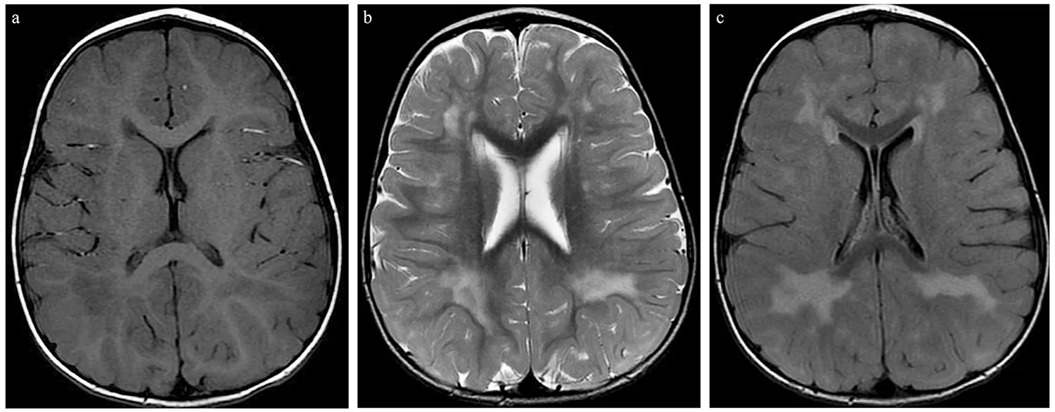
A 2-month-old girl presented facial dimorphism, including frontal bossing, hypertelorism, a broad nasal bridge,low-set ears, hypotonia with mild spasticity, and mild thrombocytopenia. Her head circumference was 40.8 cm(75–90 percentile). There was no relevant perinatal history. Whole-exome sequencing with copy number variation analysis revealed a de novo heterozygous deletion on chromosome 11q (arr[hg19] 11q24.1q25 (11123396858-134257553) x1) of approximately 10.8 Mb, indicative of Jacobsen syndrome (JS; #MIM 147791) [ 1]. Initial brain magnetic resonance imaging (MRI) at 9 months revealed diffuse high signal intensities in the bilateral periventricular and peripheral white matter (WM), prominent trigonal areas on T2-weighted images, and low signal intensity on T1-weighted image (Fig. 1). As WM myelination progressed, no significant differences between the previous MRI findings and that observed after 1-year follow-up was observed with WM myelination progression (Fig. 2).She achieved developmental milestones normally with her age.
Some reports have not clarified the classification of JS cases as hypomyelinated or demyelinated in the classification of leukodystrophy [ 2– 4]. This case suggested that WM lesions result from delayed myelination or demyelination.The genes associated with this phenotype require further investigation, but candidate genes includedFEZ1
,RICS
,and HEPACAM
(involved in axon growth and brain development) [ 2, 5]. The presence of WM lesions usually indicates the possibility of underlying metabolic diseases. However, as in this case, the possibility of JS should also be considered.
Fig. 1 Brain magnetic resonance imaging performed at 9 months of age. T1-weighted image ( a) showing diffuse low signal intensities in the bilateral periventricular white matter. T2-weighted image ( b) and FLAIR ( c) showing diffuse high signal intensities in the bilateral periventricular and peripheral white matter

Fig. 2 Brain magnetic resonance imaging performed at 1 year 9 months of age. No interval changes are noted in the 1-year follow-up MRI. Due to the progression of white matter myelination, diffuse high signal intensities are noted in bilateral corona radiata, periventricular and peripheral white matter, prominent trigonal areas on T2-weighted image ( b) and FLAIR ( c), with low signal intensities in the same area on T1-weighted image ( a)
Author contributions
YML and HL conceptualized the study, coordinated and supervised data collection, and critically reviewed and revised the manuscript. HL and HIL wrote the paper, YML, HL, and JHN reviewed and studied literature about the disease. HL and HIL contributed equally to this paper. All authors approved the final manuscript as submitted and agree to be accountable for the content of the work.Funding
None.Declarations
Ethical approval
This study was carried out in accordance with the recommendations of the Institutional Review Board of Gangnam Severance Hospital, Yonsei University College of Medicine with written informed consent from the patient.Conflict of interest
No financial or nonfinancial benefits have been received or will be received from any party related directly or indirectly to the subject of this article. World Journal of Pediatrics2022年2期
- World Journal of Pediatrics的其它文章
- Diagnostic performance of serum interleukin-6 in pediatric acute appendicitis: a systematic review
- Association between HLA alleles and sub-phenotype of childhood steroid-sensitive nephrotic syndrome
- Variations in length of stay among survived very preterm infants admitted to Chinese neonatal intensive care units
- Epidemiology and region-specific risk factors for low Apgar scores in China: a nationwide study
- Ursodeoxycholic acid prevention on cholestasis associated with total parenteral nutrition in preterm infants: a randomized trial
