
Histopathology of hepatocellular carcinoma - when and what
2022-05-05 08:41DoreenMariaGisderAndreaTannapfelIrisTischoff
Hepatoma Research 2022年1期
Doreen Maria Gisder, Andrea Tannapfel, Iris Tischoff
Institut der Pathologie, Ruhr Universität Bochum, Bochum 44789, Germany.
Abstract When do you need to take biopsies of the liver, and what information will you get is the topic of this review on hepatocellular carcinoma (HCC). If, clinically, the differential diagnosis of HCC after imaging is suggested, a biopsy has become obligatory as a diagnostic confirmation of HCC in the non-cirrhotic liver prior to definitive therapeutic interventions, as well as in a palliative therapy concept. In the case of hepatic lesions with an uncharacteristic contrast uptake, a biopsy should be performed immediately to confirm the diagnosis of HCC. After diagnosing HCC, a treatment strategy is evaluated. Further, the biopsy, or in case of surgical treatment, the resected tissue,shows us the different subtypes of HCC, with the steatohepatitic subtype being the most common and the lymphocyte-rich subtype being the least common. Further, the histological grade of HCC is determined according to the grading system of the WHO or the Edmonson and Steiner System. Through biopsies, HCC can be differentiated from intrahepatic cholangiocarcinoma or combined hepatocellular-cholangiocarcinoma or metastases of other malignant tumors, especially metastases of the gastrointestinal tract. In summary, biopsies are fundamental in the diagnosis of HCC.
Keywords: Hepatocellular carcinoma, biopsies, histology
INTRODUCTION, DEFINITION, AND EPIDEMIOLOGY
Hepatocellular carcinoma (HCC) is a primary liver tumor with hepatocytic differentiation. HCC arises from the hepatocyte in various stages of differentiation. The incidence of HCC in Europe and the United States will continue to increase with the progression of hepatitis C, nonalcoholic steatohepatitis, and obesity with diabetes mellitus and metabolic syndrome[1,2]. In Germany, around 9000 new cases of HCC occur each year,with an incidence of 6.4/100,000 for men and 1.5/100,000 for women[3-5]. Clinically, the tumor marker for HCC is alpha-fetoprotein (AFP). However, it has significant limitations in terms of sensitivity (39% to 64%)and specificity (76% to 91%). AFP levels greater than 20 ng/mL, as well as lower but slowly increasing levels,are a serious indication for HCC. Levels greater than 200 ng/mL are highly suspicious for diagnosis of HCC if positive on imaging. However, AFP levels do not correlate very closely with the size of the HCC. Other specific and sensitive HCC markers have been searched for decades. Several alternative markers have been investigated, for example, des-gamma-carboxyprothrombin, glypican-3, AFP fractions, but they are not yet used in clinical practice[6-8]. In almost all cases, prior to therapy, a precise histopathological diagnosis is mandatory, especially in tumors without cirrhosis. The histological HCC subtyping should be done according to the latest World Health Organization (WHO) classification. Next to tumor typing (i.e., HCC determination, subtyping, and ruling out intrahepatic cholangiocarcinoma), the grade of tumor differentiation (“grading”) is necessary. There are three grading systems for the HCC. The HCC-specific grading of Edmondson and Steiner described by Edmondson and Steiner[9], the grading according to Nzeakoet al.[10,11], and the International Union Against Cancer (UICC) grading[12]. The general UICC grading is not entity-specific, is less detailed, and is explicitly rarely applied in the context of HCC. Grading,according to Edmondson and Steiner, is recommended by UICC in HCC[12]. Nzeakoet al.[10,11]grading is more commonly used in the European and American regions. This grading is based purely on nuclear features. Staging of HCC must be performed according to the current Malignant Tumours (TNM)classification[13]. Another point of histopathological diagnosis is the early detection of precancerous lesions in cirrhotic and non-cirrhotic liver.
WHEN-BIOPSY?
Due to the increase of HCC to the 5th most common cancer in Europe and worldwide, there has been an update of the guidelines on treatment strategies, especially concerning when to take biopsies[14,15]. In 2013, it was recommended that a biopsy should only be obtained if it has a therapeutic consequence. In 2021, biopsy has been strengthened as an important diagnostic measure. The diagnostic algorithm has changed so that lesions greater than 1 cm in size should now receive an imaging examination rather than only lesions greater than 2 cm. If a lesion shows the characteristic contrast enhancement, a curative or palliative therapy concept can be initiated depending on the size and extension of the HCC. Interestingly, the detection rate of small lesions has increased in recent years, leading to new therapeutic strategies[16-18]. In particular, imageguided tumor ablation for early-stage HCC gained attention as a therapeutic strategy. This therapy is a nonsurgical treatment that provides local tumor control, and the patients have more favorable survival benefits.Before ablation, a biopsy is taken for the histopathological examination to confirm the diagnose of HCC.The biopsy should be taken prior to ablation to reduce necrosis in the tissue to be examined[19-21]. All in all,biopsy has become obligatory as a diagnostic confirmation of HCC in the non-cirrhotic liver before definitive therapeutic interventions, as well as in a palliative therapy concept. In the case of hepatic lesions with an uncharacteristic contrast uptake, a biopsy should be performed immediately to confirm the diagnosis of HCC. Further, if a palliative therapy concept is considered, a biopsy should be performed to exclude other differential diagnoses and to confirm HCC[22]. In the circumstance of a curative concept,further imaging should be performed. If this second imaging continues to show uncharacteristic contrast enhancement, a biopsy is an obligatory procedure. The interpretation of the biopsy and the diagnosis of HCC can be made in many cases by conventional histology. Depending on the histopathological appearance, further examinations, in particular immunohistochemistry, can be used to confirm the diagnosis[23-25].
For an adequate histopathological interpretation of the biopsy, the specimen must be taken from the lesion and must be of sufficient size[26]. To obtain a relevant sample of a focal liver lesion, image guidance, with either real-time ultrasound, computer tomography, or magnetic resonance imaging, is necessary to place the needle accurately within the lesion[27]. Further studies claim that a longer sample size increases the chance of a possible adequate histopathological interpretation[28-30]. The guidelines on the use of liver biopsies in clinical practice from the British Society of Gastroenterology, the Royal College of Radiologists, and the Royal College of Pathology recommend a sample that should be at least 20 mm[26,31]. In general, only one pass is required if an adequate sample is obtained. Nevertheless, studies have demonstrated a more accurate diagnose with an increased number of needle passes. Three or more passes should not be performed as this significantly increases the risk of complications and morbidity[32-34]. The current state of knowledge doesn’t specify if a lesion biopsy should be taken through adjacent liver tissue to minimize the risk of so-called“punch channel tumor spreading”. How biopsies should be taken depends on the case and the localization of the lesion, and they should be taken with the lowest impact on the liver and patient. In consensus with our hepatologists, a biopsy should be taken as directly as possible. The complication rate of biopsy of HCC is low and includes the risk of hemorrhage and so-called “punch channel tumor spreading”. Minor bleeding requires no treatment and occurs in approximately 3%-4% of cases. Bleeding requiring transfusion is rare(0.5%)[35]. Meta-analyses have shown that “punch channel tumor spreading” is also rare (2.7%). They typically occur late, approximately after a mean of 17 months, and are usually well treatable and have no negative impact on survival as well as on the success of therapeutic measures[36-38]. Another indication for a biopsy can be a portal vein thrombosis. Portal vein thrombosis may be either portal vein tumor thrombosis or benign portal vein thrombosis. It is important to distinguish between them as they are relevant to patient outcome and therapeutic strategies[39-41]. In particular, when HCC occurs in a cirrhotic liver, the differentiation can be difficult, as benign portal vein thrombosis often occurs in cirrhotic livers. The first diagnostic procedure for differentiation is imaging[42-44]. However, if imaging does not fulfill the characteristic criteria of portal vein tumor thrombosis or benign portal vein thrombosis, an ultrasoundguided fine-needle aspiration biopsy can be performed for differentiation[44,45].
In any case, a malignant liver tumor cannot be diagnosed without a histological examination since the classification of malignant liver tumors is based on histological criteria. Overall, histopathology is the basis of HCC diagnosis and its differential diagnoses. If any concerns occur concerning a clinical diagnose of HCC, a biopsy should be considered. The criteria for the current diagnosis of malignant liver tumors are summarized in the 5th edition of the WHO Classification of Tumors of the Digestive Tract[46-48]. If the patient’s history does not include information regarding the underlying liver disease, an additional biopsy of liver tissue could be obtained to evaluate whether fibrosis or cirrhosis is presented. This information is essential for further therapeutical decision-making and patient outcome. In this case, the Royal College of Pathologists updated guidelines suggesting a minimum adequacy requirement for samples being at least 10 mm long and containing six portal tracts. New studies underline that a biopsy including fewer than 11 portal tracts per tissue section might underestimate the degree of fibrosis and inflammation[31,49].
WHAT YOU GET AFTER BIOPSY OR RESECTION
Not only the histological subtype, the primary resectability or the degree of liver cirrhosis are important factors to evaluate the prognosis and outcome for the patient. It is further important to look at the macroscopic appearance of the tumor. HCCs occur as solid, green to yellow nodes of various sizes in the liver. The green to yellow color is caused by the bilious imbibition and the fat content of the tumor.Compared to the non-neoplastic liver tissue and especially in cirrhotic liver, HCCs appear as a soft tumor.In cirrhotic livers, they have additionally a pseudocapsule. Little has been reported on the role of macroscopic classification of HCC and the prognostic role. However, the macroscopic classification of HCC might have a strong correlation with long-term prognosis after hepatectomy[50]. Historically, four types of HCC were described by Eggel in 1901. He divided the neoplasms into the expansive (nodule), infiltrative,mixed (expansive and infiltrative), and diffuse types. The prevalence among cases are: 20% nodule, 33%infiltrative, 42% mixed, and 5% diffuse. The data published by Eggel are based on HCCs arising in cirrhosis[51]. Nowadays, the exclusive morphological classification by Eggel has been mostly left behind since it does not allow conclusions to be made concerning the etiology, clinical course, or prognosis[52,53]. Besides this, the macroscopic description of the HCC still has an important clinical impact when it comes to describing the size of the primary tumor, the size of additional tumor nodes with correlation to the primary tumor, and tumor vessel invasion. For example, for tumor size, there was a significant difference between tumors smaller than 5 cm and those larger than this. In tumors measuring less than 5 cm, intrahepatic metastasis, portal vein involvement, and lymphogenous and hematogenous metastasis were not common.However, these frequencies became significantly higher if the tumor exceeded 5 cm. Comparing the percentages of tumors less than 5 cm and greater than 5 cm, intrahepatic metastases were observed in 60.6%and 95.7%, portal vein involvement in 40% and 74.5%, lymph node metastases in 0% and 40.4%, and hematogenous metastases in 6.7% and 51.1%, respectively[53]. Further, the macroscopic description of whether there is vessel invasion is relevant to TNM. HCCs often appear hypervascular and have close contact with the vessels [Figure 1A-D][54,55]. If a second tumor occurs in the liver, the distance to the first and biggest one must be described. Additionally, it must be ensured that they were separated by non-neoplastic liver tissue. Whether this second tumor is an intrahepatic metastasis or a second synchronic or heterochinic tumor is still discussed[56-59]. Tumors arising within a radius of less than 2 cm to the primary tumor, and if they are smaller than the primary tumor, are called satellite nodules. Another special anatomic appearance of HCC is the pedunculated HCC. It is a rare subtype, which can be further categorized by its presence or absence of a pedicle attaching it to the liver[60-62]. Especially the macroscopic described tumor size is relevant for the WHO grading system. This grading system for HCC proposed by the WHO takes not only the tumor size but also the architecture and the extent of cell and nuclear pleomorphism (including sarcomatoid or anaplastic morphology) into account[12,48].
HISTOLOGICAL SUBTYPES
There are various numbers of different subtypes of HCC. They differ by morphology and/or immunohistochemistry. All different types of HCC show cytological atypia varying from minimal to marked, and the tumor cells show an increased proliferation. Characteristic cellular changes of HCC include bile production, lipofuscin deposits, glycogen accumulation leading to clear cell changes, and fatty changes[Figure 2A-C]. The tumor cells can develop inclusions such as hyaline bodies, Mallory-dense bodies, or pale bodies [Figure 2D]. Further, they have a loss of typical hepatic architecture, such as loss of portal tracts or reduction/loss of the normal reticulin framework. As described in the macroscopic appearance, they show histologically an increased arterialization with sinusoidal capillarization and aberrant arterioles. The capillaries of the tumor are lined by a single layer of endothelium, which differs from non-neoplastic capillaries. The endothelium of the tumor shows an immunohistochemical expression of CD34, factor VIII,and type IV collagen. Electron microscopy shows a basement membrane between tumor cells and endothelial cells.
There are four principal growth patterns of HCCs: trabecular, solid (syn.: compact), pseudoglandular (syn.:pseudocarina), and macrotrabecular [Figure 3A-C]. Half of all resected HCCs have mixed patterns. It is mostly a combination of trabecular plus one or two others. The pathologist may recognize the growth pattern, but it must not be described since it does not have any clinical relevance. However, in a recently published study suggests there might be an association between the macrotrabecular pattern and a worse prognosis. A special pattern is nodule-in-nodule growth. In this case, a nodule of poor differentiation arises within an existing HCC[63].

Figure 1. (A) Vascular invasion of hepatocellular carcinoma. (B) Invasion in a small lymph vessel of portal tract of hepatocellular carcinoma. (C) Hepatocellular carcinoma with perineural invasion. (D) Hepatocellular carcinoma invasion in an intrahepatic bile duct.
The histological grading system of the WHO is based on H&E staining and compares the differentiation of the tumor cell with the benign hepatocytes. The WHO proclaims a three-stage system. There are welldifferentiated (G1), moderately differentiated (G2), and poorly differentiated (G3) HCCs [Table 1,Figure 4A-C]. Undifferentiated tumors are excluded from this system because they have no compelling evidence for being either hepatocellular or biliary and are not a grade of HCC [Table 1][12]. Some HCC may have intratumoral heterogeneity concerning the grade. In this case, the predominant grade or a combination of the worst can be reported, due to the fact that the worst grade tends to drive the prognosis.A uniform worldwide used grading system remains to be developed. One example of a currently often used grading system is the Edmonson and Steiner System, which favors a three-tiered grading system [Table 2].It was developed by Edmonson and Steiner in 1954 and is based on nuclear-cytoplasmic ratio, the grade of acidophilic, chromatin content, the bile production, and the histological architecture[9].
Of the HCCs, 35% can be further classified into eight distinct subtypes, which will be described below. All subtypes except the fibrolamellar, which only occurs in non-cirrhotic livers, have been described in cirrhotic and non-cirrhotic liver. The most common type is the steatohepatitic HCC[64,65].Immunohistochemically, HCCs show an antibody reaction againstcarbamoylphosphat-synthetase-1(HEP PAR1). The poorer the degree of differentiation of HCC, the lower the expression ofHEP PAR1[66]. Further,an expression ofCD10,cytokeratin-8, andcytokeratin-18can be obtained[67]. There is no expression ofcytokeratin-20as well asepithelial membrane antigen(EMA). Cytokeratin-7 and -19 usually are not expressed but may be observed in high-grade HCC.AFPexpression usually indicates malignancy and can be seen in 50% of cases. Further,glypican 3can be used as a diagnostic marker for differentiating between HCC from non-malignant liver lesions. A highglypican 3positive staining significantly correlates with later tumor stage, higher tumor grade, presence of vascular invasion, shortened overall survival, and shortened disease-free survival. Shafizadehet al.[68]reported an expression ofglypican 3in 9/16 (56%) well, 15/18 (83%)moderately, and 22/24 (89%) poorly differentiated HCC. A diffuse expression can be seen of the 70 kilodalton heat shock proteins (HSP 70) and glutamine synthetase[68-71]. In routine clinical work,immunohistochemistry is of limited value in the differential diagnosis. In most cases, the diagnosis of HCC can be based on the H&E staining.
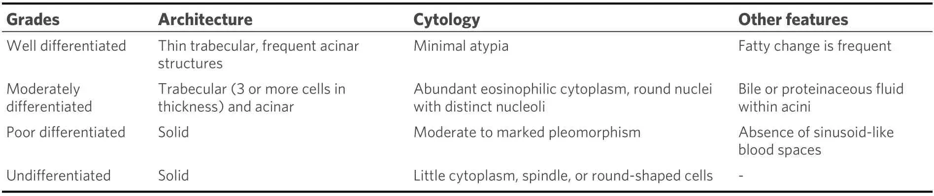
Table 1. World Health Organization[9]

Table 2. Edmondson and Steiner[9]
Steatohepatitic hepatocellular carcinom
The steatohepatitic subtype is the most common, with 5%-20% prevalence among HCCs. The tumor cells show histological features of steatohepatitis, such as containing fatty vacuoles, an intratumoral stroma reaction, and inflammation. Further, it is often associated with metabolic syndrome and steatohepatitis or even cirrhosis of the liver [Figure 5A-C][72-75].
Clear cell hepatocellular carcinoma
With 3%-7% prevalence, it is the second most common subtype. Of the tumor cells, 80% show a clear cell morphology with a light cytoplasm and minor nuclear atypia. The “brightness” of the cells occurs because the tumor cells accumulate glycogen [Figure 6A-C][76-78].
Macrotrabecular (massive) hepatocellular carcinoma
To classify this subtype, there must be at least 50% of a macrotrabecular growth pattern[79,80]. The tumor occurs in 5% of the cases and is associated with a worse prognosis. Clinically, a high serum AFP is seen [Figure 3C][81].
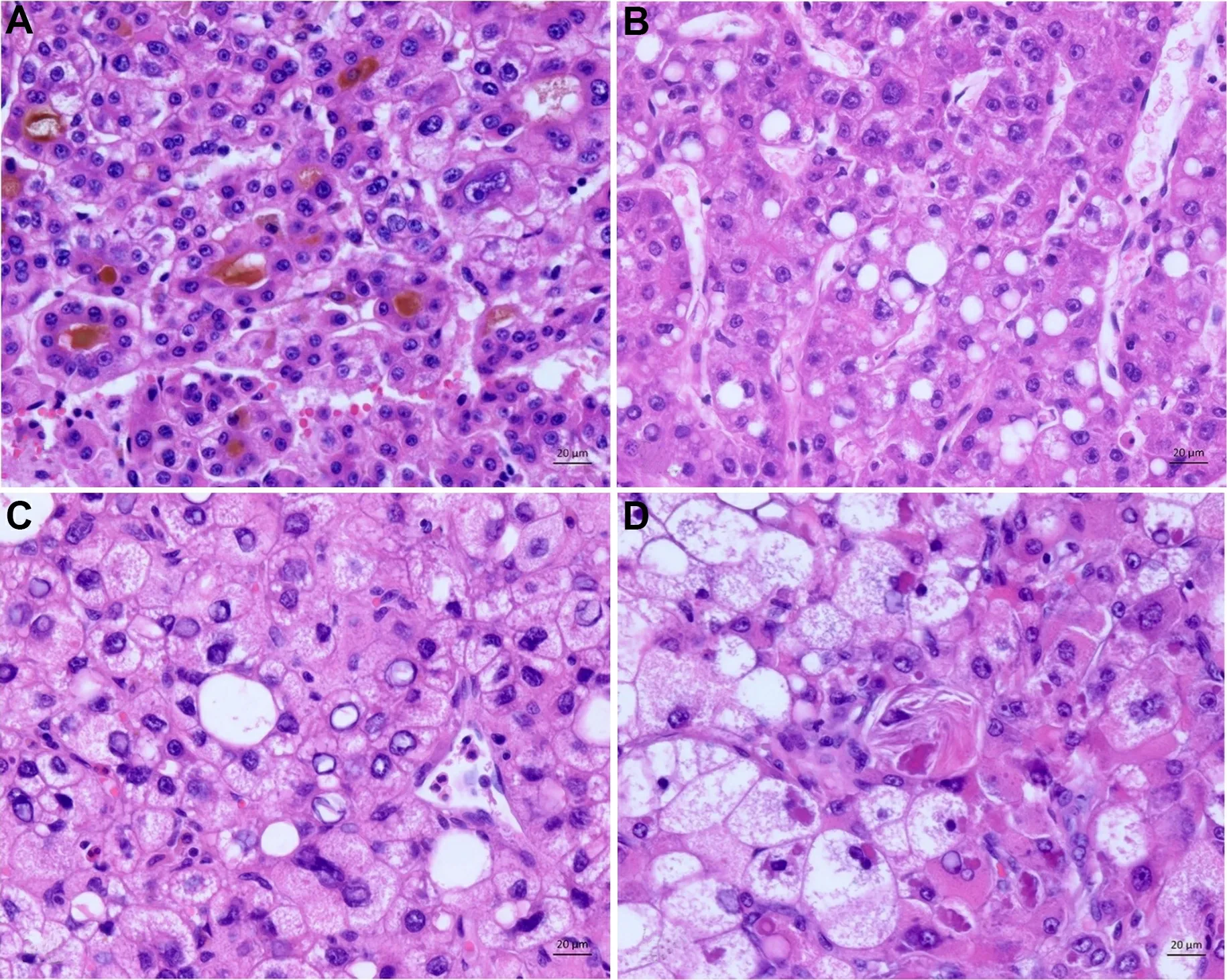
Figure 2. Characteristic cellular changes in hepatocellular carcinoma. (A) Tumor cells with bile production. (B) Fatty changes with intracytoplasmic accumulation of lipids. (C) Glycogenated nuclei. (D) Hyaline bodies (Mallory Denk bodies): eosinophilic cytoplasmic inclusions.

Figure 3. Histological growth pattern. (A) Trabecular pattern: thin tumor trabeculaes with not more than ten cells in thickness. (B)Pseudoglandular pattern: glandular-like or acinus-like structures with minimal atypia. (C) Macrotrabecular Pattern: trabecular structures with more than 10 cells in thickness, increased nuclear:cytoplasma ratio.
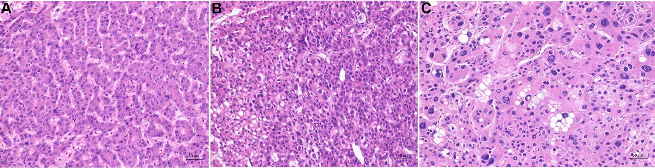
Figure 4. Histological grading of hepatocellular carcinoma according to World Health Organization. (A) Well differentiated hepatocellular carcinoma: thin trabecular structures and minimal atypia of tumor cells resembling mature hepatocytes. (B) Moderately differentiated hepatocellular carcinoma: trabecular pattern with at least 3 cells in thickness, moderate nuclear atypia with increased nuclear:cytoplasma ratio, microvesicular fatty changes (lower left). (C) Poorly differentiated hepatocellular carcinoma: marked pleomorphic tumor cells with fatty changes, prominent nucleoli und anaplastic giant cells.

Figure 5. Steatohepatitic subtype. (A) Overview of the steatohepatitic tumor. The tumor cells show histological features of steatohepatitis by containing fatty vacuoles. Further inflammation and an intratumoral stroma reaction is seen. (B) Higher magnification of the tumor. Tumor shows features of steatohepatitis tumor cells with fatty vacuoles and tumor cell ballooning. (C) The tumors characteristic intratumoral inflammation and hyaline bodies (Mallory Denk bodies) are seen here.
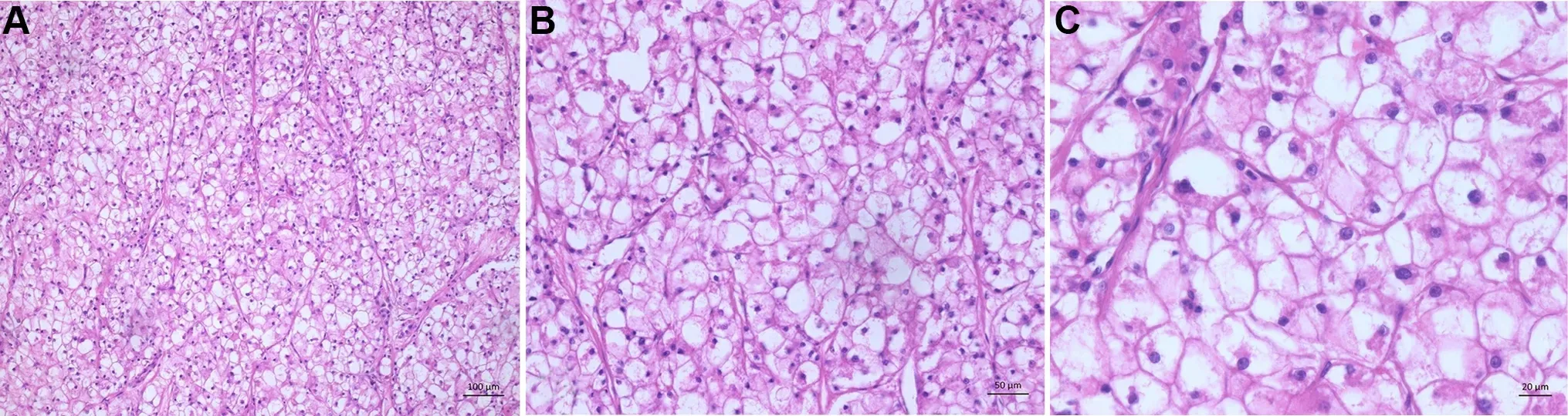
Figure 6. Clear cell subtype. (A) Overview of a clear cell subtype. (B) Tumor trabeculaes with clear cell cytoplasm of tumor cells accumulating glycogen. (C) The tumor cells show a clear cell morphology with a light cytoplasm and minor nuclear atypia.
Scirrhous hepatocellular carcinoma
Four percent of all HCCs show a scirrhous subtype. This tumor subtype shows dense intratumoral fibrosis of greater than 50%, which arouses especially in association with the sinusoidal capillarization leading to an atrophy of the tumor trabecular[60,82-84]. After chemotherapy or radiation of HCC, the tumor may mimic this subtype. Clinically, this subtype mimics a cholangiocarcinoma on imaging [Figure 7B][60,82,85].
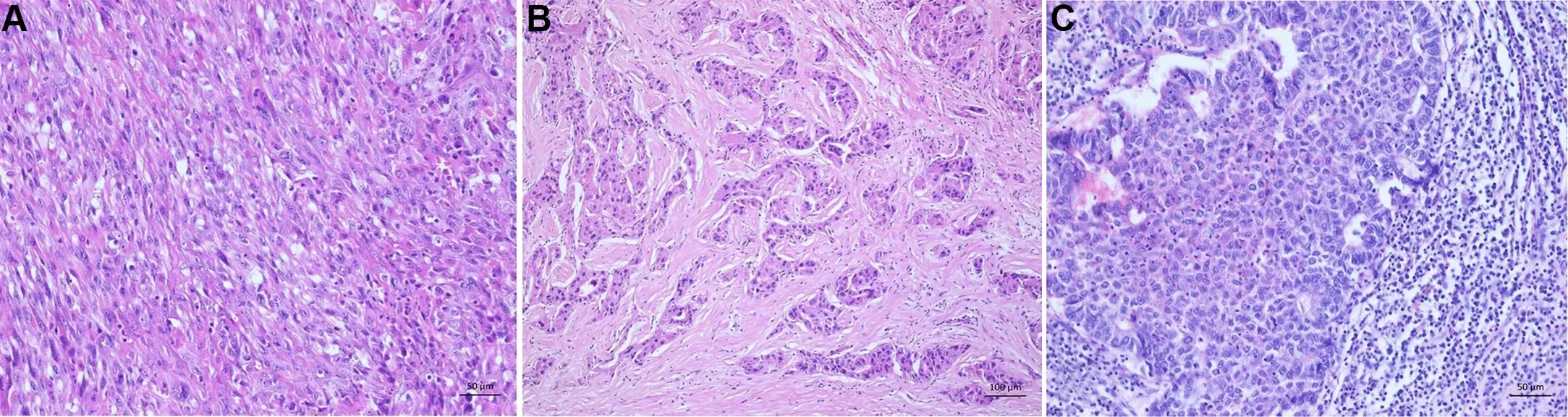
Figure 7. Rare histological subtypes. (A) Sarcomatoid subtype: poorly differentiated HCC with spindle cell morphology resembling sarcoma. (B) Scirrhous subtype: dense intratumoral fibrosis. (C) Neutrophil-rich subtype: poorly differentiated hepatocellular carinoma with numerous intratumoral neutrophils.
Chromophobe hepatocellular carcinoma
Three percent of the subtypes show a light cytoplasm with outlined cell borders. The tumor nuclei are mainly bland. However, focal areas have striking nuclear atypia, such as two nuclei within one tumor cell.Further, giant cells can be found.
Fibrolamellar hepatocellular carcinoma
Of all subtypes, 0.5%-1% are fibrolamellar HCCs. Compared to the other subtypes, they often occur in young patients with a median age of 25 years and only occur in non-cirrhotic livers[86-88]. So far, no association with chronic liver disease or specific risk factors has been described[89-91]. Due to their macroscopic appearance with a central scar and the fact that they are more common in women, they can be misdiagnosed as focal nodular hyperplasia[88,92,93]. The tumor cells are large and eosinophilic with prominent nuclei. Another characteristic for diagnosing this subtype is dense intratumoral fibrosis consisting of acellular collagen and pale bodies. The pale bodies are characteristic but not specific, and occur in the classic HCC as well[94][Figure 8]. In some cases, glandular tumor cell configurations occur, which may lead to the misdiagnosis of combined hepatocellular cholangiocarcinoma [Figure 9][80].
Neutrophil-rich hepatocellular carcinoma
This tumor has an incidence of less than 1% and mainly occurs in older individuals. It shows a histomorphologically significant intratumoral neutrophil-rich inflammation and can have sarcomatoid areas (for an illustration of sarcomatoid morphology refer to Figure 7A). The tumor’s key molecular finding is the production of granulocyte colony-stimulating factor, leading to dense areas of infiltrates with neutrophils. Further, some clinical findings, such as elevated leukocyte counts, C-reactive protein, and IL-6,support the diagnosis of a neutrophil-rich HCC [Figure 7C][64,95].
Lymphocyte-rich hepatocellular carcinoma
To diagnose this rare tumor, there must be more lymphocytes than tumor cells in most fields of the H&E staining. Characteristically, the cords of tumor cells lie in a dense lymphoid stroma. An association with Epstein-Barr virus has been frequently described, but new evidence shows no Epstein-Barr virus relationship, and most cases are negative for Epstein-Barr virus[95].
HISTOLOGICAL DIFFERENTIAL DIAGNOSIS
Histologically, HCC must be differentiated from combined hepatocellular-cholangiocarcinoma[Figure 9A-C], intrahepatic cholangiocarcinoma [Figure 10A-C], and metastases of other malignant tumors especially hepatoid adenocarcinoma, neuroendocrine tumors, non-keratinizing squamous carcinomas,urothelial carcinomas, or amelanotic melanomas [Figure 11A-E][12]. The tumor microenvironment is of limited value in the differential diagnosis of HCC. In contrast to intrahepatic cholangiocarcinoma, the tumor surrounding stroma is rare and mostly absent. However, if cirrhosis exists, the pre-test probability increases in favor of a primary malignant liver tumor (e.g., HCC or intrahepatic cholangiocarcinoma).Metastases from distant organs are rarer events in a cirrhotic liver. In certain circumstances, such as a cancer of unknown primary origin with poor differentiation, immunohistochemistry could help to identify and differentiate HCC from metastases of other origins. In this case, the markers thyroid transcription factor 1 (TTF 1) (for lung and thyroid), caudal-type homeobox transcription factor 2 (CDX 2) (for colorectum), GATA binding protein 3 (GATA3) (for breast and urothelium), NK3 homeobox 1 (NKX3.1)(for prostate), paired-box-protein 8 (PAX8) (for gynecological tumors, kidney, and thyroid gland), special AT-rich sequence-binding protein 2 (SATB2) (for enteric differentiation), and SRY-box transcription factor 10 (SOX10) (for melanoma) can be used[96,97]. Nevertheless, in almost all cases, this differential diagnosis could be ruled out by using H&E stains. Molecular markers did not have value until now[65].
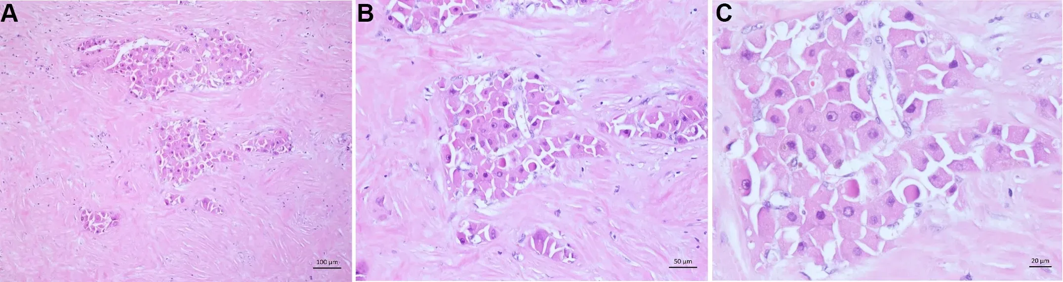
Figure 8. Fibrolamellar subtype. (A) Overview of a fibrolamellar subtype. Scattered tumor cells with eosinophilic cytoplasm in a dense intratumoral fibrosis. (B) The dense intratumoral fibrosis consists of acellular collagen. Some tumor cells contain pale bodies. (C) The tumor cells are large and eosinophilic with prominent nuclei and some tumor cells contain pale bodies.
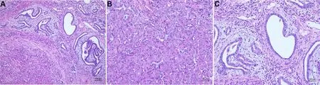
Figure 9. Combined hepatocellular-cholangiocarcinoma. (A) Typical form with moderately differentiated hepatocellular carcinoma(lower left) and well differentiated cholangiocarcinoma (upper right). Between both tumor components a fibrotic border is seen. (B)Hepatocellular carcinoma - component with trabecular pattern and moderate atypia. (C) Cholangiocarcinoma -component with tubular structures and mild atypia.

Figure 10. Intrahepatic cholangiocarcinoma. (A) Overview of a moderately differentiated adenocarcinoma with tubular pattern infiltrating a dense fibrous stroma. (B) Infiltrative irregular glands with a prominent fibrodesmoplastic stromal reaction. (C) Higher magnification of the tumor showing the moderately pleomorphic tumor cells with eosinophilic cytoplasm.
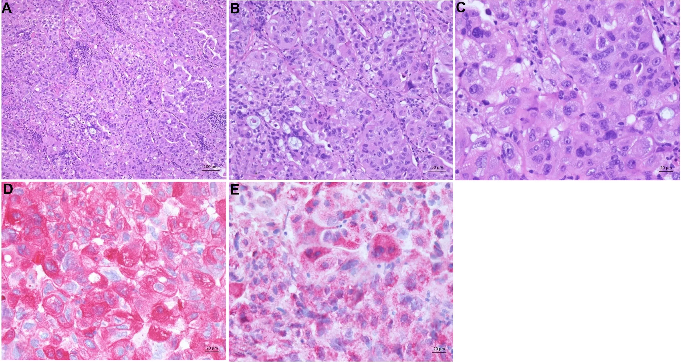
Figure 11. Hepatoid adenocarcinoma. (A) The tumor shows hepatocyte-like tumor cells and lymphoplasmacytic infiltrates localized to stroma and tumor parenchyma. (B) Moderate differentiated tumor cells forming thick trabeculae in the tumor. (C) Hepatocyte-like tumor cells with round nuclei and eosinophilic cytoplasm. (D) Tumor cells are strongly positive for CK7. (E) HepPar1 is also expressed.
CONCLUSION FOR THE CLINIC
Due to the increasing incidence and the histological variety of HCC, the histopathological diagnosis is essential for diagnosis and therapy of the tumor. Biopsy indications are well established in the diagnostic algorithm of HCC, even in cirrhotic liver. A biopsy should always be used in the palliative situation and when non-invasive methods are unequivocal. Especially if there is any doubt about the diagnosis of HCC, a biopsy should be considered in the first place.
DECLARATIONS
Authors’ contributions
Made substantial contributions to conception and design of the script: Gisder DM Made substantial contributions to conception and design of the script: Tannapfel A Made substantial contributions to conception and design of the script and selected the case images:Tischoff I
Availability of data and materials
Not applicable.
Financial support and sponsorship
None.
Conflicts of interest
All authors declared that there are no conflicts of interest.
Ethical approval and consent to participate
Not applicable.
Consent for publication
Not applicable.
Copyright
© The Author(s) 2022.

- Hepatoma Research的其它文章
- Robotic donor hepatectomy: a niche advancement or the way forward? A perspective from the world’s largest center
- Radiological imaging and non-surgical local treatments for cholangiocarcinoma
- Advances in Y-90 radioembolization for the treatment of hepatocellular carcinoma
- Molecular mechanisms of liver carcinogenesis related to metabolic syndrome