MiR-98-5p inhibits invasion and migration of thyroid cancer cells by targeting STAT3
Yongcun Liu ,Liping Wu ,Yuan Shao ,Jiaona Sun
Abstract Objective:To investigate the effects of microRNA-98-5p(miR-98-5p)on invasion and migration of thyroid tumor cells by targeting signal transducer and activator of transcription 3(STAT3).Methods:The expression levels of miR-98-5p and STAT3 mRNA in thyroid tumors and adjacent tissues were detected.SW-579 cells were divided into Control group,mimic NC group,miR-98-5p mimic group and miR-98-5p mimic+pC-STAT3 group.Control group was not treated,while the other three groups were transfected with mimic NC,miR-98-5p mimic and miR-98-5p mimic+pC-STAT3,respectively.The expression levels of miR-98-5p and STAT3 mRNA in SW-579 cells in each group were detected.The miRNA target gene prediction software and dual luciferase assay were performed to test targeting effect of miR-98-5p on STAT3.The cell invasion and migration in each group were detected.The levels of p-STAT3,STAT3,VEGF,MMP-2 and MMP-9 protein were detected by Western blotting.Results:The expression level of miR-98-5p mRNA in thyroid tumor tissues was significantly lower than that in adjacent tissues,while the expression level of STAT3 mRNA was significantly higher(P<0.01),and miR-98-5p was negatively correlated with STAT3 (r=-0.862, P=0.0001).The expression of miR-98-5p in miR-98-5p mimic group was significantly higher than that in Control group,while the expression of STAT3 was significantly lower (P<0.01).The target gene prediction software found that STAT3 might be a target gene of miR-98-5p.The dual luciferase assay results showed that luciferase activity in co-transfection of miR-98-5p mimics and STAT3 WT cells was significantly lower than that in co-transfection of mimic NC and STAT3 WT cells (P<0.01).Compared with mimic NC group,invasion rate and migration rate of SW-579 cells in miR-98-5p mimicgroup were significantly decreased (P<0.01).Compared with miR-98-5p mimic group,invasion rate and migration rate of SW-579 cells in miR-98-5p mimic+pC-STAT3 group were significantly increased (P<0.01).Compared with mimic NC group,the expression of p-STAT3,STAT3,VEGF,MMP-2 and MMP-9 protein of SW-579 cells in miR-98-5p mimic group was significantly decreased(P<0.01).Compared with miR-98-5p mimic group,expression of p-STAT3,STAT3,VEGF,MMP-2 and MMP-9 protein of SW-579 cells in miR-98-5p mimic+pC-STAT3 group was significantly increased (P<0.01).Conclusion:MiR-98-5p can inhibit STAT3,regulate cell invasion and the expression of EMT-related proteins,and inhibit invasion and migration of thyroid cancer cell SW-579.
Keywords thyroid tumor; miRNA-98-5p; signal transducer and activator of transcription 3; invasion;migration
Introduction
Thyroid cancer is a common malignant tumor in the endocrine system,and the incidence rate has increased rapidly in recent years,accounting for about 1% of the occurrence of malignant tumors in the whole body [1].According to histomorphology,thyroid carcinoma can be divided into papillary carcinoma,follicular carcinoma,medullary carcinoma and undifferentiated carcinoma,among which papillary carcinoma has the highest incidence [2-3].Signal transducer and activator of transcriptional 3 (STAT3)play an important role in signal transduction and transcriptional activation.Usually,STAT3 is only temporarily and rapidly activated for a few minutes to several hours to maintain the normal physiological function of cells.When STAT3 is continuously activated,it leads to abnormal expression of cell proliferation,differentiation and apoptosis genes,and makes cells transform to malignant.Therefore,STAT3 can be divided into proto-oncogenes [4-5].As a non-coding RNA,with short nucleotide sequence and regulatory function,microRNA plays an important role in the process of cell proliferation,differentiation and apoptosis [6].In this study,we analyzed the targeting relationship between miR-98-5p and STAT3,and studied the possible mechanism in the invasion and migration of thyroid cancer SW-579 cells in order to provide a new reference for targeted therapy of thyroid cancer.
Materials and methods
Generaldata
From May 2013 to May 2017,a total of 26 thyroid cancer specimens and adjacent tissues confirmed in our hospital were collected.Within 30 min,the thyroid cancer specimens were divided into two parts,one was sent to the Pathology Department to be pathologically confirmed,the other part was placed in liquid nitrogen,and thenstored in a cryogenic refrigerator at-80 ℃for testing.
Main materials and instruments
Thyroid cancer cell SW-579 (purchased from Shanghai Cell Bank of Chinese Academy of Sciences),miR-98-5p mimcs and its negative control (Shanghai GENECHEM),double luciferase activity detection kit,lentiviral vector pLV-EGFP(2A) Puro,containing U6 promoter and green fluorescent protein gene (Promega),liposome,Lipofectamine®2000 (Thermo Fisher Scientific),p-STAT3,STAT3,VEGF,MMP-2,MMP-9,GAPDH antibodies and anti-mouse/rabbit IgG (Cell Signaling Technology),RPMI 1640,DMEM high glucose cell culture medium,FBS (Gibco),TRIzol Kit (Thermo Fisher Scientific),Revertaid First Strand cDNA Kit (Thermo Fisher),SYBRGreen PCR (TAKARA),Matrigel gel (BD),high speed freezing centrifuge (Beckman Coulter),positive fluorescence microscope (Thermo),real-time fluorescence quantitative PCR(Roche).
Cell culture and transfection
SW-579 was cultured in complete RPMI 1640 medium containing 10% fetal bovine serum,100 U/mL penicillin or streptomycin,and cultured to logarithmic phase in 5% CO2humidified incubator at 37 ℃.0.25% trypsin digestive solution was treated to make cell suspension.The cultured SW-579 cells were divided into Control group,mimic NC group,miR-98-5p mimic group and miR-98-5p mimic+pC-STAT3 group.Control group was not given treatment.Mimic NC,miR-98-5p mimic and miR-98-5p mimic+pCSTAT3 were respectively transfected into SW-579 cells by Lipofectamine®2000 according to the operation instructions,and then the positive cells were observed under fluorescence microscope.If the green fluorescence signal is sent out,it is regarded as positive transfection,and the cells are collected for follow-up experiments.
The detection of mRNA expression of miR-98-5p and STAT3 in thyroid adenoma and its adjacent tissues by qPCR
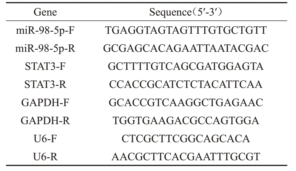
After the cells and tissues were grinded in liquid nitrogen,the total RNA was extracted and examined by ultraviolet spectrophotometer,the OD value (A260/A280) was 1.8 to 2.0,and the integrity of RNA was detected by gel electrophoresis.cDNA was synthesized according to the instructions of reverse transcription kit.Reverse transcription system(10 μL):2×miRNA reaction mixture 5 μL,0.1%BSA 1 μL,miRNA PrimeScript®RT enzyme mixture 1 μL,total RNA 0.5 μL,de-RNA enzyme ddH2O 2.5 μL.The reaction conditions were set as follows:37 ℃,60 min,85 ℃,5 s,4 ℃,30 min.PCR system 10 μL:SYBR®Prmix Ex Tap II (2×) 5 μL,upstream primer 0.4 μL,downstream primer 0.4 μL,ROX Reference Dye II (50×)0.2 μL,cDNA 1 µL,ddH2O 3 µL.See the kit instructions for detailed operation.PCR reaction parameters:50 ℃activated polymerase 5 min,95 ℃pre-denatured 30 s,95 ℃denatured 5 s,60 ℃annealing and extension 34 s,the reaction was carried out for 40 cycles.The dissolution curve was drawn as follows:95 ℃,15 s,60 ℃,60 s,85 ℃,15 s,60 ℃,15 s.Each sample hole was provided with 3 compound holes.The expression of related genes was calculated by 2-△△Ctmethod.The sequence of primers was shown in Table 1.

Table 1 The sequence of primers
The verification of the targeting relationship be⁃tween miR-98-5p and STAT3 by double lucifer⁃ase report experiment
The STAT3 3'-UTR fragment containing miR-98-5p binding site and its mutants were inserted into the downstream of luciferase reporter gene in pLV-EGFP(2A) Puro vector and named as STAT3 WT and STAT3 Mut,respectively.The constructed plasmids were sequenced,and the plasmids that met the requirements of sequencing were co-transfected into SW-579 cells with miR-98-5p mimcs and miR-control,respectively.24 h after transfection,the cells were lysed,and the luciferase activity of each group was detected by Dual-Luciferase Reporter System.
Detection of the effect of miR-98-5p targeted STAT3 on the invasion of SW-579 cells by Tran⁃swell chamber
SW-579 cells from mimic NC group,miR-98-5p mimic group and miR-98-5p mimic+pC-STAT3 group were collected and washed with PBS,and then cultured in RPMI 1640 without fetal bovine serum for 24 h.The number of cells was adjusted to 1×10⁵/mL single cell suspension.The 200 μL cell suspension was placed in the Transwell chamber covered with Matrigel glue,and three multiple holes were set up in each group.After being cultured in a 24-well plate of chamber without 500 μL 100 mL/L fetal bovine serum for 24 h,the chamber was removed and eluted with PBS.The cells and matrix glue that did not penetrate the membrane in the upper layer were gently wiped off with cotton swabs,and the filter membrane was fixed with ice formaldehyde and stained with crystal violet for 30 min.After the filter membrane was peeled off,it was fixed face down on the glass slide,and the number of cells was randomly selected under the 5 high power lens (×400).The average number of penetrating cells in each visual field was counted.The experiment was repeated for 3 times.
Detection of the effect of miR-98-5p targeted STAT3 on the migration of SW-579 cells by scratch test
The SW-579 cells of mimic NC group,miR-98-5p mimic group and miR-98-5p mimic+pC-STAT3 group were finally put into a 6-well plate.When the cells were attached to the wall and spread all over the plate,a 200 μL gun was used to draw straight horizontal scratches on the plate.After 24 h of culture,the repair of scratches was observed,and the relative area was calculated by Image-Pro plus 6.0.
Detection of the expression of p-STAT3,STAT3,VEGF,MMP-2 and MMP-9 in SW-579 cells by Western blotting
The total proteins of SW-579 cells were extracted after being cultured for 48 h,and the protein concentrations of mimic NC group,miR-98-5p mimic group and miR-98-5p mimic+pC-STAT3 group were adjusted by Bradford.The protein was adjusted by SDSPAGE gel electrophoresis,electrotransferred to formaldehyde-treated PVDF membrane,sealed for 2 hours,incubated overnight with rabbit anti-human p-STAT3,STAT3,VEGF,MMP-2,MMP-9,GAPDH primary antibody (1∶500) at 4 ℃,andrinsed with TBST for 40 min.The protein was incubated with HRP-labeled secondary antibody (1∶500) for 1 h,and rinsed with TBST for 40 min.The protein bands on the membrane were observed by ECL kit and DNR BioImaging System,and the images were collected.The gray values of the bands in each group were analyzed by gel image processing system,and statistical analysis was made according to the relative gray values.
Statistical method
The data were analyzed by GraphPad Prism and SPSS 22.0 software.The measurement data were expressed by mean±standard deviation(SD).Single factor analysis of variance was used for comparison among groups,LSD-ttest was used for pairwise comparison between groups,ttest was used for comparison between the two groups,and Spearman correlation analysis was used for correlation analysis between variables.
Results
Expression and correlation of miR-98-5p and STAT3 in thyroid carcinoma and its adjacent tis⁃sues
The expression level of miR-98-5p mRNA in thyroid tumor tissue was significantly lower than that in adjacent tissue (P<0.01),as shown in Figure 1A.The expression level of STAT3 mRNA in thyroid tumor tissue was significantly higher than that in adjacent tissue (P<0.01),as shown in Figure 1B.The results of Spearman correlation analysis showed that there was a negative correlation between miR-98-5p and STAT3 mRNA expression (r=-0.862,P=0.0001),as shown in Figure 1C.
MiR-98-5p inhibited the relative expression of STAT3
The expression of miR-98-5p in miR-98-5p mimics group was significantly higher than that in Control group (P<0.01),but there was no significant difference in the expression of miR-98-5p between mimic-NC group and Control group (P>0.05),as shown in Figure 2A.The expression of STAT3 in mimics group was significantly lower than that in Control group(P<0.01),but there was no significant difference in the expression of STAT3 between mimic NC group and Control group(P>0.05),as shown in Figure 2B.
miR-98-5p targeted STAT3
MiRNA target gene database was used to predict the target site of miR-98-5p,it was found that STAT3 was a potential target gene of miR-98-5p,and there were theoretical complementary pairing sequences in the seed region of STAT3 mRNA 3'UTR and miR-98-5p,as shown in Figure 3A.The results of double luciferase report assay showed that the luciferase activity of co-transfected miR-98-5p mimics and STAT3 WT cells was significantly lower than that of co-transfected mimic NC and STAT3 WT cells (P<0.01),but there was no significant difference in luciferase activity of co-transfected miR-98-5p mimics and STAT3 Mut cells and co-transfected mimic NC and STAT3 Mut cells(P>0.05),as shown in Figure 3B.
MiR-98-5p targeted STAT3 to regulate the inva⁃sion of SW-579 cells
The invasion rate of SW-579 cells in miR-98-5p mimic group was significantly lower than that in mimic NC group(P<0.01),and the invasion rate of SW-579 cells in miR-98-5p mimic+pC-STAT3 group was significantly higher than that in miR-98-5p mimic group(P<0.01),as shown in Figure 4.
MiR-98-5p targeted STAT3 to regulate the migra⁃tion of SW-579 cells
The migration rate of SW-579 cells in miR-98-5p mimic group was significantly lower than that in mimic NC group(P<0.01),and the migration rate of SW-579 cells in miR-98-5p mimic+pC-STAT3 group was significantly higher than that in miR-98-5p mimic group(P<0.01),as shown in Figure 5.
MiR-98-5p targeted STAT3 to regulate the ex⁃pression of invasion and migration-related pro⁃teins in SW-579 cells
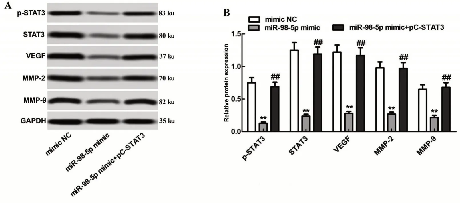
The protein expression levels of p-STAT3,STAT3,VEGF,MMP-2 and MMP-9 in SW-579 cells in miR-98-5p mimic group were significantly lower than those in mimic NC group (P<0.01).The protein expression levels of p-STAT3,STAT3,VEGF,MMP-2 and MMP-9 in miR-98-5p mimic+pC STAT3 group were significantly higher than those in miR-98-5p mimic group(P<0.01),as shown in Figure 6.

Figure 1 The expression and correlation of miR-98-5p and STAT3 in thyroid carcinoma and its adjacent tissues.A:mRNA expression of miR-98-5p in thyroid carcinoma and adjacent tissues.B:mRNA expression of STAT3 in thyroid carcinoma and adjacent tissues.C:The correlation relationship between the expression of miR-98-5p and STAT3 mRNA.

Figure 2 MiR-98-5p inhibited the relative expression of STAT3.A:Expression of miR-98-5p in cells of each group after transfection.B:The expression of STAT3 in cells of each group after transfection.**P<0.01 vs.Control group.

Figure 3 miR-98-5p targeted the study of STAT3.A:Gene sequence.B:Luciferase activity.**P<0.01 vs.mimic NC.

Figure 4 The results of Transwell test and the rate of cell invasion in each group.A:Results of Transwell test of cells.B:Thecell invasion rate in each group.**P<0.01 vs.mimic NC;##P<0.01 vs.miR-98-5p mimic group.

Figure 5 Results of cell scratch test and cell migration rate in each group.A:Results of scratch healing test of cells.B:The cell migration rate in each group.**P<0.01 vs.mimic NC;##P<0.01 vs.miR-98-5p mimic group.

Figure 6 Expression of invasion and migration of proteins in each group of cells.A:Representative graphs for p-STAT3,STAT3,VEGF,MMP-2 and MMP-9 protein expression by Western blotting.B:Quantitative analysis demonstrated the levels of p-STAT3,STAT3,VEGF,MMP-2 and MMP-9.Results were normalized to GAPDH.**P<0.01 vs.mimic NC;##P<0.01 vs.miR-98-5p mimic group.
Discussion
MiRNA is a kind of RNA,with the function of regulating gene expression,which is no more than 26 amino acid sequences in length.At present,most of the members of microRNA have the characteristics of high conservatism,timing and tissue-cell specificity,and play unique roles in the process of cell differentiation,proliferation and apoptosis [7-8].MiRNA needs to activate or inhibit the transcriptional activity of target genes by binding to the 3' or 5' UTR regions of downstream target genes.Basically,microRNA is involved in the occurrence and development of all malignant tumors.According to the different functions of its genes,it can be divided into tumor suppressor genes and tumor promoting genes[9].Previous studies have shown that miR-98-5p is involved in the occurrence and progression of many tumors,and its level can be used as an index to judge the prognosis of triple negative breast cancer [10].Low levels of miR-98-5p may predict the shortening of survival in patients with lung cancer [11].Some studies have also shown that the expression of miR-98-5p is significantly decreased in ovarian cancer tissues [12].It is suggested that miR-98-5p plays the role of tumor suppressor gene in the process of malignant tumor.In this study,we detected the expression of miR-98-5p in thyroid cancer and adjacent tissues by qPCR,and found that the expression of miR-98-5p was significantly down-regulated,while the expression of STAT3 was significantly increased.At the same time,the correlation analysis showed that there was a negative correlation between them,and the correlation was high,suggesting that miR-98-5p may play the role of tumor suppressor gene in the process of thyroid cancer.STAT3 is an important member of the STAT family,which can form a signal pathway with the tyrosine kinase JAK to regulate the occurrence and development of tumor,and many anticancer drugs play their anticancer role by inhibiting the JAK/STAT signal pathway [13].In order to further study the function of miR-98-5p in thyroid carcinoma,we overexpressed miR-98-5p in SW-579 cells by transfecting miR-98-5p mimic.It was found that the expression of STAT3 decreased significantly,indicating that there may be a targeted relationship between miR-98-5p and STAT3.We found that miR-98-5p and STAT3 may have binding sites by using miRNA target gene data.At the same time,double luciferase report experiment further confirmed that miR-98-5p could specifically inhibit the expression of STAT3.Then we also found that overexpression of miR-98-5p could reduce the invasion and migration of SW-579 cells,while co-expression of miR-98-5p and STAT3 could restore this cell characterization,indicating that miR-98-5p inhibits cell invasion and migration by down-regulating STAT3.Liu et al.[14] found that miR-98-5p can not only promote the apoptosis of lung cancer A549 cells by reducing the level of STAT3,but also inhibit the invasion and migration of A549 cells,which is consistent with the results of this study.
In order to analyze the possible mechanism of miR-98-5p targeting STAT3 in regulating cell invasion and migration,we detected the expression of invasion and migration-related proteins by Western blotting.It was found that the protein expression of VEGF,MMP-2 and MMP-9 decreased significantly after overexpression of miR-98-5p,while the protein expression levels of VEGF,MMP-2 and MMP-9 were restored after co-expression of miR-98-5p and STAT3.It is suggested that miR-98-5p targeting STAT3 may inhibit the invasion and migration of cancer cells by regulating the protein expression of VEGF,MMP-2 and MMP-9.As a vascular growth factor,VEGF can not only promote the growth of tumor vascular endothelial cells,but also increase the permeability of blood vessels,resulting in fibrin deposition in the surrounding tissue,which is conducive to the formation of tumor matrix and the invasion of neovascularization,metastasis and invasion of tumor[15].MMP-2 and MMP-9,as members of the MMPs family,are important ECM proteolytic enzymes,mainly by hydrolyzing IV collagen in the basement membrane,destroying the integrity of the basement membrane,thus promoting tumor metastasis[16].Therefore,the possible mechanism of miR-98-5p targeting STAT3 to regulate cell invasion and migration is that miR-98-5p inhibits the phosphorylation of STAT3,weakens the signal transduction of downstream signaling factors,and reduces the expression of downstream elements such as VEGF,MMP-2,MMP-9 and other proteins,resulting in block of vascular endothelial cell growth and weakening of epithelial-mesenchymal transformation,thus reducing the ability of invasion and migration of cancer cells.
In conclusion,miR-98-5p can inhibit the expression of STAT3 and the protein expression of downstream effector proteins such as VEGF,MMP-2 and MMP-9,and then reduce the invasion and migration ability of thyroid cancer cells.The results of this study provide a theoretical basis for miR-98-5p as a site for targeted therapy of thyroid cancer.
AcknowledgmentsThis study was funded by Basic Research Program of Natural Science in Shaanxi Province(No.2018JM7108096).

