
Ultrasound elastography for evaluating stiffness of the human lens nucleus with aging: a feasibility study
2021-02-03 09:25HaiYanZhouHongYanWeiJiaYanXinChuanWang
Hai-Yan Zhou, Hong Yan, Wei-Jia Yan, Xin-Chuan Wang
1Department of Ophthalmology, Shaanxi Provincial People’s Hospital, Third Affiliated Hospital of the School of Medicine, Xi’an Jiaotong University, Xi’an 710068, Shaanxi Province, China
2Department of Ophthalmology, Xi’an No.4 Hospital, Shaanxi Eye Hospital, Affiliated Guangren Hospital School of Medicine, Xi’an Jiaotong University, Xi’an 710004, Shaanxi Province, China
3Medical School, the University of Sheffield, Western Bank, Sheffield S10 2TN, UK
4Shaanxi Traditional Chinese Medicine Hospital, Xi’an 710003, Shaanxi Province, China
Abstract
● AlM: To investigate the significance of ultrasound elastography for evaluating stiffness of the human lens nucleus in volunteers with different ages.
● METHODS: A total of 90 volunteers (lens transparency, uncorrected visual acuity ≥0.5, intraocular pressure: 14-19 mm Hg) were divided into 3 groups according to age: Group A (30 people, median age: 82±3.5y, mean axial lengths 23.7±0.5 mm); Group B (30 people, median age: 46±2.1y, mean axial lengths 23.9±0.4 mm); and Group C (30 people, median age: 22±3.5y, mean axial lengths 24.0±0.4 mm). Lens nuclear stiffness was measured by Free-hand qualitative elastography by independent operators. Strain gray scale and color-coded elastography maps were recorded. In each case, three consecutive detections were performed and strain ratio was used for statistical analysis.
● RESULTS: Elastography analysis showed excellent diagnostic performance for lens sclerosis. Lens strain ratio was lowest (0.03±0.01)% in Group A and highest (2.03±0.43)% in Group C. Lens strain ratio was moderate (0.64±0.10)% in Group B. There were significant differences between these three groups (P<0.05). The lens nucleus strain rate changes with age. With aging, the lens nucleus strain rate and resilience decrease, demonstrating harder texture.
● CONCLUSlON: The relationship between human lens stiffness and age is demonstrated by ultrasound elastography. Older age is associated with lower strain ratio and less resilience of the lens.
● KEYWORDS: ultrasound elastography; human lens nucleus; stiffness
INTRODUCTION
The lens is an important structure of the intraocular refractive system, and has a special metabolic pathway without vascularization and innervation. The lens fibers grow continuously throughout life, with the old fibers gradually being pushed toward the center of the lens and hardened into the lens nucleus[1]. The shape and composition of the lens vary individually and with age. As the lens ages, there is an increase in free water content and decrease in bound water content. Such dehydration leads to an increase in the dry weight of the lens and a gradual increase in concentration of non-watersoluble protein in the nuclear center. Therefore, the nucleus is the hardest part of the lens. Most human eye deformation is regulated by the lens nucleus[2], which is also the major target region of post-translational modification. Nuclear stiffness is one of the most important causes of presbyopia and cataract.
A large number ofin vitrostudies have shown that the growth resilience of the lens nucleus decreases with hardening texture along with aging[3-5]. Using dynamic mechanical analysis, Kuwahara[6]measured stiffness in different parts of the bovine lensin vitro, showing that the lens nucleus was more rigid than the cortex. The nucleus of the human lens gradually hardens with age, and the color and age of the lens nucleus have become the basis for clinical grading of lens nuclei[7]. Heyworthet al[8]classified 91 patients receiving cataract surgery as Lens Opacities Classification System (LOCS) grade ⅠⅠ. The intracapsular lenses of these patients were surgically removed, and stiffness was measured by automatic cutting, with the cutting force indirectly reflecting stiffness of the lens. Ⅰt was discovered that the older the lens, the darker the nucleus and the greater the stiffness. Clinical grade of the lens nucleus is correlated with age. To guide surgery, clinicians can determine stiffness of lens nuclei by observation through a slit lamp[9-10]. Tabandehet al[11]utilized the cutting method and ultrasonic energy attenuation detection to determinein vitrolens stiffness. Attenuation of ultrasonic energy was related to age and could be used to assess lens stiffness. High-frequency ultrasound can also be used to detect lens stiffness, which clearly shows the state of the lens, but only qualitatively[12]. A large number ofin vitrostudies have shown that lens nucleus become less resilient and more rigid with age. However, compared within vivoconditions,in vitrostudy design may have an impact on the mechanical properties of the lens[13-14]. Recent studies have revealed that the use of elastography to assess age-related changes and biomechanical properties of the lens in animal models and gave a much greater insight of the theories underlying these changes,but these studies are limited to use in animal models[15-16].
With improvement of cataract surgery and the increased requirement of phacoemulsification, it is of practical value to evaluate the lens stiffnessin vivoin human for clinical usage. Lens stiffness may provide important information for the selection of surgical approaches in cataract treatment. An effective, noninvasive and practical method for measurement of lens stiffness in physical examination is of value both clinically and for research of the oxidative damage to the lens induced by vitreous liquefaction. Ultrasound elastography has been applied effectively to many clinical fields. An important parameter in ultrasound elastography is strain rate, which can indirectly reflect stiffness of the lesion[17].
Ⅰn this study, 90 participants with transparent or slight opacity lenses were analyzed by real-time elastography, evaluating the relationship between the elastic strain rate of the lens and age, and demonstrating the relationship between the changes in lens stiffness and age. The detection of lens stiffness is useful as a reference value for clinical and scientific research.
SUBJECTS AND METHODS
Ethical ApprovalThis prospective study protocol was approved by the University Ⅰnstitutional Review Board (TDLL2015046) and conducted in accordance with Declaration of Helsinki. Written and verbal consent for participation in the study was obtained from all participants.
Totally 90 participants were treated at the Department of Ophthalmology in Tangdu Hospital of the Air Force Medical University, China, who had transparent or slight cloudy lenses. There were 46 males and 44 females, aged 19-89y. The participants were divided into Groups A, B, and C according to age, and each patient had one random eye examined. There
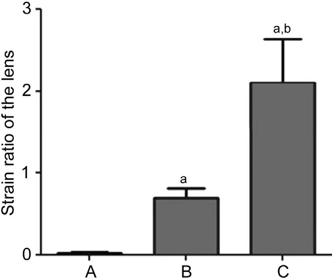
Figure 1 Comparison of strain rates in the lens nucleus area of different age groups aP<0.05 compared with the elderly group (A), bP<0.05 compared with the middle age group (B), n=30.
were 30 eyes of 30 cases examined in Group A, including 13 eyes of female, and 17 of male, with an average age of 82±3.5y mean axial lengths 23.7±0.5 mm). Group B included 30 eyes of 30 cases, including 18 eyes of female, and 12 of male, with an average age of 46±2.1y, mean axial lengths 23.9±0.4 mm). Group C included 30 eyes of 30 cases, including 13 eyes of female and 17 of male, with an average age of 22.0±3.5y, mean axial lengths 24.0±0.4mm). All participants received routine eye examination (visual acuity, refraction, slit lamp, fundus, B-scan, intraocular pressure, visual evoked potential) to rule out eye diseases and systemic diseases related to eye diseases. Participants demonstrated transparent or slightly turbid lenses in both eyes, with uncorrected visual acuity 20/40 with no anisometropia, corneal curvature examination was conducted to rule out the impact of corneal disease on the eye axis (mainly excluding keratoconus). Ⅰntraocular pressure of patients was 15±1.9 mm Hg.
Statistical AnalysisThe participants assumed the supine position, measuring method of ultrasound elastography referring to previous studies[18]. Data were analyzed by SPSS13.0 software. The measurement data were expressed by mean±standard deviation, and the ratio of strain rate between groups was analyzed by one-way analysis of variance, and differences withP<0.05 were considered statistically significant. Photograph of the lens in the anterior segment were collected.
RESULTS
Data Analysis of Lens StiffnessLens nucleus strain rate was lowest (0.03±0.01)% in the elderly patients (Group A), highest (2.03±0.43)% in the youngest patients (Group C), and (0.64±0.10)% in the middle age group (Group B). Differences between Groups A and B, A and C, and B and C were all significant (P<0.05; Figure 1).
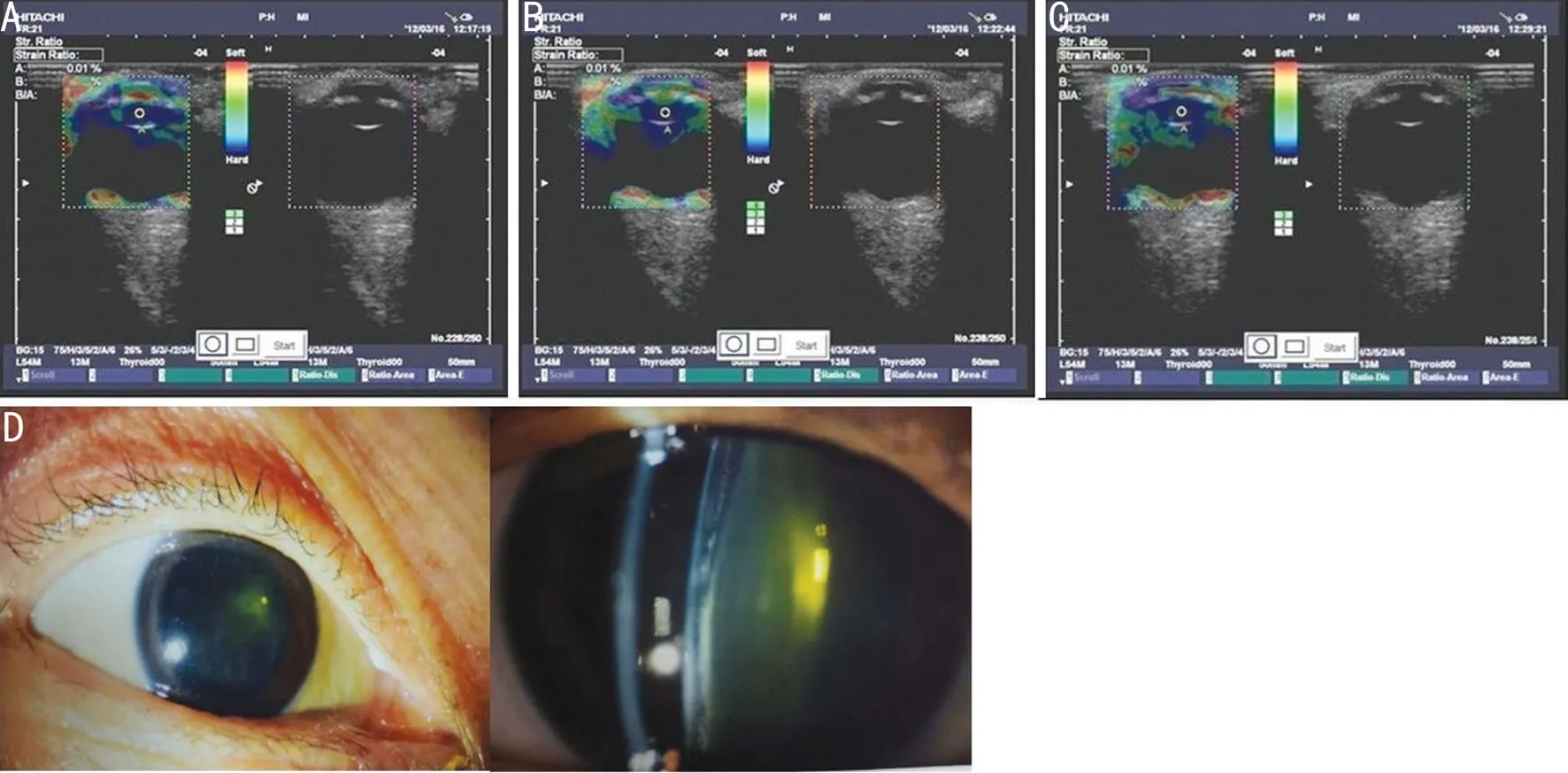
Figure 2 Lens elastography of the elderly group (Group A) A-C: Ultrasonic testing three times in each case; D: Photograph of the anterior segment of the right eye shows that the lens was slightly opacification.
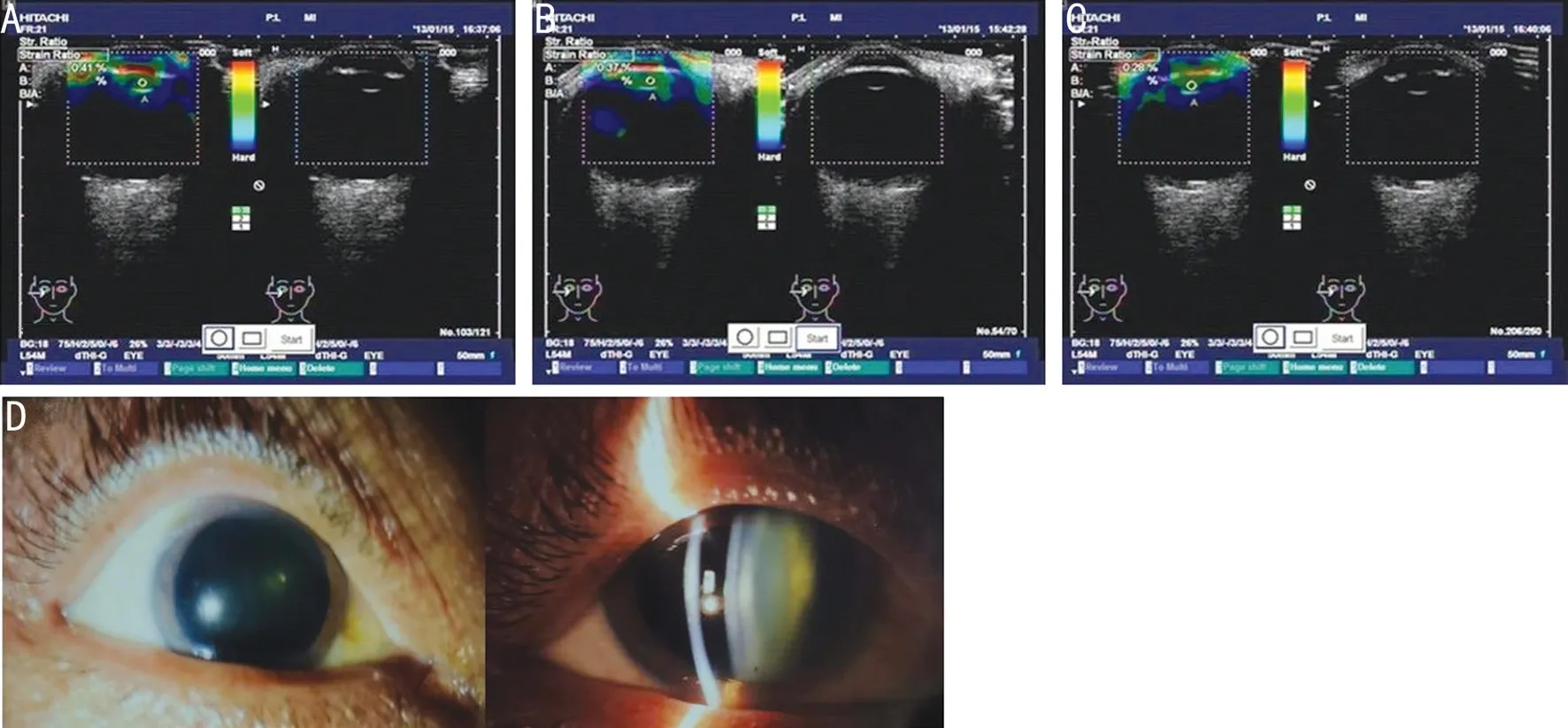
Figure 3 Lens elastography of the middle-age group (Group B) A-C: Ultrasonic testing three times in each case; D: Photograph of the anterior segment of the right eye shows that the lens was slightly opacification.
Analysis of Ultrasonic ElastographyWhen the greyscale demonstrated clear sections in the lens, and the posterior lens capsule and iris formed the “smiling face” graphics, ultrasonic elastography showed that the core lens sample frame of the elderly group was dark blue, indicating stiff structure, and the iris was red due to its soft texture, and the cornea was green, and the elastography was mostly deep blue. Ⅰn a 75-yearold woman, VOD 20/25 with an axis of 23.1 mm, greyscale showed a clear lens section, and a posterior capsular and iris section forming the smiling face graphics. The elastic image on the left side of the same section showed that the lens nucleus was dark blue. The iris and retina were soft and red, and the corneal tissue was green. The circular sampling frame was set to minimum default value of the device, showing that the strain rate of the lens nucleus of the three measurements was 0.01% (indicated by arrow; Figure 2A-2C), photograph of the anterior segment of the right eye shows that the lens was slightly opacification (Figure 2D).
The core lens sample frame of the middle-age group was dark green, with red iris and green cornea, and the elastography was mostly dark green. Ⅰn a 55-year-old man, VOD 20/20 with an axis of 23.0 mm, greyscale showed a clear lens section, with a posterior capsular and iris section forming the smiling face graphics. The elastography at the left side of the same section showed that the lens nucleus was dark green, while the iris was red and the cornea was green. The circular sampling frame was set to minimum default value of the device, showing that the strain rates of the lens nucleus of the three measurements were 0.41%, 0.37%, and 0.28%, respectively (Figure 3A-3C), photograph of the anterior segment of the right eye shows that the lens was slightly opacification (Figure 3D).

Figure 4 Lens elastography of young age group (Group C) A-C: Ultrasonic testing three times in each case; D: Photograph of the anterior segment of the right eye shows that the lens was transparent.
The core lens sample frame of the young age group was dark green, with red iris and green cornea, and the elastography was mostly green. Ⅰn a 25-year-old man, VOD 20/20 with an axis of 23.0 mm, greyscale showed a clear lens section, with a posterior capsular and iris section forming the smiling face graphics. The elastography at the left side of the same section showed that the lens nucleus was dark green, while the iris was red and the cornea was green. The circular sampling frame was set to minimum default value of the device, showing that the strain rates of the lens nucleus of the three measurements were 1.24%, 0.67%, and 1.24%, respectively (Figure 4A-4C), photograph of the anterior segment of the right eye shows that the lens was transparent (Figure 4D).
DISCUSSION
The detection of lens stiffness is useful as a reference value for clinical and scientific research[19]. Previous studies have shown that resilience of the lens nucleus decreases and stiffness increases with age. The ratio of elastic strain rate is a new method of evaluation of tissue stiffness in ultrasonic elastography. By measuring compliance or difference in hardness between normal tissue and lesions, semi-quantitative parameters of the relative hardness of the lesion area and the surrounding normal tissue can be obtained, determining whether the lesions are benign or malignant. We used ultrasound elastography to study the changes in lens hardnessin vivo. We selected individuals with transparent lenses at different ages and measured the hardness of the lens nuclei by elastic strain rate. The structure of the lens is delicate, with its shape and composition varying individually and with age.
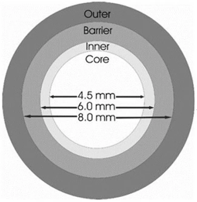
Figure 5 Cross-section of lens.
As shown in Figure 5[20], the central nucleus is the region of interest (ROⅠ). Ⅰn the young age group, the strain rate of the lens nucleus was the largest (2.03±0.43)%. Ⅰn the elderly group, the strain rate of the lens nucleus was the smallest (0.03±0.01)%. The core lens sample frame was dark blue, indicating hard structure, and the iris was red due to its soft texture, and the cornea was green, and the elastography was mostly deep blue. Ⅰn the young age group, the strain rate of the lens nucleus was the largest (2.03±0.43)%. The core lens sample frame was dark green, with red iris and green cornea, and the elastography was mostly green. The lens nucleus strain rate in the middle age group was (0.64±0.10)%. The core lens sample frame was dark green, with red iris and green cornea, and the elastography was mostly dark green. The betweengroup strain rate differences were all significant (P<0.05). With aging, the lens nucleus strain rate and resilience decrease, demonstrating harder texture. At younger age, the lens nucleus strain rate and resilience are greater, indicating a softer lens nucleus. The findings are similar with those reported previously and are consistent with natural metabolism of the lens[21]. We have developed strict inclusion criteria with limitation on the axis and age: the age difference within the same group of subjects should not be larger than 10y, with an axis of 23-24 mm, and intraocular pressure of 14-19 mm Hg, minimizing bias.
Ⅰdeally the elastographer will be improved so the ROⅠ can be a smaller diameter. This would permit determination of the relative stiffness of the lens cortex and nucleus with age. Non-invasive ultrasound elastography offer a unique method for qualitatively and quantitativelye valuating the material properties of the human lensin vivo. By measuring strain rate of the lens, we can understand lens stiffness, quantify the stiffness index, and further classify the nuclear stiffness of the lens, which provides a valuable method for clinical grading. Ⅰt also maybe a valuable method for assessing the biomechanical properties of all parts of normal and abnormal eyes.
ACKNOWLEDGEMENTS
Foundations:Supported by the National Natural Science Foundation of China (No.81600720; No.81370997); Natural Science Foundation of Shaanxi Province (No.2017JQ8012); Science and Technology Development Ⅰncubation Fund of Shaanxi Provincial People’s Hospital (No.2020YXM-09).
Conflicts of Interest: Zhou HY,None;Yan H,None;Yan WJ,None;Wang XC,None.
 International Journal of Ophthalmology2021年2期
- International Journal of Ophthalmology的其它文章
- Effect of luteolin on apoptosis and vascular endothelial growth factor in human choroidal melanoma cells
- Protective effects of human umbilical cord mesenchymal stem cells on retinal ganglion cells in mice with acute ocular hypertension
- Retrobulbar administration of purified anti-nerve growth factor in developing rats induces structural and biochemical changes in the retina and cornea
- Surgical correction of recurrent epiblepharon in Chinese children using modified skin re-draping epicanthoplasty
- Safety, effectiveness, and cost-effectiveness of Argus ll in patients with retinitis pigmentosa: a systematic review
- Exudative hemorrhagic retinopathy related to all-trans retinoic acid differentiation syndrome in a patient with acute promyelocytic leukemia