Identifi cation and characterization of endosymbiosis-related immune genes in deep-sea mussels Gigantidas platifrons*
LI Mengna , CHEN Hao , WANG Minxiao , ZHONG Zhaoshan , ZHOU Li , LI Chaolun , ,
1 Center of Deep Sea Research and Key Laboratory of Marine Ecology & Environmental Sciences (CODR and KLMEES), Institute of Oceanology, Chinese Academy of Sciences, Qingdao 266071, China
2 Laboratory for Marine Ecology and Environmental Science, Qingdao National Laboratory for Marine Science and Technology, Qingdao 266237, China
3 Center for Ocean Mega-Science, Chinese Academy of Sciences, Qingdao 266071, China
4 University of Chinese Academy of Sciences, Beijing 100049, China
Abstract Deep-sea mussels of the subfamily Bathymodiolinae are common and numerically dominant species widely distributed in cold seeps and hydrothermal vents. During long-time evolution, deep-sea mussels have evolved to be well adapted to the local environment of cold seeps and hydrothermal vents by various ways, especially by establishing endosymbiosis with chemotrophic bacteria. However, biological processes underlying the establishment and maintenance of symbiosis between host mussels and symbionts are largely unclear. In the present study, Gigantidas platifrons genes possibly involved in the symbiosis with methane oxidation symbionts were identifi ed and characterized by Lipopolysaccharide (LPS) pull-down and in situ hybridization. Five immune related proteins including Toll-like receptor 2 (TLR2), integrin, vacuolar sorting protein (VSP), matrix metalloproteinase 1 (MMP1), and leucine-rich repeat (LRR-1) were identifi ed by LPS pull-down assay. These fi ve proteins were all conserved in either molecular sequences or functional domains and known to be key molecules in host immune recognition, phagocytosis, and lysosome-mediated digestion. Furthermore, in situ hybridization of LRR-1, TLR2 and VSP genes was conducted to investigate their expression patterns in gill tissues of G. platifrons. Consequently, LRR-1, TLR2, and VSP genes were found expressed exclusively in the bacteriocytes of G. platifrons. Therefore, it was suggested that TLR2, integrin, VSP, MMP1, and LRR-1 might be crucial molecules in the symbiosis between G. platifrons and methane oxidation bacteria by participating in symbiosis-related immune processes.
Keyword: Gigantidas platifrons; endosymbiosis; innate immunity; pull-down assay; immune recognition; methane oxidation bacteria
1 INTRODUCTION
Deep-sea cold seeps and hydrothermal vents are characterized by darkness, high-pressure and high concentration of sulfur and methane (Corliss et al., 1979; Kennicutt II et al., 1985). However, flourished chemosynthesis-driven ecosystems where chemolithoautotrophs act as primary producers have been continuingly found in these harsh environments (Sievert and Vetriani, 2015). Besides these chemolithoautotrophs living on hydrogen sulfi de and methane, diversity of invertebrates including mussels, squat lobsters and tubeworms were also found thriving there with high biomass. Moreover, some of them have evolved with specifi c strategies to cope with the extreme environments (Brooks et al., 1987; Danovaro et al., 2014, 2017). And among these strategies, the intimate symbiosis with chemosynthetic microorganisms is of most interest and suggested as the key trait to thrive in deep-sea extreme environments (Van Dover et al., 2002).
Bathymodiolin mussels are common and numerically dominant macrofauna widely distributed in cold seeps and hydrothermal vents (Sibuet and Olu, 1998). They could obtain most of nutrients needed from methane-oxidizing bacteria (MOB) and/or sulfi de-oxidizing bacteria (SOB) that are hosted in specialized epidermal cells of their gills, bacteriocytes (Dubilier et al., 2008). Besides, the symbiotic relationship could also shelter both hosts and symbionts from other environmental stresses (Sayavedra et al., 2015; Ponnudurai et al., 2017). Given their biological and ecological characteristics, Bathymodiolin mussels have therefore been regarded as a model organism for investigating the symbiotic interaction of deep-sea macrofauna and chemosynthetic microorganisms, as well as the adaptation of invertebrates in deep-sea. Meanwhile, the biological processes underlying the symbiosis have been clearer due to massive efforts made in recent years. For example, it was found that the chemosynthetic symbionts could be acquired from the surrounding environment through horizontal transmission during the juvenile stage of Bathymodiolin mussels (Fontanez and Cavanaugh, 2014). Meanwhile, an epithelial-wide distribution of symbionts could also be observed in juvenile mussels instead of exclusive distribution in gill bacteriocytes of adult hosts (Won et al., 2003; Wentrup et al., 2013). After the establishment of symbiosis, it was suggested that host mussels can obtain nutrients from either directly digesting symbionts via lysosome (“farming” hypothesis) (Fiala-Médioni et al., 2002) or indirectly assimilating the metabolic products secreted by symbionts (“milking” hypothesis) (Kádár et al., 2008). Nevertheless, molecules involved in above processes have been largely uncovered to date.
Gigantidas platifrons (previously referred to as Bathymodiolus platifrons) is a widely distributed species in the hydrothermal vents and methane seeps of Western Pacifi c Ocean (Fujiwara et al, 2000; Barry et al., 2002; Feng et al., 2015). As known, G. platifrons can only harbor methane-oxidizing bacteria in bacteriocytes of gills (Barry et al., 2002). The specifi c and robust symbiosis between G. platifrons and MOBs is suitable for studying how hosts interact with symbionts in deep-sea. As known, the selective establishment of symbiosis between hosts and symbionts depends largely on the innate immunity. The immune recognition of symbionts mediated by pattern recognition receptors (PRRs) could be the fi rst step of symbiosis (Chow et al., 2010; Chu and Mazmanian, 2013). Moreover, endocytosis or phagocytosis of bacteria could also play an important role in the establishment of intracellular symbiosis. Recently, multiple immune related genes have been found positively selected or highly expressed in deep-sea mussels, suggesting their participation in symbiosis (Bettencourt et al., 2009, 2010; Zheng et al., 2017). However, none of them were verifi ed or further characterized due to noncultivable symbionts and difficulties in sampling. In the present study, lipopolysaccharides (LPS) pull-down assay along with in situ hybridization (ISH) were conducted to isolate G. platifrons proteins potentially involved in the symbiosis with symbiotic methane oxidation bacteria. Moreover, possible roles of above genes were also investigated for better understanding how symbiosis was established and maintained.
2 MATERIAL AND METHOD
2.1 Mussels collection and preparation
The G. platifrons mussels were collected from the Formosa ridge cold seep of the South China Sea (22°06′N, 119°17′E), using remotely operated vehicle (ROV) Faxian ( Discovery in Chinese) operated from R/V Kexue during cruise 2017 dive 149. Mussels were transferred to the deck with thermo-insulated Biobox to avoid temperature variation. Once onboard the R/V Kexue ( Science in Chinese) mussel gills from fi fty individuals were immediately dissected and stored in liquid nitrogen for pull-down assay. For ISH, freshly collected gills were fi xed in cold 4% paraformaldehyde at 4°C overnight. The gills were washed 3 times in phosphate buffer saline (PBS) and dehydrated in 75% ethanol, and stored in -20°C before use.
2.2 LPS pull-down assay
Given that symbiotic MOBs are Gram-negative bacteria with LPS structure and cannot be cultured in vitro, LPS pull-down assay was conducted to isolate host proteins that potentially binding with symbionts according to the method reported previously (Xu et al., 2016). Briefly, 5 mg/mL LPS was fi rstly coupled to an Epoxy-activated Sepharose 6B column. After washing away unbound LPS, a total of 3-mg gill proteins (1 mg/mL in PBS with phenylmethanesulfonylfluoride (PMSF)) were added to the column and incubated overnight. Subsequently, the column was thoroughly washed with PBS buffer to remove nonspecifi cally bound gill proteins, and then the protein samples were eluted with LPS (7.5 mg/mL) and urea (8 mol/L). Host gill proteins binding with LPS were fi nally separated by sodium dodecyl sulfatepolyacrylamide gel electrophoresis (SDS-PAGE) and further characterized by lipid chromatographytandem mass spectrometry (LC-MS/MS). Protein annotation was conducted given genome information released before (Wong et al., 2015; Sun et al., 2017) and updated by our lab (unpublished data).
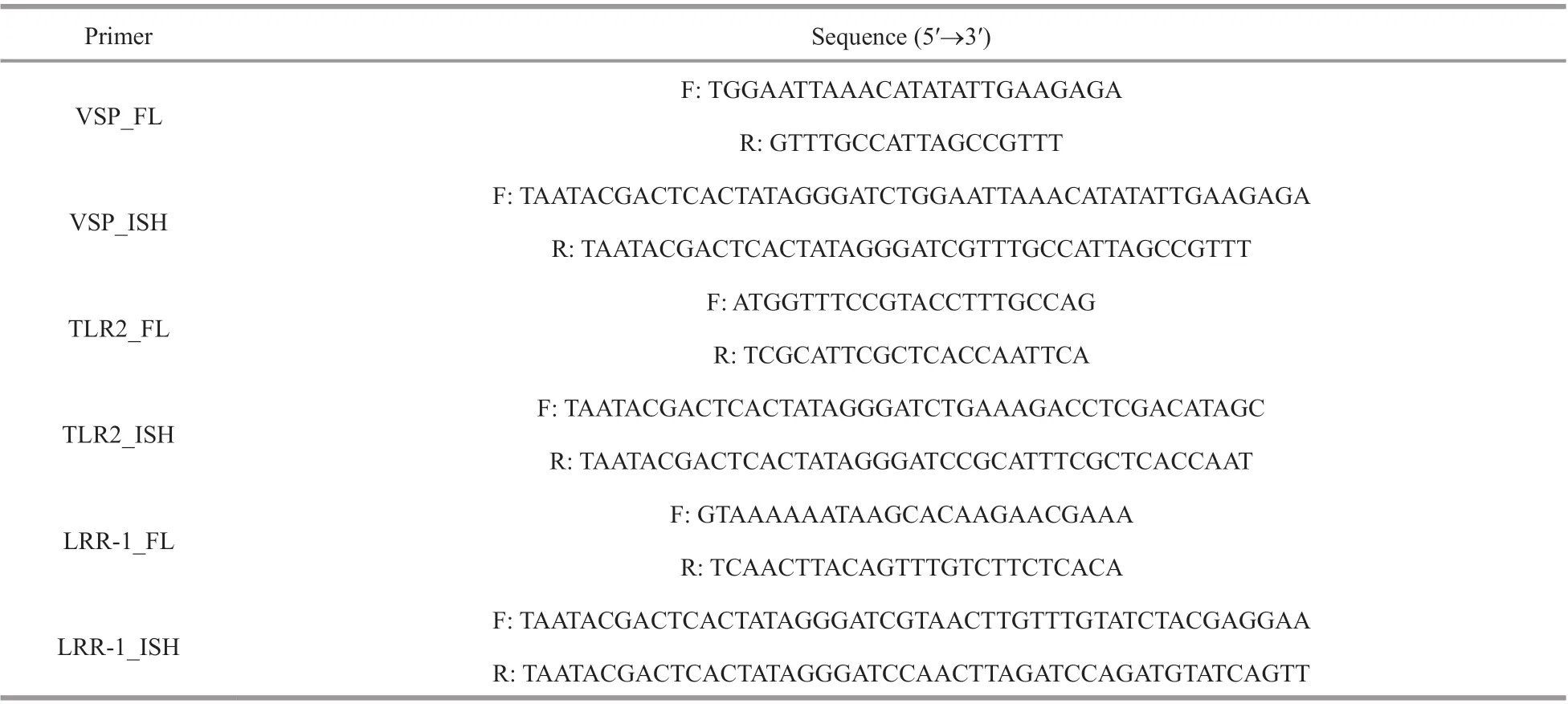
Table 1 Primers used in this study
2.3 Gene cloning and bioinformatics analysis
For ISH, full-length cDNA of vacuolar sorting protein (VSP), toll-like receptor 2 (TLR2), and leucine-rich repeat (LRR-1) were cloned using cDNA template from gill tissues, and the primers were designed based on the genome information of G. platifrons (Table 1). The PCR reactions were performed using exTaq (TaKaRa) according to the manual. The PCR program was set as follow: 94°C for 5 min, 35 cycles of 94°C for 20 s, 50–65°C (determined by Tm of primers) for 30 s and 72°C for 30 s–1 min (1 min of extension time per 1 kb amplicon), followed by the fi nal extension at 72°C for 10 min. The PCR products were purifi ed and then connected into the pMD19-T vector for sequence verifi cation. Full-length cDNA was translated into protein sequence and subjected to protein motif and structure prediction. The motif features were predicted by SMART (http://smart.embl-heidelberg.de) and the presumed tertiary structure of identifi ed proteins was conducted by Swiss-Model (http://swissmodel.expasy.org/interactive). The signal peptides were predicted by SignalP-5.0 (http://www.cbs.dtu.dk/services/SignalP/).
2.4 In situ hybridization (ISH)
Based on the results of pull-down experiments, VSP, TLR2, and LRR-1 genes were selected as representatives and subjected to ISH to investigate their expression patterns in gills. The synthesis of ISH probes and ISH assay were conducted using the method described by Halary et al. (2008). In brief, the DNA fragments of above genes were fi rst amplifi ed using specifi c primers pair (Table 1) as probe templates. Digoxigenin-labeled ssRNA probes including antisense probes and sense probes were synthesized using DIG RNA Labeling kit (Roche). Meanwhile, gills stored in 75% ethanol were dehydrated and embedded with paraffin. After sectioned at 7 μm thickness, gill samples were fi rstly treated with 10 μg/mL proteinase K for 10 min at 37°C to increase permeability, and pre-hybridized in hybridization buffer (50% formamide, 5×saline sodium citrate (SSC), 50 μg/mL Heparin sodium, 0.5 mg/mL fi sh sperm DNA, and 0.01% Tween 20) for 3 h at 37°C. Subsequently, gill sections were hybridized for 12–16 h at 37°C with 0.5 ng/μL probes in hybridization buffer. Afterward, the slides were washed in phosphate buffered saline with Tween 20 (PBST) and 4×SSC twice for 10 min each, and incubated with 2% bovine serum albumin (BSA) for 30 min along with 1‰ anti-digoxigenin-AP Fab fragments for 4 h. After washed by PBST, sections were incubated in development buffer pH 9.5 (100 mmol/L Tris-HCl, 100 mmol/L NaCl, 50 mmol/L MgCl2) for 10 min and stained with 5-bromo-4-chloro-3-indolyl-phosphate/nitro-blue-tetrazolium (BCIP/NBT) for 6–12 h. The reaction was then terminated by 0.5 mol/L ethylenediaminetetraacetic acid (EDTA) and imaged with microscopy after staining nucleic acids with 4’,6-diamidino-2-phenylindole (DAPI).

Fig.1 The eluted proteins were isolated by LPS pull-down assay and identifi ed by LC-MS/MS
3 RESULT
3.1 Isolation and identifi cation of G. platifrons proteins binding with LPS
LPS pull-down assay was conducted to isolate G. platifrons proteins with LPS-binding activity in gills. As a result, massive proteins with molecular weight ranging from 15–20 kDa and 40–75 kDa were obtained in the LPS pull-down elution. A total of 208 host gill proteins were then identifi ed by LCMS/MS (Supplementary Table S1). Among them, fi ve immune genes including LRR-1 (ID evm.TU.Super-Scaffold_12.16), TLR2 (ID evm.TU.Super-Scaffold_2293.4), integrin (ID evm.TU.Super-Scaffold_1642.4), VSP (ID evm.TU.scaffold307_size820992_obj.18) and matrix metalloproteinase 1 (MMP1, ID evm.TU.Super-Scaffold_1699.3) were identifi ed from LPS elution and urea elution. In detail, 3 fragments of TLR2, 7 fragments of VSP, 5 fragments of MMP1, and 18 fragments of integrin were also identifi ed in LC-MS/MS (Fig.1).
3.2 Structure characteristics of host gill proteins binding with LPS
Among the LPS binding genes described above, the structure characteristics and function of LRR-1 have been verifi ed by Chen et al. (2019). The protein structures of TLR2, integrin, MMP1 and VSP proteins were herein analyzed. For TLR2, the full length of its coding region was 1 023 bp and the open reading frame encoded a polypeptide of 340 amino acids residues with molecular weight approximately 38.75 kDa (Supplementary Table S2). The protein sequence had a typical tollinterleukin 1 receptor (TIR) domain but lacked LRR tandem repeats (Fig.2a). At the same time, the TLR2 protein exhibited a three-dimensional structure with a total of six α-helix and fi ve β-sheets (Fig.3a). For VSP, the full length of coding region was 816 bp and the open reading frame encoded a polypeptide of 271 amino acids residues with molecular weight approximately 31.40 kDa (Supplementary Table S1). Two low-density lipoprotein receptor class A (LDLa) domains were annotated (Fig.2b) and a signal peptide (Sec/SPI) was predicted in the VSP protein sequence. At the same time, the VSP protein exhibited a three-dimensional structure with two β-sheets (Fig.3b). For MMP1, the full length of coding region was 1 260 bp, and it could encode a polypeptide of 420 amino acids residues with molecular weight 48.25 kDa (Supplementary Table S1). A typical ZnMc domain and three tandem hemopexin-like repeat (HX) domains were annotated (Fig.2c). In addition, a total of fi ve α-helix and 22 β-sheets were predicted in its three-dimensional structure (Fig.3c). For integrin, the full length of coding region was 594 bp, which can encode a polypeptide of 197 amino acids residues with molecular weight 22.24 kDa (Supplementary Table S1). The protein sequence contained an integrin β subunit domain (Fig.2d), and exhibited a threedimensional structure with a total of four α-helix and eight β-sheets (Fig.3d).
3.3 Expression patterns of TLR2, VSP, LRR-1 in gill tissue
To further investigate the potential role of the above gene products in the symbiosis between hosts and MOBs, the expression patterns of LRR-1, TLR2, and VSP were analyzed by ISH assay in the gill tissues. As a result, mRNA of LRR-1, TLR2, and VSP was exclusively detected in bacteriocytes of gills where the symbionts occurred instead of ciliated cells (Figs.4–6). What’s more, combined with the localization of nuclei in gill cells by DAPI staining, majority of ISH signals were observed near the nuclei of the bacteriocytes (Fig.7).
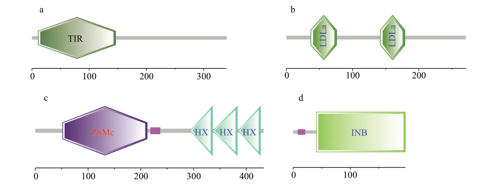
Fig.2 Protein domains of PRRs were annotated by SMART
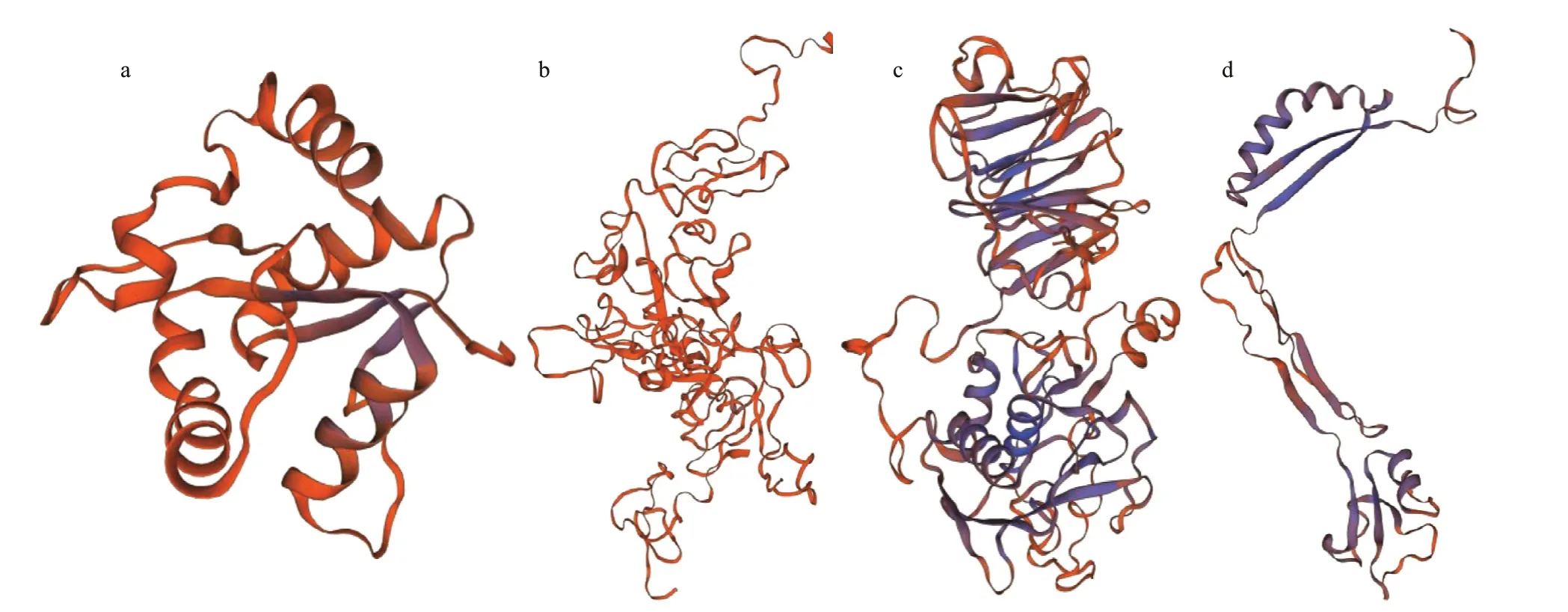
Fig.3 Tertiary structures of PRRs were built by Swiss-Model
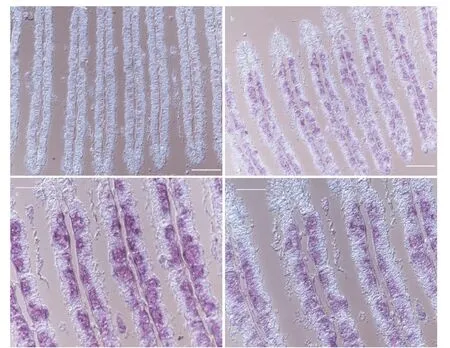
Fig.4 ISH of VSP in the gills of G. platifrons
4 DISCUSSION
4.1 LPS pull-down as an efficient method to identify symbiosis-related immune gene products
Regardless of massive efforts made in recent years, the lack of cultivable symbiotic MOBs has greatly hindered further research into the molecular mechanisms of the interaction between Bathymodioline and MOBs. Pull-down assay has been proven as an efficient method for verifying molecules involved in pathogen or symbionts recognition (Wu et al., 2011; Li et al., 2016). Given that symbiotic MOBs are γ-proteobacteria with LPS (constituted by lipid A, core oligosaccharide and O polysaccharide) in the outer membrane, LPS pull down assay was herein conducted to capture gill proteins that potentially bound with MOBs. Consequently, 208 proteins ranging from 15–75 kDa were identifi ed, among which fi ve immune genes were also identifi ed demonstrating the reliability of the method. What’s more, further verifi cation showed that LRR-1 was likely to play crucial roles in the immune recognition of MOBs (Chen et al., 2019), suggesting the credibility and applicability of the method in Bathymodioline research.
4.2 Immune recognition of symbionts
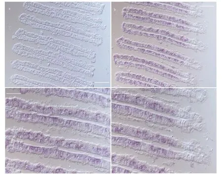
Fig.5 ISH of LRR-1 in the gills of G. platifrons
As suggested, PRRs mediated immune recognition of symbionts could be the fi rst step in initializing symbiosis. To date, multiple PRRs including Toll-like receptors (TLRs), immunoglobulin superfamily members (IgSF), peptidoglycan recognition proteins (PGRPs), lectins, and complement molecule (C1q) have been reported in Bathymodioline mussels and suggested their irreplaceable role in symbiosis (Bettencourt et al., 2009; Martins et al., 2014; Détrée et al., 2017; Zheng et al., 2017). What’s more, it was reported that G. platifrons can maintain a specifi c symbiotic relationship with MOBs. However, invertebrates are in lack of acquired immunity or immunoglobulins (Cerenius and Söderhäll, 2013). It was therefore speculated G. platifrons might recognize its symbionts through the arrangement and combination of multiple PRRs, which is worthy further investigation. However, none of them was further verifi ed either in vitro or in vivo. Two candidate PRRs including LRR-1 that has been verifi ed by Chen et al. (2019) and TLR2 in present study were isolated by LPS pull-down assay and further characterized by analyzing their structures and performing ISH.
LRR-1 is a classic member of leucine-rich repeat family with diverse functions in host immunity. As found by Chen et al. (2019), LRR-1 might serve as an intracellular recognition receptor for the symbionts by binding with LPS structure. Here, the distribution of LRR-1 was further confi rmed by ISH. In consistent with previous conclusions, ISH results showed that LRR-1 was expressed exclusively in bacteriocytes, further demonstrating that LRR-1 could play an important role in immune recognition symbiotic MOBs.
Besides, TLR2 is known as a typical member of TLR family that locates on the cell membrane, and could play important roles in the either pathogen recognition or symbionts recognition by binding with microbe-associated molecular patterns (Akira and Takeda, 2004). Genomic and transcriptome information has showed that TLR family, including TLR2 members, was highly expanded or expressed in G. platifrons and might be crucial in immune recognition of symbionts (Wong et al., 2015; Sun et al., 2017). In consistency, TLR2 was herein found exclusively expressed in bacteriocytes with signifi cant binding activity of LPS. As reported, TLR usually consists of an extracellular LRR domain and an intracellular TIR domain, and can interact with MyD88 to activate downstream intracellular signaling pathways (Kawai and Akira, 2010). In the present study, G. platifrons TLR2 was only found encoding a typical TIR domain which is believed to further trigger downstream signals in bacteriocytes and activates symbiosis related immune response.
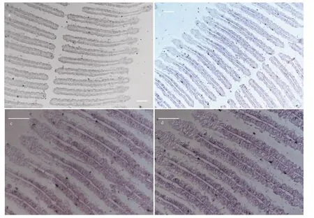
Fig.6 ISH of TLR2 in the gills of G. platifrons
4.3 Phagocytosis of symbionts
After host immune recognition of symbionts, phagocytosis or endocytosis of symbionts was suggested to be initiated. In our study, two immune genes involving in phagocytosis processes including integrin and VSP were identifi ed by pull-down assay. Integrins are transmembrane receptors that facilitate cell-extracellular matrix (ECM) adhesion. Upon ligand binding, integrins can activate signal transduction pathway that initiate cell phagocytosis (Giancotti and Ruoslahti, 1999). Integrins usually consist of two subunits, α subunit, and β subunit. It was suggested that α subunit could bind with divalent cations that regulate integrin activity. Meanwhile, β subunit usually has four cysteine-rich repeats and that could promote cell adhesion or cell-cell interactions (Humphries, 2000). The integrin protein isolated by LPS pull-down was found containing a typical β subunit. It is therefore speculated that the integrin protein might activate the phagocytosis of symbiotic MOBs through binding with LPS structure.
VSP is known to be involved in the intracellular sorting and delivery of soluble vacuolar proteins, which is essential for late endosome and lysosome assembly and for numerous endolysosomal trafficking pathway (Kolesnikova et al., 2009). In our study, VSP protein, with two LDLa domains and a signal peptide, was also identifi ed in LPS pull-down assay. Though the binding site of VSP against LPS remained unknown, ISH assay demonstrated unique expression pattern of VSP in bacteriocytes, implying its participation in the phagocytosis and transport of symbiotic MOBs in bacteriocytes.
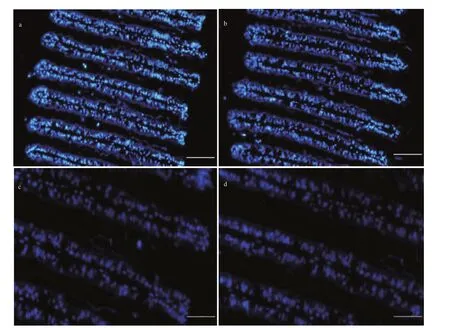
Fig.7 FISH with DAPI, showing the distribution of nuclei of gill cells
4.4 Digestion of symbionts
As demonstrated, the Bathymodioline mussels could obtain majority of nutrient needed from their endosymbionts via either “farming” or “milking” way (Fiala-Médioni et al., 2002; Kádár et al., 2008). Though the exact way remains largely unknown, lysosome mediated protein digestion is irreplaceable. Here, MMP1, a key proteinase involving in digestion of proteins, was identifi ed by LPS pull-down assay. MMPs are calcium-dependent zinc-containing endopeptidase, which have been widely identifi ed in vertebrates, invertebrates and plants (Verma and Hansch, 2007). These enzymes can directly digest various extracellular matrix proteins and other bioactive molecules, and play essential roles in cell differentiation and motility, as well as in inflammation and wound healing (Van Lint and Libert, 2007). The MMPs usually contain three conserved domains including pro-peptide domain, catalytic domain and hemopexin-like C-terminal domain. The MMP molecule isolated by LPS pull-down was identifi ed as MMP1, which has a typical ZnMc domain and three tandem HX domains. Given the crucial role of MMPs in the digestion of extracellular matrix in multiple processes, it is inferred that MMP1 may be responsible for the degradation of symbionts directly or indirectly and further promoting the nutrient transferring into G. platifrons cells.
5 CONCLUSION
In this study, through LPS pull-down assay, we identifi ed four new candidate immune genes that may participate in the symbiosis between G. platifrons and its symbiotic MOBs in addition to LRR-1: VSP, TLR2, MMP1, and integrin. ISH assay was further conducted to investigate the expression patterns of LRR-1, TLR2, and VSP. Consequently, it was found that LRR-1, TLR2 and VSP were expressed exclusively in gill bacteriocytes where symbionts are harbored. Meanwhile, host immune recognition, phagocytosis and lysosome-mediated digestion were suggested to be modulated by above genes. Collectively, the present study has proposed an effective method for screening symbiosis-related immune genes in G. platifrons considering the lack of culturable symbiotic MOBs. Moreover, results obtained herein have also shed new insights into the biological process of symbiosis between G. platifrons and symbiotic MOBs.
6 DATA AVAILABILITY STATEMENT
All data generated and/or analyzed during the study are available from the corresponding author on reasonable request.
7 ACKNOWLEDGMENT
We thank all the crews onboard the R/V Kexue for their assistance in sample collection and all the laboratory staff for continuous technical advice and helpful discussions.
 Journal of Oceanology and Limnology2020年4期
- Journal of Oceanology and Limnology的其它文章
- Geological, physical, and chemical characteristics of seafloor hydrothermal vent fi elds*
- Rediscovery of the abyssal species Peniagone leander Pawson and Foell, 1986 (Holothuroidea: Elasipodida: Elpidiidae): the fi rst record from the Mariana Trench area*
- Gametogenesis and reproductive traits of the cold-seep mussel Gigantidas platifrons in the South China Sea*
- Dynamic features of near-inertial oscillations in the Northwestern Pacifi c derived from mooring observations from 2015 to 2018*
- Arctic multiyear sea ice variability observed from satellites: a review*
- Fabrication of anodized superhydrophobic 5083 aluminum alloy surface for marine anti-corrosion and anti-biofouling*
