Simplified Modeling and Mathematical Analysis in Cardiac Electric Field Propagation for Human and Animals
Ping Lang and Lei Han
(1. School of Information and Electronics, Beijing Institute of Technology, Beijing 100081, China;2. School of Mechatronical Engineering, Beijing Institute of Technology, Beijing 100081, China)
Abstract: The different characteristics of cardiac electric field (CEF) radiation in humans and other animals are presented in this paper. Physical modeling and mathematical analysis are developed to comprehensively unveil the properties of CEF, based on typical heartbeat waveforms. Our numerical simulation results demonstrate that the frequency bandwidths and the cycle durations of CEF are different for healthy humans versus humans on the verge of death and for humans versus other animals. The results indicate that the present study may extensively contribute towards recognizing human beings or other animal targets quickly and accurately with CEF in dangerous situations or in other applications.
Key words: mathematical analysis;distinguishing standards;cardiac electric field;targets identification
Over the past few years, noncontact techniques for detecting humans (NTDHs) have been developing rapidly, given the increasing requirements for NTDH in anti-terrorism, anti-riot, local urban war, security, and other fields[1−3]. For example, in city street wars, NTDH can discover hidden enemies, significantly improving soldiers’survivability. NTDH also plays an important role in modern warfare. The police, for example, can determine the location of criminals and hostages with NTDH, and execute effective measures to save hostages. In addition, security guards can detect whether containers contain stolen items or stowaways, using NTDH. Furthermore, NTDH plays a particularly vital role in lifesaving efforts during fires, underground cave or mine emergencies, and earthquakes, vastly decreasing rescue costs and casualties while improving rescue efficiency.
Electrostatic detection (ED) is a novel method to obtain the object’s information, which detects the electric field originating from an object’s surface charge or heartbeat[4−6]. This paper studies the characteristics of the cardiac electric field propagation using physical modeling and mathematical analysis based on previous works[7−8]. The technique used is effective, practical, and simple. The field is a time-varying electric field, which is suitable for transmitting through an obstacle, different from the fields used in the previous studies. The main difference between this work and previous works is establishment of reasonable and practical cardiac electric field models, which contribute to presenting recognition standards that can easily distinguish between humans and other animals based on frequency spectrum analysis.
1 Model Description
1.1 Human cardiac electric field model
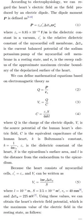

Since the heart is located in the chest, the cardiac electric field sequentially radiates through organic tissue from inside to outside the body:heart, lungs, skeletal muscle, subcutaneous fat,skin, and so on. Impedance attenuation occurs during the transmission of the electric field due to the impedance of organic tissues. In this work,a reasonable heart-torso model is necessary to calculate the impedance of organic tissue and deduce the transfer function from intra- to outerbody. We can obtain an equal-impedance-coupled circuit model from the heart–torso model and a transfer function from the equal-impedancecoupled circuit model. Then, the cardiac electric field of the human body’s surface can be obtained based on the transfer function and the heart’s electric field potential shown in Eq.(6).
Different electrocardiogram (ECG) studies use a variety of heart modeling methods[11−12], including concentric ball models, eccentricity models, cylindrical models, and CT and MRI scans,to devise a heart–torso model[13]. In this work, a simple heart–torso model that contains the heart,lungs, skeletal muscle, subcutaneous fat, and skin is adopted. The heart and lungs are modeled with the ball model, and the torso uses the elliptical column model, as shown in Fig.1. The center of the heart and the lungs coincide with the center of the torso for the model and calculations; in the present work, we only consider the organic tissues that have a significant impact on transmission of the electric field. We make the following assumptions:

Fig. 1 Heart–torso model and cross section.
① The organic tissues are homogeneous structure;
② The heart’s electric field undergoes isotropic transmission in organic tissue.
The typical parameters of the heart–torso model are as follows: the long axis of the torso’s cross-sectional area is 400 mm, the short axis is 200 mm, and the height is 600 mm. The thickness of the skin is 2 mm, the thickness of subcutaneous fat is 8 mm, the thickness of the skeletal muscle is 10 mm, the radius of the endocardium is 40 mm, the radius of the epicardium is 50 mm,and the radius of the lung is 80 mm[14−15].
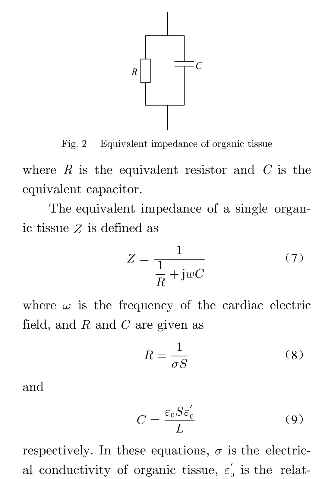
The equivalent impedance of a single organic tissue can be modeled by an equivalent resistor in parallel with an equivalent capacitor, as shown in Fig.2.

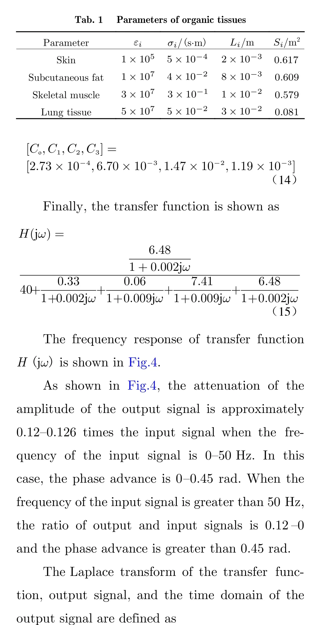

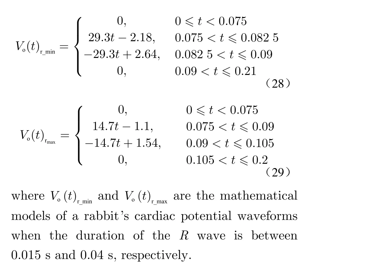
1.2 Mathematical model of the cardiac electric field for humans and animals

Fig. 4 Frequency response of transfer function H(jω)


2 Mathematical simulation results
Fig.6 shows the waveform of a cardiac electric field of the body’s surface for a healthy human in the intra-body and a human on the verge of death. As shown, the frequency bandwidth and the cycle of the heart’s electric field from the intra-body to the body’s surface are approximately constant, whereas the amplitude of the electric field is dramatically reduced. The cardiac electric field of a human on the verge of death is approximately zero and the frequency is mainly between 0–8Hz. As shown in Fig.7, the maximum potential is 3.3 mV and the cycle time is 0.83 s when the duration of a human cardiac cycle’s electric field is 0.04 s–0.07 s. In addition, the frequency bandwidth range of the electric field is approximately 30 Hz–47 Hz and the center frequency bandwidth is 40 Hz, which demonstrates that the cardiac electric field of a human is at an ultralow frequency and has a weak signal. In the literature[23], the frequency spectrum range for ECG is 0–40 Hz (±7 Hz), based on the analysis and statistics from the European ST-T Database,which agrees with the results of this work.

Fig. 6 Waveform of the cardiac electric field for a healthy human at the body’s surface, in the intra-body, and on the verge of death

Fig. 7 Waveform of a human cardiac electric field over the course of a cycle

Fig. 8 Time domain waveform
Fig.8 shows the experimental results of several Japanese scholars[4−6]. (Image from Koichi K.et al.[6]). As shown, there is noQwave, which indicates that a detector cannot detect the electric field generated by this weak wave. The simplification of the waveform of a human cardiac electric field to just theRwave in the noncontact detection, presented in this work, is consistent with the method used in the Japanese scholars’experiment. The frequency spectrum over the course of a cycle is shown in Fig.9. (Data from Koichi K. et al.[6]).
The frequency of a human heart’s electric field primarily stays between 0 and 40 Hz, which agrees with the experimental results of this paper.
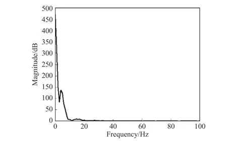
Fig. 9 Frequency spectrum
In summary, the mathematical model of the human body's electric field established in this paper is reasonable and feasible.
The radiation results of different animals’cardiac electric fields are shown in Figs.10–12.The error between the maxima of the two curves in the time domain graph is due to an approximation caused by the software and can be ignored.

Fig. 10 Waveform of a dog’s cardiac electric field

Fig. 11 Waveform of a guinea pig’s cardiac electric field
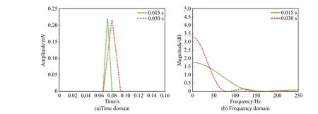
Fig. 12 Waveform of a rabbit’s cardiac electric field
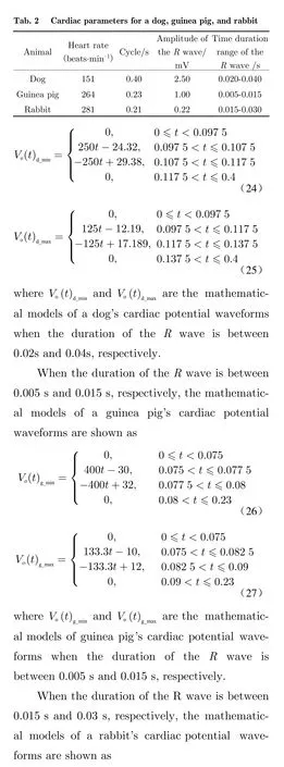
Figs.10– 12 show the time domain waveforms and the frequency spectra of the heart’s electric field of noncontact detection for a dog,guinea pig, and rabbit, respectively. As shown in Fig.10, the maximum of the dog’s potential is approximately 2.5 mV, the cycle time is 0.4 s, and the bandwidth range of the dog’s cardiac electric field is 50 Hz–90 Hz. As shown in Fig.11, the maximum of the guinea pig’s potential is approximately 1 mV, the cycle time is 0.23 s, and the bandwidth range of the guinea pig’s cardiac electric field is 120 Hz–500 Hz. As shown in Fig.12,the maximum of the rabbit’s potential is approximately 0.22 mV, the cycle time is 0.21 s, and the bandwidth range of a rabbit’s cardiac electric field is 70 Hz–120 Hz.
The distinguishing standards of object identification, based on the aforementioned analysis of the cardiac electric field for human and other animals, obtained by noncontact detection technology are as follows:
①There are three different types of human cardiac electric fields based on the frequency bandwidth. The bandwidth range of a healthy human heart’s electric field is 30 Hz–47 Hz, with a center frequency of 40 Hz. The average bandwidth of a cardiac electric field of a human on the verge of death is 8 Hz, and the bandwidth of a dead heart’s electric field is 0 Hz.
②We can identify humans and other animals by the frequency bandwidth, cycle duration,and amplitude of the heart’s electric field. The bandwidth ranges of the heart’s electric field for a healthy human, dog, guinea pig, and rabbit are 30 Hz–47 Hz, 50 Hz–90 Hz, 120 Hz–500 Hz, and 70 Hz–120 Hz, respectively, and the cycle durations are 0.83 s, 0.4 s, 0.23 s and 0.21 s, respectively. Therefore, we can distinguish between human and other animal targets quickly and accurately based on the time and spectral analysis of the cardiac electric field detected.
In summary, this section concludes a distinguishing standard based on characteristics of the cardiac electronic field. This standard can be used to identify a normal human body, a human body on the verge of death, and other animal targets quickly and accurately, based on a time domain and frequency domain analysis of the detected target’s cardiac electric field. The research results maybe assist with a precision strike, a rescue, anti-terrorism, or other actions quickly, and when engaged in dangerous situations, such as city street warfare, earthquake relief, mine disaster relief, rescuing hostages, border security,thereby greatly improving the rescue or relief efficiency.
3 Conclusion
This work develops the characteristics analysis of cardiac electric field for humans and other animals, using modeling and mathematical analysis in a non-interference environment, which can be applied in target identification. The data analysis may provide some theoretical basis for guiding the design of the detector described in this paper. The present results demonstrate the tremendous potential of heartbeat measurement,biological identification technology, healthcare monitoring, and especially living target identification in engineering applications, although deeper theoretical and experimental studies are required to validate the predictions and conclusions of this study.
 Journal of Beijing Institute of Technology2020年2期
- Journal of Beijing Institute of Technology的其它文章
- Anisotropic Total Variation Regularization Based NAS-RIF Blind Restoration Method for OCT Image
- 2D Augmented Coprime Array Geometry Based on the Difference and Sum Coarray Concept
- Research on Measurement Method of Muffler Performance under High Temperature andHigh-Speed Airflow Conditions
- Design of a 70 MPa Two-Way Proportional Cartridge Valve for Large-Size Hydraulic Forging Press
- Object Recognition Algorithm Based on an Improved Convolutional Neural Network
- Research of Current Mode Atomic Force Microscopy (C-AFM) for Si/SiC Heterostructures on 6H-SiC(0001)
