Anisotropic Total Variation Regularization Based NAS-RIF Blind Restoration Method for OCT Image
Xuesong Fu, Jianlin Wang,, Zhixiong Hu, Yongqi Guo, Kepeng Qiu and Rutong Wang
(1. College of Information Science and Technology, Beijing University of Chemical Technology,Beijing 100029, China;2. Division of Medical and Biological Measurement, National Institute of Metrology, Beijing 100029, China)
Abstract: Based on anisotropic total variation regularization (ATVR), a nonnegativity and support constraints recursive inverse filtering(NAS-RIF) blind restoration method is proposed to enhance the quality of optical coherence tomography (OCT) image. First, ATVR is introduced into the cost function of NAS-RIF to improve the noise robustness and retain the details in the image.Since the split Bregman iterative is used to optimize the ATVR based cost function, the ATVR based NAS-RIF blind restoration method is then constructed. Furthermore, combined with the geometric nonlinear diffusion filter and the Poisson-distribution-based minimum error thresholding, the ATVR based NAS-RIF blind restoration method is used to realize the blind OCT image restoration. The experimental results demonstrate that the ATVR based NAS-RIF blind restoration method can successfully retain the details in the OCT images. In addition, the signal-to-noise ratio of the blind restored OCT images can be improved, along with the noise robustness.
Key words: optical coherence tomography(OCT) image;blind image restoration;cost function;nonnegativity and support constraints recursive inverse filtering(NAS-RIF)
Optical coherence tomography (OCT) is widely used in biomedical applications, clinical medicine, and nondestructive testing[1]owing to the characteristics such as noncontact, noninvasive, and real-time imaging. However, the OCT system is susceptible to charge-coupled device(CCD) integration effects and noise during imaging. This problem leads to the degradation of OCT images[2]and low noise robustness[3], making it difficult to distinguish target details in OCT images and affecting the accuracy of OCT image analysis. Therefore, the accuracy of OCT image analysis will be improved by achieving OCT image restoration, improving the signal-tonoise ratio (SNR) of OCT images, and obtaining restored OCT images with clear structures and obvious edges.
The main image restoration methods consist of the non-blind restoration method[4]and the blind restoration method[5]. The non-blind restoration method obtains clear image and realizes image restoration via deconvolution of the blurred image and the point spread function (PSF). Given the difficulty of obtaining the accurate PSF of images in practical applications, it is difficult to obtain a high-quality restored image via the nonblind restoration method[6]. When the PSF is unknown or not fully known, the blind restoration method obtains a better estimate of the restored image by adding the constraints of nonnegativity,the target intensity, and the size of support[7].The main blind restoration methods include iterative blind deconvolution (IBD)[8]and nonnegativity and support constraints recursive inverse filtering (NAS-RIF)[9]. The IBD method achieves blind restoration by alternately imposing energyconservation and nonnegativity constraints on the PSF and image in the frequency domain and spatial domain, and it exhibits lower computational complexity. Tsumuraya et al.[10]introduced the Lucy-Richardson (LR) algorithm into the IBD method to maintain the nonnegativity and energy-conservation of the restored image during image restoration, which could improve the reliability and convergence stability of the IBD method. However, the problem of PSF constraints remained unresolved. Li et al.[11]reconstructed OCT speckle images using the IBD method to maximize the speckle contrast at different depths. However, the reliability and convergence of this method were unsatisfactory, and it was not possible to guarantee the uniqueness of the solution. Furthermore, the method was highly dependent on the initial estimation of OCT images.
The NAS-RIF method achieves image restoration by constraining the nonnegativity and support of the target image. It does not heavily rely on prior knowledge or accurate estimation of the PSF and has advantages such as a simple structure, few iterations, and good convergence. Additionally, the uniqueness of the solution is guaranteed.Yet the image restoration effect of the NASRIF method is susceptible to noise. With regard to the noise-sensitive problem, Zhang et al.[12]added low-pass filtering to the iteration of the NAS-RIF method based on the pre-processing of the image using a smooth-edge filter to enhance the noise robustness. Compared with the restoration result of the LR method, the improved NASRIF method exhibited a higher SNR of restoration image. Gao et al.[13]used a curvelet filter to pre-process the image and introduced the target edge preservation term in the cost function of the NAS-RIF method. Guo et al.[14]proposed a spaceadaptive and regularized NAS-RIF method by combining NAS-RIF with regularization. Two spatial adaptive weighting terms were introduced in the cost function to suppress noise, improving the robustness of the image-restoration process to noise. Subsequently, concerning the detail loss of restored image caused by the image pre-processed, Zhang et al.[15]initially pre-processed the image via adaptive pseudo-median filtering and introduced a correction term based on the target information to the cost function,which was optimized using a conjugate gradient method and a logarithmic function. Consequently,the image edge retention effect and peak SNR were improved. Li et al.[16]introduced isotropic total variation regularization in the cost function and used an iterative method of optimization minimization. Although the image SNR was significantly improved, the isotropic total variation did not effectively use the information in different directions of the target structure, which could cause structural blurring. Furthermore, the solution process of iterative optimization minimization was complex. Due to the advantages of NAS-RIF method in blind image restoration, Jan et al.[17]used the NAS-RIF method for blind OCT image restoration. The blind OCT image restoration quality of NAS-RIF was better than that of the constrained iterative restoration method. However, the NAS-RIF method did not consider the effects of actual noise. Although the above improved NAS-RIF methods are investigated to improve the SNR and blind OCT image restoration effects, they do not preserve the detailed image information or fully utilize the image information to improve the cost function.The cost function and its optimization are crucial for blind image restoration. The improvement and optimization of the cost function of the NAS-RIF method can effectively improve the noise robustness of the blind OCT image restoration, reduce the complexity of the cost function solution, and preserve the target details of OCT images.
To address the problems of low noise robustness and loss of target details in OCT image for the blind OCT image restoration using the NASRIF method, an anisotropic total variation regularization (ATVR)based NAS-RIF blind restoration method is proposed to enhance the quality of OCT image. First, ATVR employs the L1 norm of the gradients inxandydirections to characterize the information inxandydirections, respectively. Hence, ATVR is sensitive to the difference in the directionality of the edges and can effectively protect the target edges and details.Therefore,ATVR was introduced into the cost function of NAS-RIF to improve the noise robustness and OCT image detail information protection ability of NAS-RIF. The split Bregman iterative was used to optimize the ATVR based cost function. On this basis, the ATVR based NAS-RIF blind restoration method was constructed. Then, the noise in the OCT image was partially filtered via the geometric nonlinear diffusion filter (GNLDF), and the Poisson-distribution-based minimum error thresholding was used to estimate the support of the OCT image. Finally, the ATVR based NAS-RIF blind restoration method was used for the blind OCT image restoration. The effectiveness of the proposed method was verified by blind restoration experiments of various OCT images.
1 NAS-RIF Blind Restoration
The NAS-RIF blind restoration achieves restoration by limiting the inverse filtering iterative process to predict the clear image[9]. The blurred imageg(x,y) (xandyare the coordinates of image pixel) is input into a two-dimensional variable coefficient finite impulse response (FIR) filteru(x,y) to obtain an estimate of the clear imagefˆ(x,y). Additionally,fˆ(x,y) is input into the nonlinear filter NL to obtain the projected imagefˆNL(x,y). The FIR filter coefficient is continuously corrected according to the cost function which is obtained fromfˆ(x,y) andfˆNL(x,y). The conjugate gradient is used to reduce and gradually converge the cost function. The restoration of the degraded image is realized whenfˆNL(x,y)gradually approach the clear image. A block diagram of the NAS-RIF blind restoration is shown in Fig.1.

Fig. 1 Block diagram of the NAS-RIF blind restoration
The nonlinear filter NL is defined as follows

whereDsuprepresents a set of pixels in the region of support,Dsuprepresents a set of pixels beyond the region of support which is the background,andLBrepresents the mean pixel value of the background. The support is the region that includes the target. In the NAS-RIF method,the support is represented by the smallest rectangular containing complete targets. The background is the area without the target.
The cost function is given by

The expanded form is given by
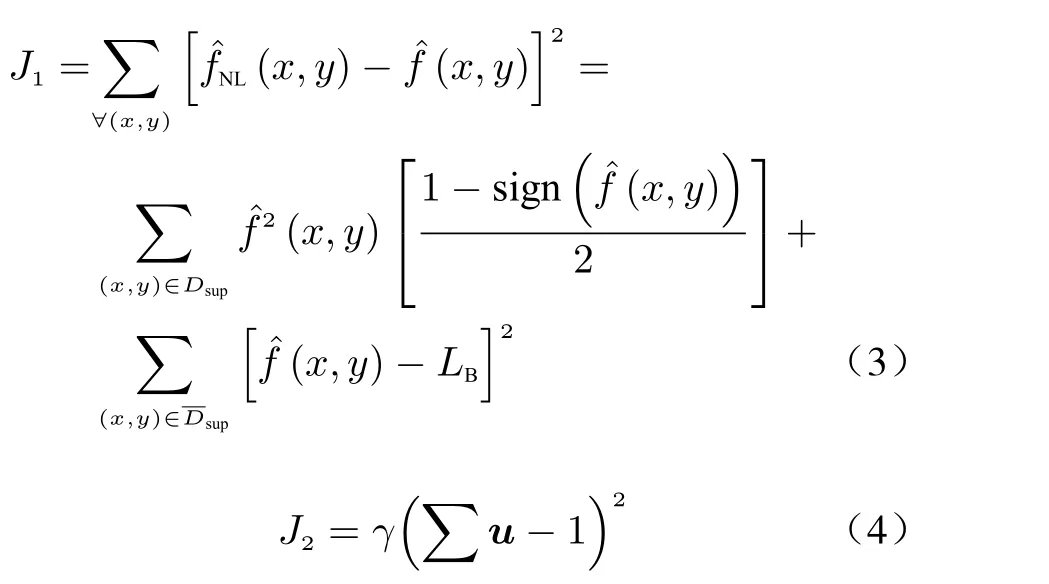
The first part inJ1is used to limit the negative pixels in the support, and the second part inJ1is used to limit the pixels outside the support that are not equal to the background mean pixel value. sign(·) is a symbolic function that is defined as followsJ2is used to constrainu(x,y) away from the trivial all-zero global minimum. Thus,γis nonzero only whenLBis zero.

The NAS-RIF blind restoration can effectively improve the image quality, but its restoration process is easily affected by noise. There is a large amount of speckle noise in the OCT image,which affects the blind OCT image restoration effect of the NAS-RIF blind restoration. Therefore,it is necessary to improve the noise robustness of the NAS-RIF blind restoration, make the structure edges of the OCT image more clear after restoration, and improve the SNR of the OCT image after restoration.
2 ATVR Based NAS-RIF Blind Restoration Method
Noise is commonly present in the imaging process. The small disturbance caused by noise in the degraded image reduces the accuracy of image estimation and leads to significant deviations in the estimated image. The NAS-RIF has low robustness to noise. Thus,it was enhanced by improving the cost function and optimization.First, an ATVR based cost function was constructed by introducing ATVR of estimated image to the original cost function. The robustness to noise in the restoration process was improved by limiting the ATVR to suppress the noise. To maintain the detailed information and edge integrity of the image, the smoothing effect was not applied to the target. Subsequently, the split Bregman iterative was used to optimize the ATVR based cost function with the L1 term.Auxiliary variables, quadratic penalty function,and Bregman iteration were introduced to optimize the ATVR based cost function via a simplified solution process, which improved the numerical solution stability of the method.
2.1 ATVR based cost function


2.2 Optimization of cost function based on the split Bregman
The cost function of the NAS-RIF is a convex function optimization solution aboutu(x,y),which is generally converged to the global minimum via the conjugate gradient. However, the ATVR based Eq. (11) corresponds to the optimization for the L1 regularization term, which cannot be solved via the conjugate gradient. The split Bregman iterative can transform the optimization problem with the L1 regularization term into a series of unconstrained optimization problems by introducing auxiliary variables, quadratic penalty function, and Bregman iteration.Then,the numerical solution process can be optimized by alternately solving sub-problems[18]. This optimization method improves the stability of the numerical solution process and simplifies the solution of the ATVR based cost function.


2.3 ATVR based NAS-RIF blind restoration method


Fig. 2 Block diagram of the ATVR based NAS-RIF blind restoration method
3 ATVR Based NAS-RIF Blind Restoration Method for OCT Image
3.1 Implementation of ATVR based NAS-RIF blind restoration method for OCT image
OCT system is interfered by noise such as photoelectricity noise and speckle noise during the imaging process. The NAS-RIF blind restoration exhibits poor noise resistance, affecting the cost function and leading to a deviation in the estimated image. The GNLDF[19]utilizes the global noise characteristic information and considers the local information of the neighborhood characteristics of the pixel to be processed. It exhibits good local diffusivity, a high diffusion speed, and a good edge preservation effect. Therefore, an OCT image was pre-processed via the GNLDF,part of the noise was filtered, while the original information of the OCT image was preserved.Hence, the effect of noise on the restoration process was decreased, meanwhile, the quality of the OCT restored image was improved.
For NAS-RIF blind restoration, the optimization of the cost function is affected by support.Therefore, the accuracy of support will affect the optimization of the cost function, and then affect the OCT image restoration effect. The support of NAS-RIF is a rectangular frame containing the target, but the most target shapes are non-rectangular. Hence, the rectangular support will affect the extraction effect of the target area and then reduce the OCT image restoration effect. When the image is divided into target and background, the pixel value distribution of the image conforms to a Poisson distribution. Therefore, the Poisson-distribution-based minimum error thresholding[20]was used to estimate the target support,which could accurately segment targets from images. And all the segmented target regions were the support in our method.
The processing steps of ATVR based NASRIF blind restoration method for OCT image are shown in Fig.3. First, the OCT image is input and denoised via the GNLDF. The Poisson-distribution-based minimum error thresholding is used to estimate the support of the OCT image. The ATVR based cost function is then constructed,and the split Bregman iterative is used to optimize the ATVR based cost function. The FIR filter coefficients are adjusted via iteration such that the projected image is close to the clear image to achieve blind restoration of the OCT image.
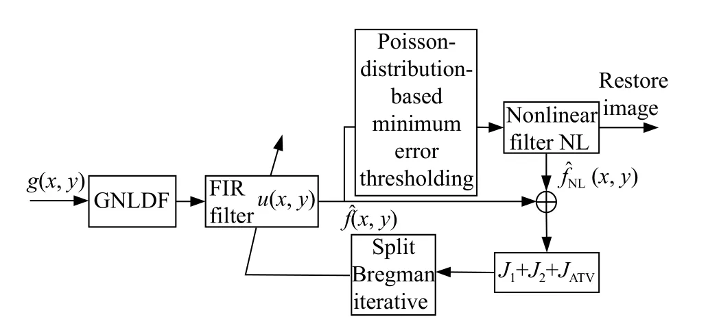
Fig. 3 Block diagram of ATVR based NAS-RIF blind restoration method for OCT image
3.2 ATVR based NAS-RIF blind restoration algorithm for OCT image
3.2.1 Algorithm flow
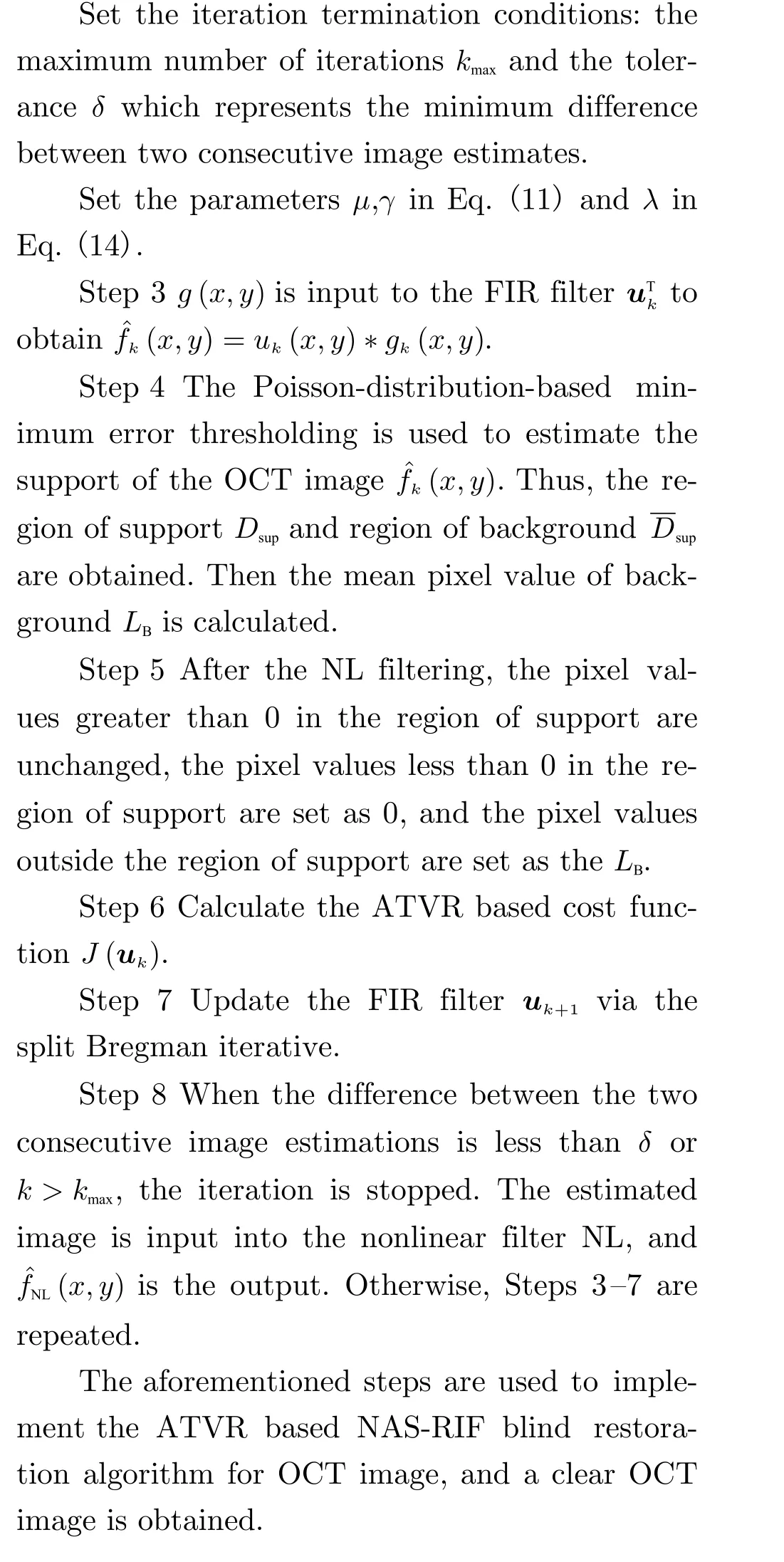
The OCT image is pre-processed via the GNLDF. The Poisson-distribution-based minimum error thresholding is used to accurately estimate the support of the OCT image. Subsequently, the ATVR based NAS-RIF blind restoration method is used to blindly restore the OCT image. The implementation process of ATVR based NAS-RIF blind restoration algorithm for OCT image is shown in Fig.4.


4 Results and Analysis of Experiments
OCT images obtained by a swept-source OCT (SS-OCT) system and a spectral-domain OCT (SD-OCT) system were used to blind restoration experiments.
The SS-OCT system used a Santec HSL-2100 light source with a center wavelength of 1 310 nm, a bandwidth of 170 nm, and a scan frequency of 20 kHz. The axial resolution of the system was 10 μm. Orange tissue was scanned by the SS-OCT system, and the obtained B-scan image is shown in Fig.5a. The partial enlargement of the orange tissue OCT image is shown in Fig.6a. The SD-OCT system used a light source with a center wavelength of 840 nm and a bandwidth of 100 nm. The system acquisition frequency was 50 kHz. And the axial resolution of the system was 6 μm. A human retina was scanned via the SD-OCT system, and the obtained B-scan image is shown in Fig.7a. Additionally, the open-access lesion retinal OCT image[21]was used, as shown in Fig.8a. The specifications of the graphics data workstation were listed as follows: Intel Core i7-4720HQ, 8 GB of random-access memory, MATLAB R2017b.µ=200 when dealing with the orange OCT image, andµ=300 when dealing with the normal and lesioned retinal OCT images.γ=10 when background is all black, otherwise,γ=0,λ=0.5,kmax=20 ,δ=0.001 in three experiments. Those values are obtained through multiple experiments.

Fig. 5 Results of the orange tissue OCT image processed via different algorithms
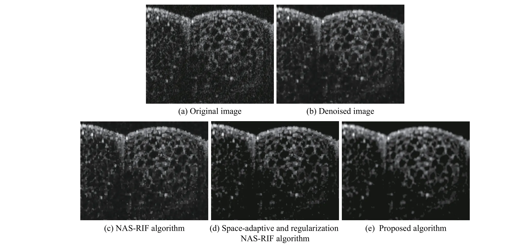
Fig. 6 Enlarged detail results of orange tissue OCT image processed via different algorithms
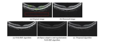
Fig. 7 Results of human retinal OCT image processed via different algorithms

Fig. 8 Results of the lesioned human retinal OCT image processed via different algorithms
The Metrics —SNR, contrast-to-noise ratio(CNR)[22], and average equivalent number of looks (ENL)[23], which are given in Eq.(17)—were used to evaluate the OCT image quality. SNR measured the signal-to-noise ratio of the image feature region, CNR measured the contrast between the image feature region and the background noise region, and the ENL measured the smoothness of the image feature region.

whereIrepresents the pixel value of the image,Rrepresents the number of target regions, andµrandσrrepresent the mean and standard deviation of therth target region, respectively. Additionally,µbandσbrepresent the mean and standard deviation of the background region, respectively.
As shown in Figs.5a, 7a and 8a, three target areas and one background area were selected in the OCT images for the calculation of SNR, CNR and ENL. The target areas were marked with a green square, and the background area was marked with a red square. The blind restoration results for the orange tissue OCT image are shown in Fig.5. To explain the restoration results, a partial enlargement of the orange tissue is shown in Fig.6. The results of the blind restoration for the human retinal OCT image are shown in Fig.7. The results of the blind restoration for the lesioned retinal OCT image are shown in Fig.8. The three kinds of OCT images were preprocessed via the GNLDF, and the Poisson-distribution-based minimum error thresholding was used to estimate the support of OCT images.The ATVR based NAS-RIF blind restoration method was compared with the NAS-RIF and the space-adaptive and regularization NASRIF[14].
The blind restoration results of orange tissue OCT image are shown in Fig.5. Fig.5a shows the original image. The noise is severe, and the edges of the orange structure are distorted by noise. Fig.5b shows the pre-processed image.Some of the noise in the image is filtered out,and the edge of the orange structure is clearer.Fig.5c shows the result of the NAS-RIF. The image is dark overall owing to the energy loss. The noise is still severe, and the edge of the orange structure is unclear. Fig.5d shows the result of the space-adaptive and regularization NAS-RIF.Compared with the NAS-RIF, the noise is suppressed, and the edge of the orange structure is clear. However, the image energy loss is severe,the effect of noise suppression is poor, and the CNR of the method is lower than that of the NAS-RIF. Fig.5e presents the result of the proposed algorithm. As shown in this figure, the noise and energy attenuation are suppressed, and the edge of the orange structure is clear. The regularization term of noise suppression is added to the cost function; thus, the restored orange structure is relatively smooth. To further compare the restoration effect, the local tissue of the orange in Fig.5 is enlarged, as shown in Fig.6. The clarity of the tissue structure for our method is better than that of the other methods. The NAS-RIF and the space-adaptive and regularization NASRIF yield speckle noise in the orange tissue structure, and the effect of orange tissue structure restoration is poor. The evaluation metrics are compared in Tab.1. The SNR, CNR, and ENL are significantly improved for the proposed algorithm compared with those of the other two algorithms.
The blind restoration results of human retinal OCT image are shown in Fig.7. Fig.7a shows the original image.The noise is severe, and the edges of the retinal tissue are blurred. Fig.7b shows the pre-processed image.Some of the noise in the image is filtered out, and the edges of the image are well maintained. Fig.7c shows the result of the NAS-RIF. The image energy loss and noise are still severe. Additionally, the retinal layered structure is unclear. Fig.7d shows the result of the space-adaptive and regularization NASRIF. Although the noise is suppressed and the edges of the target are clear,there is image energy loss, a limited noise-suppression effect, and poor structural protection of the retinal layer.Fig.7e presents the results of the proposed algorithm. As shown in this figure, the noise and energy attenuation are suppressed, and the edges of the retinal tissue structure are clear. The regularization term of noise suppression is added to the cost function; hence, the restoration image is relatively smooth. The evaluation metrics are compared in Tab.2. The SNR, CNR, and ENL are significantly improved for the proposed algorithm compared with those of the other two algorithms.
The blind restoration results of the lesion human retinal OCT image are shown in Fig.8.Fig.8a shows the original image.The diseased tissue is covered by noise, and the structure edge is blurred. Fig.8b shows the pre-processed image.Some of the noise is filtered out, and the edge of the structure is adequately retained.Fig.8c shows the result of the NAS-RIF. The image energy loss is severe, and the structure of the lesion is unclear, which would affect the diagnosis of the lesion. Fig.8d shows the result of the space-adaptive and regularization NAS-RIF.Compared with the NAS-RIF, the noise is suppressed, and the retinal edges are clear. However,image energy loss occurred, and noise suppression is limited. Additionally, the clarity of the diseased tissue structure is poor. Fig.8e presents the result of the proposed algorithm. As shown in the figure, the noise and energy attenuation are suppressed. The edge of the diseased tissue structure is clear. The lesion structure is clear and prominent owing to the smoothing ability of ATVR. The evaluation metrics are compared in Tab.3. The SNR, CNR, and ENL are significantly improved for the proposed algorithm compared with those of the other two algorithms.Thus, the effectiveness of the proposed algorithm for the restoration of noisy OCT images is validated. The noise in the OCT image is suppressed. Additionally, the edge and the detailed information of the OCT image are preserved, and the blind OCT image restoration is realized.

Tab. 1 Comparison of orange tissue OCT image restoration evaluation metrics

Tab. 2 Comparison of human retinal OCT image restoration evaluation metrics

Tab. 3 Comparison of lesioned human retinal OCT image restoration evaluation metrics
5 Conclusion
To address the problems of low noise robustness and loss of target details in the OCT image for the blind OCT image restoration using the NAS-RIF method, an ATVR based NAS-RIF blind restoration method is proposed to enhance the quality of OCT image. The noise in the OCT image is partially filtered via GNLDF, and the Poisson-distribution-based minimum error thresholding is used to estimate the support of the OCT image. Finally, based on ATVR, the NAS-RIF blind restoration method is presented for the blind OCT image restoration. Experimental results demonstrated that the proposed method retains the details in the OCT image and enhance the SNR of the OCT image. Thus, the method can achieve blind OCT image restoration with high noise robustness.
 Journal of Beijing Institute of Technology2020年2期
- Journal of Beijing Institute of Technology的其它文章
- 2D Augmented Coprime Array Geometry Based on the Difference and Sum Coarray Concept
- Research on Measurement Method of Muffler Performance under High Temperature andHigh-Speed Airflow Conditions
- Design of a 70 MPa Two-Way Proportional Cartridge Valve for Large-Size Hydraulic Forging Press
- Object Recognition Algorithm Based on an Improved Convolutional Neural Network
- Research of Current Mode Atomic Force Microscopy (C-AFM) for Si/SiC Heterostructures on 6H-SiC(0001)
- Experimental and Numerical Study on the Gas Explosion in Urban Regulator Station
