
Hepatoid carcinoma of the pancreas: A case report and review of the literature
2020-04-25 05:00ShaoXiongZengSiWeiTanChristJonathanTsiaHinFongQiongLiangBinLiangZhaoKeLiuJiaXiangGuoJinTao
World Journal of Clinical Cases 2020年5期
Shao-Xiong Zeng,Si-Wei Tan,Christ-Jonathan Tsia Hin Fong,Qiong Liang,Bin-Liang Zhao,Ke Liu,Jia-Xiang Guo,Jin Tao
Shao-Xiong Zeng,Si-Wei Tan,Christ-Jonathan Tsia Hin Fong,Ke Liu,Jia-Xiang Guo,Jin Tao,Department of Gastroenterology,the Third Affiliated Hospital of Sun Yat-Sen University,Guangzhou 510630,Guangdong Province,China
Qiong Liang,Department of Pathology,the Third Affiliated Hospital of Sun Yat-Sen University,Guangzhou 510630,Guangdong Province,China
Bin-Liang Zhao,Department of Radiology,the Third Affiliated Hospital of Sun Yat-Sen University,Guangzhou 510630,Guangdong Province,China
Abstract
BACKGROUND
Hepatoid carcinoma(HC) is an extremely rare neoplasm that is morphologically similar to hepatocellular carcinoma.HC has been described in various organs;however,HC of the pancreas is extremely rare.To our knowledge,only 38 cases have been reported.We present a case of HC of the pancreas in a 36-year-old male patient.
CASE SUMMARY
A 36-year-old cachexic man with no significant past medical history was transferred to our hospital with a history of painless jaundice,elevated blood glucose and significant weight loss.Lab tests showed elevated serum transaminases,bilirubin and alpha-fetoprotein levels.Magnetic resonance imaging of the upper abdomen showed a diffusely enlarged pancreas,appearing“sausage-shaped”.Magnetic resonance cholangiopancreatography showed upstream ductal dilation secondary to stricture of the main pancreatic duct and the common bile duct,which were not visible.Immunohistochemistry of biopsied tissue from a percutaneous pancreatic biopsy showed tumor cell positivity for HepPar1,polyclonal carcinoembryonic antigen and CK19,suggestive of HC of the pancreas.The characteristics of 39 patients with HC of the pancreas were reviewed.
CONCLUSION
HC of the pancreas is more prevalent in males,and patients have a median age of 57 years.It is most commonly asymptomatic or presents as abdominal back pain,and the pancreatic tail is the most common location.At the time of diagnosis,liver metastasis is often present.
Key words:Hepatoid carcinoma;Pancreas;Case report;Review
INTRODUCTION
Hepatoid carcinoma(HC) is a primary rare tumor that grows outside the liver with similar serological,morphological and immunohistochemistry features to hepatocellular carcinoma(HCC).First described by Ishikuraet al[1]in 1985 in the stomach,where it is most commonly found,HC may involve any part of the gastrointestinal tract[2-5],lungs[6],and genitourinary tract[7-10].HC of the pancreas is extremely rare.The clinical features,diagnosis,management and prognosis of HC of the pancreas have yet to be clearly studied because of its rarity and the limited number of case reports in the literature.In this paper,we present a case of hepatoid carcinoma of the pancreas in a 36-year-old cachexic male patient with painless jaundice,elevated blood glucose and weight loss,as well as a review of the current literature focusing on clinical presentation,management and prognosis.
CASE PRESENTATION
Chief complaints
Painless jaundice and emaciation for the past 2 mo.
History of present illness
A 36-year-old man was transferred to the Third Affiliated Hospital of Sun Yat-Sen University with a recent history of painless jaundice,elevated blood glucose and a weight loss of approximately 10 kg for the past 2 mo with no complaints of diarrhea or vomiting.
History of past illnesses
The patient's past medical and surgical histories were nonsignificant.He was previously diagnosed with autoimmune pancreatitis in another institution and had no response to steroid treatment.
Personal and family history
He had a 10 pack-year history of smoking.He denied any other specific personal or family history of other diseases.
Physical examination upon admission
The patient appeared cachexic and was mildly jaundiced.A nontender epigastric mass of approximately 5 cm was palpable,with a soft nondistended abdomen and normal bowel sounds.
Laboratory examinations
Laboratory tests showed a normal white blood cell count(9.32 × 10E9cells/L),mild anemia(118 g/L) and an elevated platelet count(476 × 10E9cells/L).Liver function tests showed elevated transaminases(ALT 97 U/L and AST 46 U/L),alkaline phosphatase(377 U/L),gamma-GT(337 U/L),total bilirubin(107.6 µmol/L),direct bilirubin(77.64 µmol/L),indirect bilirubin(30.2 µmol/L) and a mild decrease in albumin(31.8 g/L).Autoimmune antibodies such as ANA and rheumatoid factor were negative,and IgG4(0.333 g/L),amylase and lipase levels were normal;the tumor marker panel showed elevated levels of alpha-fetoprotein(AFP)(475.6 ng/mL)and carbohydrate antigen 125(77.1 U/mL) but normal serum levels of carcinoembryonic antigen(CEA)(2.2 µg/L) and carbohydrate antigen 19-9(15.94 U/mL).Markers for hepatitis B and C and human immunodeficiency virus serology were negative.
Imaging examinations
Magnetic resonance imaging of the upper abdomen showed a diffusely enlarged pancreas,appearing “sausage-shaped”,with loss of pancreatic lobular structure;the lesion was isointense to hypointense on the T1-weighted image,isointense to hyperintense on the T2-weighted image,and had a mixed signal on DWI,with central necrosis.In addition,there was a distinct hyperintense rim surrounding the mass,which demonstrated delayed enhancement and a capsule appearance.Magnetic resonance cholangiopancreatography showed upstream ductal dilation secondary to strictures of the main pancreatic duct and common bile duct,which were not visible(Figure 1).
Further diagnostic work-up
A percutaneous pancreatic biopsy was performed under ultrasound guidance.
Pathological examination
Histopathological analysis revealed heteromorphic neoplastic cells arranged in glandular,nested or striped patterns.Immunohistochemistry showed tumor cell positivity for HepPar1(a hepatocyte-specific antigen),polyclonal CEA and CK19(Figure 2).The morphological and immunohistochemistry features were suggestive of hepatoid carcinoma of the pancreas.
FINAL DIAGNOSIS
The final diagnosis of the presented case was hepatoid carcinoma of the pancreas.
TREATMENT
The patient finally underwent exploratory laparotomy,during which a large mass of the whole pancreas,approximately 6 cm × 7 cm,was found invading the coeliac trunk,the root of the transverse mesocolon,and the upper mesojejunum.Therefore,no radical surgery was performed,and palliative jejunostomy and cholecystostomy were performed.
OUTCOME AND FOLLOW-UP
At 4 mo after diagnosis and refusing palliative chemotherapy,the patient died of the disease.
DISCUSSION
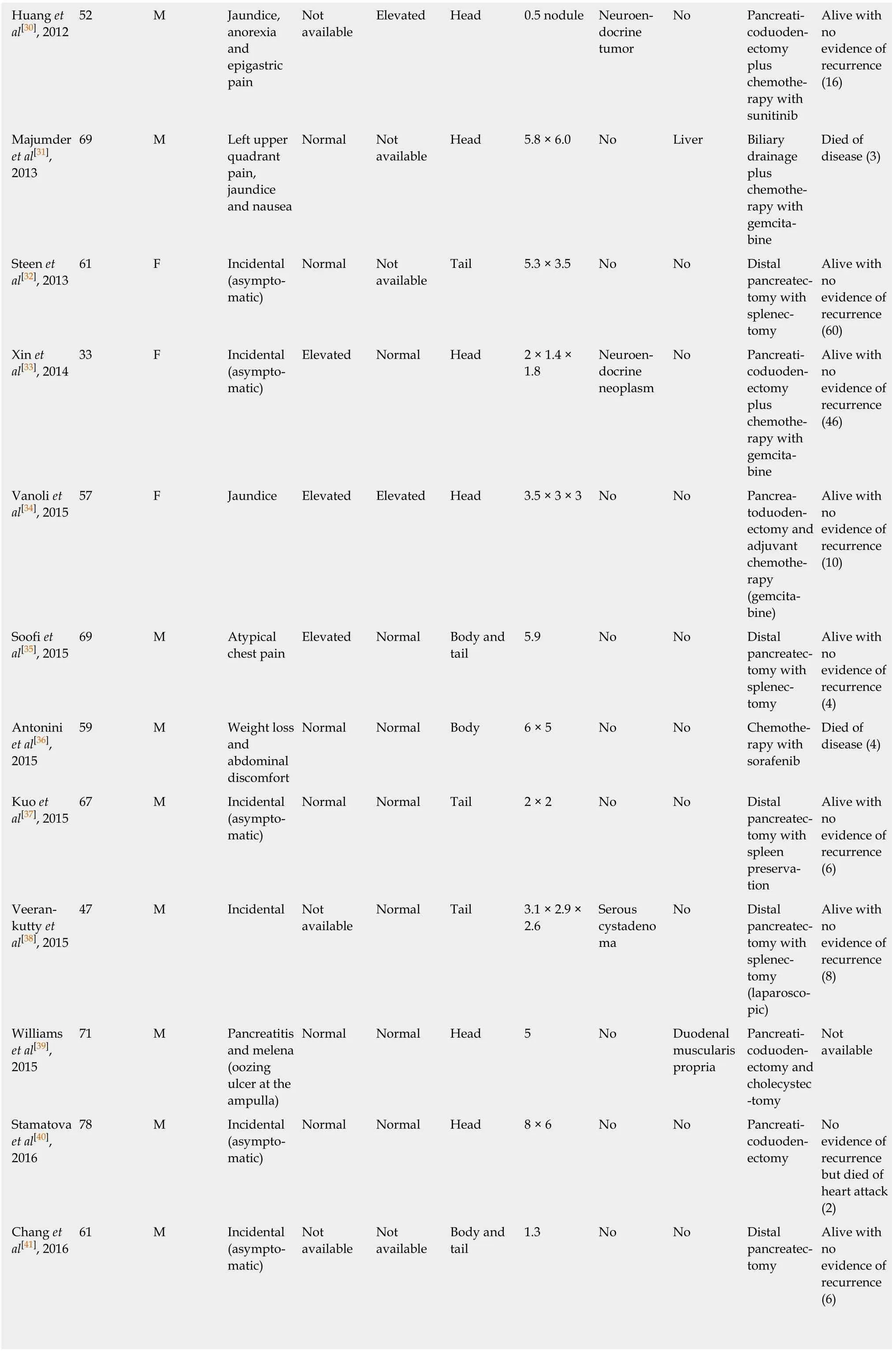
HC was first described in 1987 by Hrubanet al[11],and we report the 39thcase of hepatoid carcinoma of the pancreas diagnosed based on morphological and immunohistochemical features.The demographics and clinical presentation of the 39 cases are summarized in Table 1[11-45].From the review,we can establish a clear male predominance(69.3%).The ages of patients range from 21 years to 83 years,with a median age of 57 years.The sizes of tumors range from 1 cm to 12 cm,with a median size of 6 cm.HC of the pancreas can be divided into either pure HCC-like(61.54%) or mixed(38.46%) forms with other histological findings,such as neuroendocrine tumors(n= 9),pancreatic ductal adenocarcinoma(n= 3),acinar cell carcinoma(n= 1) and microcytic cystadenoma(n= 2).

Figure 1 Magnetic resonance imaging of the patient.
Table 2 outlines the main clinical features of HC of the pancreas in the reported literature,with the most common tumor site being the pancreatic tail,accounting for most of the patients who are asymptomatic or complain of abdominal/back pain.
The pathogenesis of hepatoid carcinoma of the pancreas remains to be elucidated.Three theories have been proposed: The ectopic liver tissue theory,in which HC may originate from ectopic pancreatic liver tissue[21,22,46];the pancreas-to-liver transdifferentiation theory,in which pancreatic cells can transdifferentiate into hepatocytes[47,48];and the pancreatic multipotent/stem cell theory,in which the liver and pancreas share the same embryonic derivation - the foregut endoderm - and genes controlling hepatocytic differentiation that are normally suppressed in the pancreas may be activated during carcinogenesis[12,35].
There are currently no standard criteria to establish a diagnosis of hepatoid carcinoma of the pancreas.The differential diagnosis of HC of the pancreas includes HCC or combined hepatocellular-cholangiocarcinoma,metastatic hepatoid carcinoma and other primary pancreatic tumors with eosinophilic cell cytoplasms.Its diagnosis relies on typical morphological features and immunohistochemical staining.Histopathologically,HC consists of medium to large cords of polygonal cells withabundant eosinophilic or clear cytoplasms with centrally located and vesicular nuclei in the sheet-like or trabecular portions.The presence of bile production is a more conclusive finding and is strong evidence of hepatocyte lineage differentiation[14,49].For immunohistochemistry findings,the hepatoid carcinoma cells show positive staining for immunoreactivity with polyclonal antibodies against AFP,CEA,glypican-3,and HepPar1(a hepatocyte-specific antigen),as well as albumin mRNA detection by in situ hybridization[14,20].Cytokeratin 19 positivity plays an important role in differentiating hepatoid tumors from HCC[27].HC of the pancreas with acinar differentiation should be tested with arginase-1 to exclude acinar cell carcinoma of the pancreas,which also presents with AFP elevation.As seen in our review,serum AFP is often elevated at the time of diagnosis of HC of the pancreas(41.15%),and can be used to monitor therapeutic response and recurrence[13,15,18,24].Serum protein induced by vitamin K absence or antagonist II,a specific marker used for early diagnosis of HCC,was elevated in some cases,aiding in early diagnosis and indicative of better prognoses[18].Serum CEA,which was elevated in 28.21% of cases,is a less sensitivediagnostic marker for hepatoid carcinoma of the pancreas.

Figure 2 Pathological presentation of the patient.

Table 1 Summary of clinical features of hepatoid carcinomas of the pancreas reported in the English language literature

?
?
AFP: Alpha-fetoprotein;CEA: Carcinoembryonic antigen;F: Female;M: Male.

Table 2 Outline of main features characterizing presentation of the 39 cases of hepatoid carcinoma of the pancreas
Due to its rarity,there is currently no standardized treatment for HC of the pancreas.Owing to its aggressive nature and tendency for early liver metastasis,HC of the pancreas warrants surgical resection,if possible.The effect of adjuvant therapy after surgery resection,advocated because of the metastatic potential of the tumor,is still unclear[43,44].Survival was poor in patients treated with only chemotherapy compared to those treated with surgery and chemotherapy: 5 out of 6 patients treated with chemotherapy succumbed to the disease(after 2.75-14 mo),while 3 out of 9 patients treated with chemotherapy and surgery succumbed to the disease(after 22-102 mo).Variable survival rates of 3 locally unresectable,metastatic or recurrent cases treated with surgery and adjuvant chemotherapy with mFOLFIRINOX,di-amino triazeno-imidazol carboxamide or gemcitabine have been reported in the literature,with one patient dying of the disease at 102 mo and two patients alive at 10 mo and 48 mo[14,18,44].
The prognosis of hepatoid carcinoma of the pancreas is unclear due to its rarity and possible heterogeneity.HCs of the gastrointestinal tract are associated with an unfavorable prognosis[50]since at the time of diagnosis,liver metastasis is often already present,indicating advanced stage[37].Survival outcomes mainly depend upon the extent of the disease and the completeness of resection,with greater survival rates after resection and adjuvant chemotherapy,as depicted in Figure 3,with the longest disease-free interval being 107 mo[45].Owing to the limited data,further studies with long-term follow-up are needed to standardize the treatment and to predict the natural history and prognosis of HC of the pancreas compared to those of the relatively more common gastric hepatoid carcinoma.
Our case was highly challenging due to the clinical presentation of the patient that was inconsistent with the imaging that suggested autoimmune pancreatitis(AIP).AIP commonly presents with obstructive jaundice,abdominal pain,vomiting and weight loss.Type 1 AIP is associated with high serum levels of IgG4(> 140 mg/dL),IgG4-positive plasma cell infiltration,and sclerosis,while type 2 AIP is often associated with inflammatory bowel disease[51].The patient may have been previously diagnosed with AIP due to the “sausage-shaped” appearance of the diffusely enlarged pancreas,the presence of a capsule-like rim and ductal stricture on imaging,and the lack of biopsy.Differentiating between AIP and pancreatic malignancy has become a diagnostic challenge for modern gastroenterologists because they often share overlapping clinical and imaging features.The poor response to steroid treatment prior to admission prompted reassessment of the diagnosis.The patient was cachexic,with recent onset of diabetes mellitus(DM),no sign of systemic involvement,negative autoantibodies,and non-elevated amylase and lipase,indicating malignancy.Type 3C DM,as reported in the literature,is difficult to control,requiring at least 1 IU/kg body weight of insulin[52].The prevalence of DM in patients with pancreatic cancer has been reported to be 40%,with half developing DM within 2 years[53].Interestingly,DM was reported in only 6 previously reported cases(two of which had an associated neuroendocrine component and increased glucagon levels;the length of DM history was not reported in the remaining 4 cases).In our case,it was attributed to pancreatic islet destruction resulting from advanced-stage hepatoid carcinoma with the absence of a neuroendocrine component on pathology.

Figure 3 Outcomes of the 39 patients with hepatoid carcinoma of the pancreas reported in the literature.
CONCLUSION
In summary,this review attempts to summarize the clinical characteristics,diagnostic methods,treatment and prognosis of HC based on the current literature.HC of the pancreas is an extremely rare neoplasm that resembles HCC in terms of morphology and immunohistochemistry findings.Diagnosis is mainly based on histopathological and immunohistochemical features.Elevation of serum AFP and protein induced by vitamin K absence or antagonist II may be a clue leading to the diagnosis of this tumor.Surgical resection is the mainstay of therapy and is more likely to result in long-term survival.Adjuvant chemotherapy has a role in recurrent,residual,unresectable and metastatic disease.Survival outcomes mainly depend upon the extent of the disease at diagnosis.The possibility of hepatoid carcinoma of the pancreas should be considered for diffuse lesions throughout the pancreas.
 World Journal of Clinical Cases2020年5期
- World Journal of Clinical Cases的其它文章
- Gut microbiota and nutrient interactions with skin in psoriasis: A comprehensive review of animal and human studies
- Microbiota-gut-brain axis and its affect inflammatory bowel disease:Pathophysiological concepts and insights for clinicians
- Distal esophageal spasm: Update on diagnosis and management in the era of high-resolution manometry
- Clinical course of percutaneous cholecystostomies: A crosssectional study
- Clinical characteristics and 28-d outcomes of bacterial infections in patients with hepatitis B virus-related acute-on-chronic liver failure
- Application of hybrid operating rooms for treating spinal dural arteriovenous fistula