基于Hessian矩阵和RSF模型的CT图像淋巴结分割
王鑫 严加勇 林涛


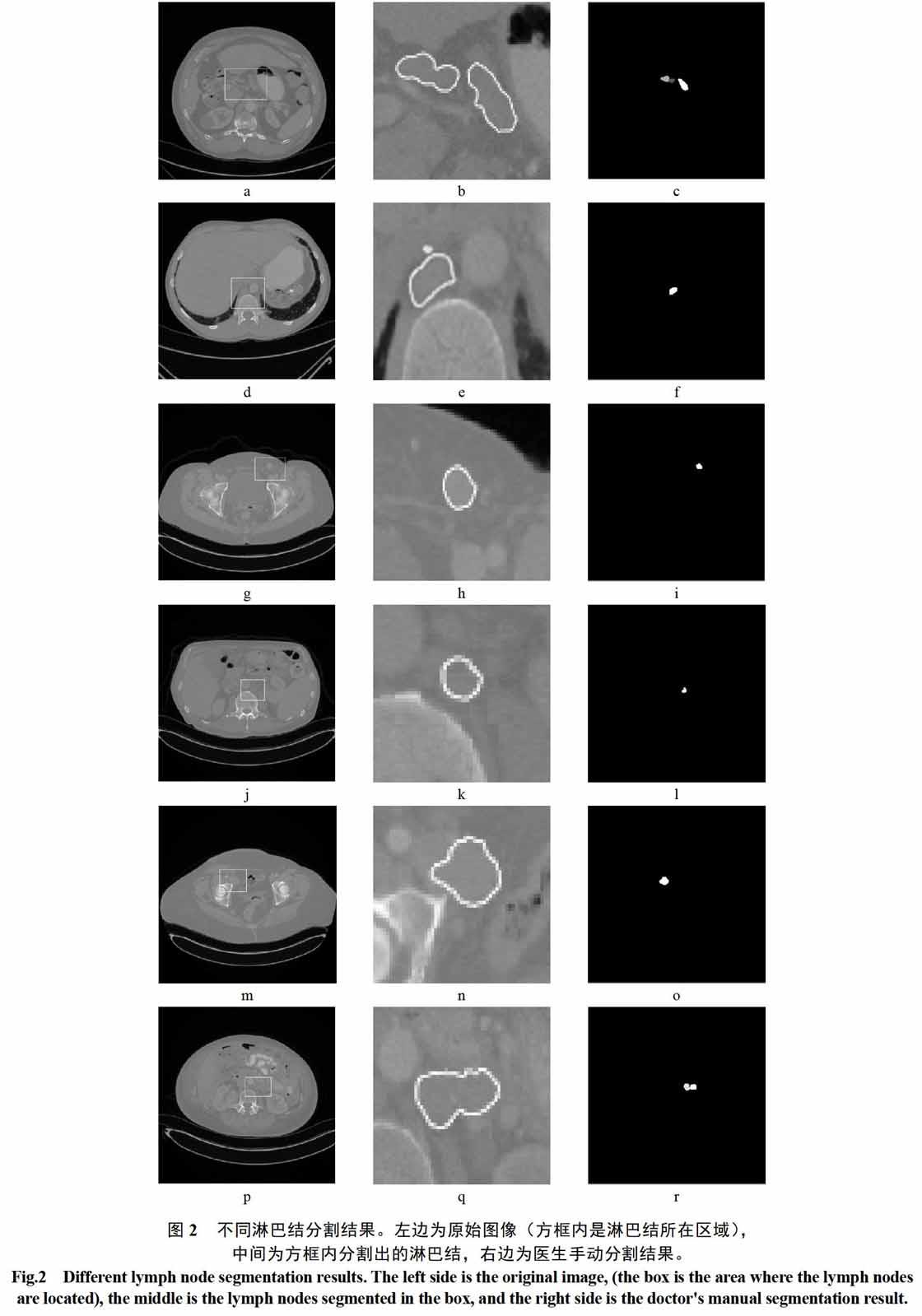
摘 要: 临床上医生分割淋巴结主要依靠手动,针对手动分割淋巴结的缺点和局限,本文提出一种基于Hessian矩阵和区域扩展拟合水平集模型(Region-Scalable Fitting,RSF)的淋巴结自动分割算法。该算法首先利用Hessian矩阵对CT图像中的淋巴结进行增强,并得到淋巴结粗略轮廓,然后把该粗略轮廓作为RSF模型的初始轮廓,并利用RSF模型对初始轮廓进行演化以实现淋巴结的有效分割。将该方法应用于6个病例的CT淋巴结图像中,初步实验結果与医生手动分割结果对比,平均重叠率93.3%,平均Hausdorff距离为3.8 mm。
关键词: 淋巴结;分割;Hessian矩阵;RSF模型;CT图像
【Abstract】: In current clinical practice, the segmentation of the lymph nodes is still performed manually by the doctors. In order to avoid the disadvantages and limitations of manual segmentation, the present paper proposes a lymph node segmentation algorithm based on Hessian matrix and Region-Scalable Fitting (RSF) model. At first, the algorithm uses Hessian matrix to enhance the lymph node in CT image and obtain the rough boundary of the lymph node. Then the rough boundary is adopted as the initial contour for the RSF model. Finally, the rough lymph node contour is evolved by the RSF model to segment the target lymph node effectively. The algorithm was applied to the lymph node CT images of 6 cases. The preliminary experimental results were compared with the doctor's manual segmentation results. The average overlap rate was 93.3%, and the average Hausdorff distance was 3.8 mm.
【Key words】: Lymph node; Segmentation; Hessian matrix; RSF model; CT image
0 引言
淋巴结检测和评估在癌症的诊断和治疗上有显著的帮助[1-4]。CT是淋巴结检测的主要成像模式,淋巴结与周围软组织CT值相近,淋巴结的图像特征也比较复杂。目前,临床上,对CT图像中淋巴结的分割主要依靠医生手动完成,手动分割容易产生误差且工作量大。近年来,淋巴结自动分割算法受到不少研究人员的关注[5-13]。Dornheim L和Dornheim J.等[5-6]提出基于一种弹簧质量模型的淋巴结分割算法,Barbu A,Suehling M,Xu X等[7-9]提出一种基于学习的方法来分割淋巴结,Johannes Feulner等[10]提出一种基于差异性学习和空间先验的分割算法,魏骏等[11]提出采用遍历阈值提取淋巴结种子点的区域生长算法,Tan,YQ等[12]提出通过动态规划和活动轮廓进行淋巴结分割,Yu,PC等[13]提出基于区域snake模型的边缘约束淋巴结分割。
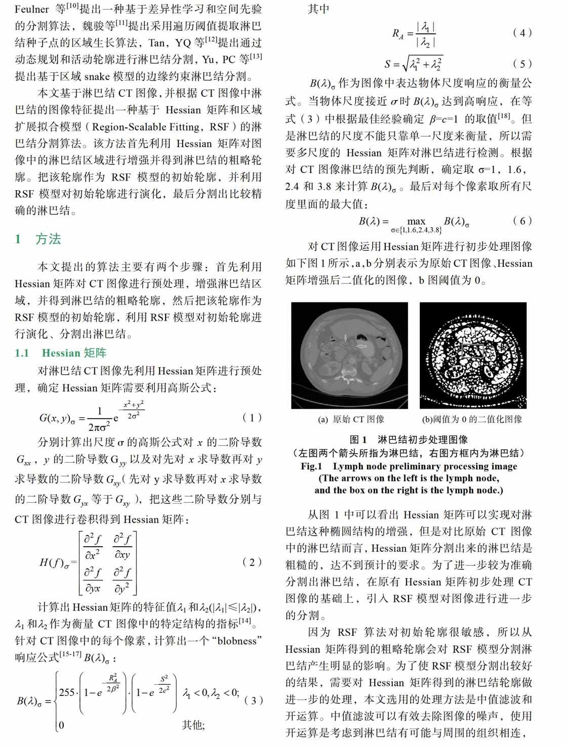
本文基于淋巴结CT图像,并根据CT图像中淋巴结的图像特征提出一种基于Hessian矩阵和区域扩展拟合模型(Region-Scalable Fitting,RSF)的淋巴结分割算法。该方法首先利用Hessian矩阵对图像中的淋巴结区域进行增强并得到淋巴结的粗略轮廓。把该轮廓作为RSF模型的初始轮廓,并利用RSF模型对初始轮廓进行演化,最后分割出比较精确的淋巴结。
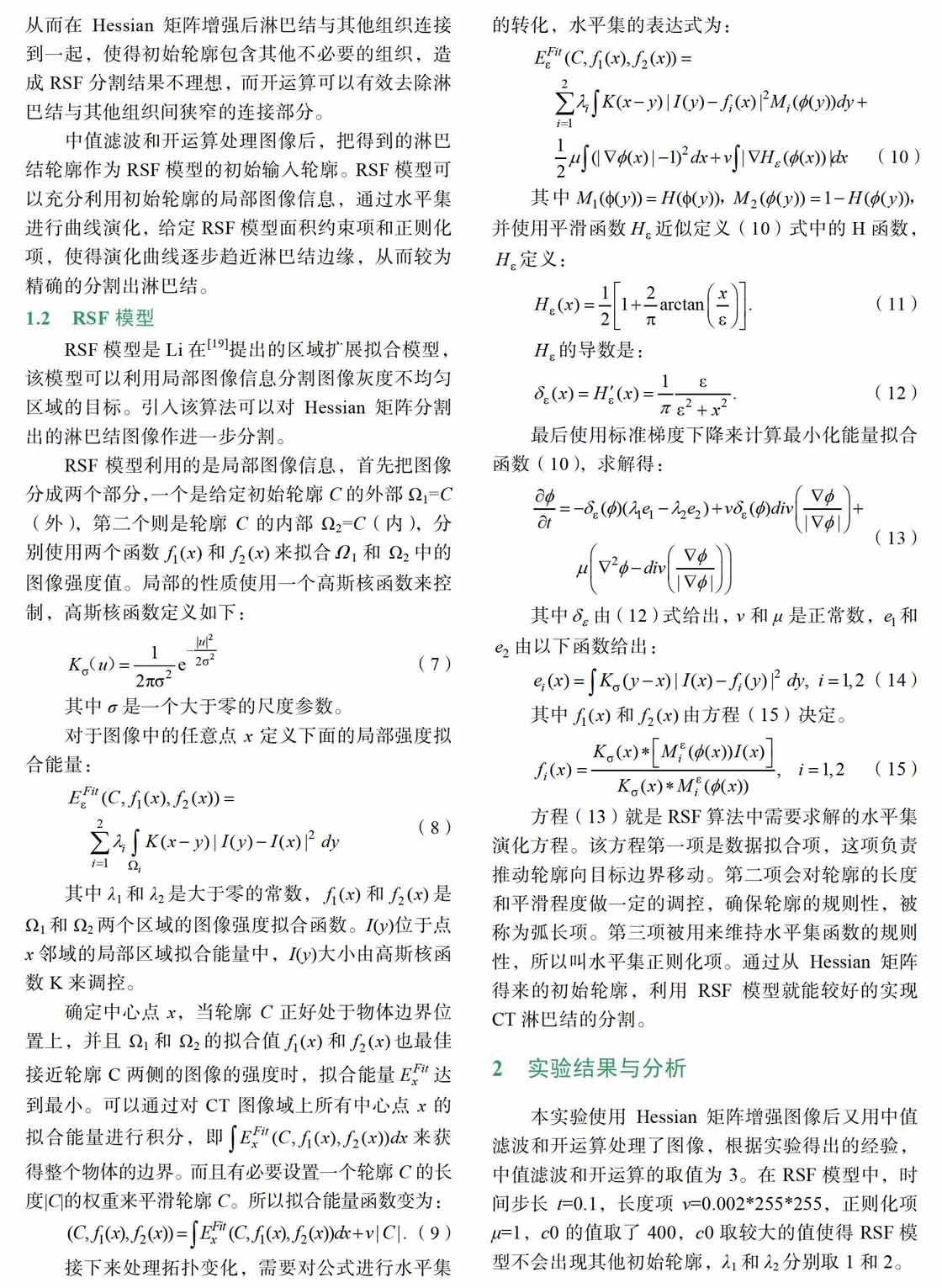
本文提出的基于Hessian矩阵和RSF模型的算法能较好实现对淋巴结的分割。在6例病例的淋巴结图像中对淋巴结的分割都能有较好的结果,从两项验证结果的指标——重叠率以及平均Hausdorff距离比上可以看出算法是有效果的,重叠率平均达到93.3%,平均Hausdorff距离3.8mm。用本文算法对淋巴结进行分割可以有效减少医生工作量,医生只需知道淋巴结的位置信息就能较好实现对淋巴结的分割,而且淋巴结的面积信息可以为医生提供病人淋巴结生长相关的信息,对医生诊断和治疗也提供了一定的帮助,在临床上有一定参考价值。
3 结束语
淋巴结的诊断和评估可以有效帮助医生诊断和治疗癌症。针对临床上医生对淋巴结分割多是手动而产生的局限性以及淋巴结的特征,本文提出一种基于Hessian矩阵和RSF模型的分割算法。本算法首先使用Hessian矩阵对图像进行增强,得到粗略的淋巴结轮廓,再利用中值滤波和开运算对粗略轮廓进行去噪和优化,把得到的淋巴结粗略轮廓作为RSF模型的初始轮廓,利用RSF模型对初始轮廓进行水平集演化,使之逐步逼近真实淋巴结边界并分割出淋巴结。本文算法对6例病例的淋巴结图像进行了实验,得到的结果与医生手动分割的结果相比较,面积重叠率及平均边界距离比都比较接近,医生只需提供淋巴结的大概位置就能较好实现淋巴结的分割,得到分割结果,而且分割结果也能为医生提供淋巴结的面积信息,为医生对淋巴结的临床诊断和评估提供帮助,提高医生工作效率。
参考文献
[1]Schwartz L H, Zhao B S, Yan J Y. Automated determination of lymph nodes in scanned images[P]. USA: US 8355552 B2, 2013-01-15.
[2]Yan J Y, Zhao B S, Curran S, et al. Automated matching and segmentation of lymphoma on serial CT examinations[J]. Medical Physics, 2007, 34(1): 55-62.
[3]Yan M, Lu Y, Lu R Z, et al. Automatic detection of pelvic lymph nodes using multiple MR sequences [C]. Proceedings of SPIE, Medical Imaging 2007, San Diego, CA, US, 2007, 6541: 65140W. 1-65140W. 10.
[4]Nystrom K. Automatic detection and segmentation in lym phoma with PET/CT[D]. Sweden: Lund University, 2009.
[5]Dornheim L, Dornheim J. Automatische Detektion von Lymphknoten in CT-Datens?tzen des Halses[C]// Bildverar beitung für die Medizin 2008, Algorithmen, Systeme, An wendungen, Proceedings des Workshops vom 6. bis 8. April 2008 in Berlin. DBLP, 2008: 308-312.
[6]Dornheim L, Dornheim J, R?ssling I. Complete fully auto matic model-based segmentation of normal and pathological lymph nodes in CT data.[J]. International Journal of Com puter Assisted Radiology & Surgery, 2010, 5(6): 565-581.
[7]Barbu A, Suehling M, Xu X, et al. Automatic detection and segmentation of axillary lymph nodes[C]. Medical Image Computing and Compter-Assisted Intervention-MICCAI 2010, Beijing, China, 2010, 6361: 28-36.
[8]Barbu A, Suehling M, Xu X, et al. Automatic Detection and Segmentation of Lymph Nodes From CT Data[J]. IEEE Transactions on Medical Imaging, 2012, 31(2): 240.
[9]Barbu A, Suehling M, Xu X, et al. Method and system for automatic detection and segmentation of axillary lymph nodes [P]. USA: US8391579 B2, 2011-03-05.
[10]Feulner J , Kevin Zhou S , Hammon M , et al. Lymph node detection and segmentation in chest CT data using discri minative learning and a spatial prior[J]. Medical Image Ana lysis, 2013, 17(2): 254-270.
[11]魏駿, 何凌, 车坤, 等. CT图像的颈部淋巴结半自动分割算法[J]. 计算机工程与设计, 2015(11): 3014-3018.
[12]Tan Y, Lu L, Bonde A, et al. Lymph node segmentation by dynamic programming and active contours[J]. Medical Phy sics, 2018.
[13]Yu P, Poh C L . Region-based snake with edge constraint for segmentation of lymph nodes on CT images[J]. Computers in Biology and Medicine, 2015, 60: 86-91.
[14]Li Q, Sone S, Doi K. Selective enhancement filters for nodules, vessels, and airway walls in two- and three-dim ensional CT scans[J]. Medical Physics, 2003, 30(8): 2040-0.
[15]Frangi, A. F., Niessen, W. J., Vincken, K. L. , and Viergever, M. A., “Multiscale vessel enhancement filtering” in [MICCAI], LNCS 1496, 130–137 (1998).
[16]Sato, Y., Westin, C. -F., Bhalerao, A., Nakajima, S., Shiraga, N., Tamura, S., and Kikinis, R., “Tissue classification based on 3d local intensity structures for volume rendering, ” IEEE Transactions on Visualization and Computer Graphics 6, 160–180 (April–June 2000).
[17]Antiga, L., “Generalizing vesselness with respect to dimen sionality and shape” The Insight Journal (July–December 2007).
[18]Feuerstein M, Deguchi D, Kitasaka T, et al. Automatic mediastinal lymph node detection in chest CT[J]. Proc eedings of SPIE-The International Society for Optical Engin eering, 2009, 7260(6): 72600V-72600V-11.
[19]Li C, Kao C Y, Gore J C, et al. Minimization of Region- Scalable Fitting Energy for Image Segmentation[J]. IEEE Transactions on Image Processing, 2008, 17(10): 1940- 1949.

