Periostin mediates epithelial-mesenchymal transition through the MAPK/ERK pathway in hepatoblastoma
Lu Chen, Xiangdong Tian, Wenchen Gong, Bo Sun, Guangtao Li, Dongming Liu, Piao Guo, Yuchao He,Ziye Chen, Yuren Xia, Tianqiang Song, Hua Guo
Department of Tumor Cell Biology, Tianjin Medical University Cancer Institute and Hospital, National Clinical Research Center for Cancer, Key Laboratory of Cancer Prevention and Therapy, Tianjin, Tianjin's Clinical Research Center for Cancer,Tianjin 300060, China
ABSTRACT Objective:The aim of the present study was to analyze the prognostic factors in patients with hepatoblastoma (HB) in our single center and to evaluate periostin (POSTN) expression in HB and its association with clinicopathological variables. In addition, the underlying mechanism of how POSTN promotes HB progression was discussed.Methods:POSTN expression was investigated in HB tumors by immunohistochemistry (IHC), immunofluorescence (IF) and Western blot (WB). The association among POSTN expression, clinicopathological features and overall survival (OS) was also evaluated. The migration and adhesion ability of HB cells were measured using chemotaxis and cell-matrix adhesion assays,respectively. Epithelial-mesenchymal transition (EMT)-associated markers and activation of the ERK pathway were detected by WB.Results:HB patients had poor prognosis which displayed lymph node metastasis, vascular invasion, POSTN and vimentin expression. POSTN expression was also associated with lymph node metastasis. Furthermore, overexpressed POSTN promoted migration and the adhesive ability of HB cells in vitro. In addition, we demonstrated that POSTN activated the MAPK/ERK pathway, upregulated the expression of Snail and decreased the expression of OVOL2. Finally, POSTN promoted the expression of EMT-associated markers.Conclusions:POSTN might modulate EMT via the ERK signaling pathway, thereby promoting cellular migration and invasion.Our study also suggests that POSTN may serve as a therapeutic biomarker in HB patients.
KEYWORDS Periostin; hepatoblastoma; EMT; MAPK/ERK
Introduction
Hepatoblastoma (HB), an embryonal tumor derived from hepatoblast cells, is the most common malignant liver cancer diagnosed in childhood1,2. HB occurs more frequently in boys than girls (ratio 2:1), and the incidence of HB peaks at 0 to 3 years of age3. Histopathologically, HB can be divided into two main histological types: epithelial (approximately 80%) and mixed (epithelial-mesenchymal differentiation)3,4.Despite improvements in the treatment of HB during the past 4 decades, a complete surgical resection offers the only chance for a cure. In addition, cisplatin-based chemotherapy has become an effective salvage therapy for patients with unresectable tumors; this has contributed to the increase in the 5-year survival rate of HB, which is greater than 70%-80%5,6. Despite the clinical advancement and high cure rate, in a portion of patients, a cure is beyond reach due to a lack of a thorough understanding of the origin and pathophysiology of the disease. According to the literature, a strong consensus has been reached regarding prognostic factors such as alphafetoprotein (AFP), complete surgical resectability, tumor stage, metastasis and histology7-9. Many basic science studies have noted genetic mutations in pathological subtypes of HB10, but few of these mutations are applied in the clinic. It is therefore necessary to discover new molecular mutations in HB and to present homologous treatment for refractory HBs.
Periostin (POSTN), which was originally cloned from a mouse osteoblastic cell line, has been reported to be associated with the grade of malignancy and the level of invasion11,12. Aberrant POSTN expression has been found in numerous cancer entities, including head and neck, lung,neuroblastoma, breast and colorectal cancers13-15. The tumor progressive properties of POSTN are believed to be due to its role in the facilitation of tumor angiogenesis, interaction with cell surface receptors and modulation of signal transduction pathways16,17. In addition, the exogenous expression of POSTN in 293T cells drove the cells to undergo epithelialmesenchymal transition (EMT)18. EMT occurs when epithelial cells lose their epithelial features and acquire mesenchymal features, which is commonly associated with the acquisition of invasive characteristics, metastatic potential and drug resistance in cancer cells19,20. The levels of invasion and metastasis are major prognostic factors of malignant tumors, and thus these associations of POSTN with oncogenesis and progression have novel implications on tumor detection and therapy.
However, the effect of POSTN expression on the progression and prognosis of HB remains largely unknown.In this study, we demonstrated that POSTN induced hepatoblastoma cells to undergo EMT, which was mediated by the MAPK/ERK pathway. Moreover, we explored the expression pattern of POSTN and the correlation between POSTN and the biological and pathological characteristics of HB. Our results revealed that POSTN might be a potential prognostic and/or predictive marker in HB.
Materials and methods
Tissue specimens and clinical materials
This study included 47 patients with HB who underwent radical surgery between December 2010 and January 2016 at the Department of Pediatric and Department of Hepatobiliary Oncology, Tianjin Medical University Cancer Institute and Hospital. Tumor tissue samples were obtained during surgery or ultrasound-guided percutaneous puncture,which were pathologically confirmed. The clinical data and tumor characteristics, including patient age, gender, survival status, survival time, lymph node metastasis, vascular invasion, E-cadherin level, vimentin level, and p-ERK level were available for all patients. This study was implemented according to the Declaration of Helsinki and was authorized by the Tianjin Medical University Cancer Institute and Hospital Ethics Committee. Informed consent was obtained from all patients.
Western blot
Fresh-frozen lymphnode specimens were broken into smaller pieces and then lysed in lysis buffer (1% SDS, 10 Mm Tris-HCl, pH 7.6, 100 mM phenylmethanesulfonyl fluoride) on ice for 15 minutes. Protein denaturation was performed at 95°C for 10 minutes, and then, lysates were centrifuged at 12,000 rpm at 4°C for 10 minutes followed by collection of the upper clear cell lysates. The protein concentration was measured by the Bradford method. Equal amounts of protein(60-90 µg/line) were loaded, separated by SDS-PAGE gels and blotted onto poly vinylidene difluoride (PVDF)membranes. GAPDH was used as an internal control.Antibodies against the following proteins were used: GAPDH(0411: sc-47724), OVOL2 (sc85803) (Santa Cruz Biotechnology, CA, USA), vimentin (2707-1) (Epitomics,Burlingame, CA, USA), E-cadherin (610181), Smad2/3(610842) (BD Biosciences, San Jose, CA, USA), Snail (3879),p-AKTSer473(4060), AKT (9272), p-Smad2/3 (8828),Smad2/3 (8685), p-ERK1/2 (Thr202/Tyr204) (4370), ERK1/2(9102) (Cell Signaling Technologies, Danvers, MA, USA),POSTN (ab14041) (Abcam, Hong Kong, China). Goat antirabbit (Santa Cruz, sc-2004), goat anti-mouse (Santa Cruz,sc-2005) and donkey anti-goat (Santa Cruz, sc-2020)secondary antibodies were incubated with the membranes.
Immunohistochemistry
Paraffin-embedded HB tissues were deparaffinized and rehydrated in xylene and graded concentrations of ethanol,respectively. Then, 3% H2O2was used to block endogenous peroxidase activity for 15 minutes, while nonspecific staining was blocked by 3% BSA (bovine serum albumin, Roche, HK,China) for 1 hour.
Incubation with the POSTN, E-cadherin, vimentin and p-ERK1/2 antibodies was performed overnight at 4°C. The following day, the tissues were maintained at room temperature for 30 minutes, at 37°C for 40 minutes and were then washed with PBS three times. Then, tissues were incubated with biotinylated secondary antibody (Zhongshan Goldbridge Biotechnology CO., Ltd, Beijing, China) at 37°C for 1 hour, and staining was visualized by DAB (3,3′-diaminobenzidine) staining. Slides were counterstained with Mayer's hematoxylin, dehydrated, mounted, dried and observed. Sections stained by IHC were qualitatively scored according to the presence of nuclear and cytoplasmic staining by two independent pathologists in a blinded manner. The scoring criteria for POSTN expression can be found in our previous study21.
Frozen tissue immunofluorescence
The tumor tissues cryopreserved at -80°C were removed from storage and warmed to room temperature for 5 minutes. The tissues were fixed in ice-cold acetone for 10 minutes, following by three washes in PBS. Then, the tissues were blocked in 3% BSA at 37°C for 1 hour and afterwards were incubated with POSTN and vimentin antibodies overnight at 4°C. The following day, the tissues were first washed three times in PBS and then stained with Alexa Fluor 488-conjugated secondary antibodies (Invitrogen) at 37°C for 1 hour in the dark. The nuclei of the tissues were counterstained with prolong gold anti-fade reagent(Invitrogen).
Cell transfection
The Lenti-PacTMHIV Expression Packaging Kit (Gene Copoeia, Rockville, MD, USA) was used to transfect the HB cells. Transfection was performed according to the manufacturer's instructions. HepG2 cells were transduced with the HIV vector at a multiplicity of infection (MOI) of 10. A POSTN-specific shRNA (sequence: CGGTGACAG TATAACAGTAAA) and a scrambled sequence of shRNA against GFP, which served as the negative control (SCR),were purchased from Genechem (Shanghai, China). POSTN cDNA was subcloned into the XhoI and NotI sites of the pCDH-CMV-MCS-EF1-puro lentiviral vector. The forward and reverse sequence were 5′-GAGAGCCACC-3′ and 5′-GCGGCCGC-3′, respectively. Stable cell lines were selected by puromycin (Gibco)-containing medium for 4 weeks, and the transfection efficiency was confirmed by WB.
Chemotaxis assay
Briefly, 30 μL DMEM medium containing 20% FBS was loaded into the lower chamber. Then, 50 μ L cells (5 × 10⁵cells/mL) suspended in DMEM medium were loaded into the upper chamber. An 8 μm polycarbonate filter was pretreated with 0.001% fibronectin, and the polycarbonate filter was treated by 0.1% fibronectin (Sigma-Aldrich) diluted in DMEM at 4°C overnight. Two chambers were used to clamp the membrane. The whole device was incubated at 5% CO2and 37°C for 12 hours, after which the membrane was washed, fixed, and stained. The number of cells that had migrated was counted in three randomly selected high-power fields (400 ×) by microscopy. All samples were tested in triplicate, and the data are expressed as the mean ± SD.
Cell-matrix adhesion
Briefly, a 12-well plate containing glass was first pretreated with 10 µg/mL fibronectin (R&D Systems Inc., Minneapolis,MN, USA). Second, 2 × 10⁵cells were plated in each well.Third, the cells were incubated at 5% CO2and 37°C for 5, 15,or 30 minutes. The cells were washed twice in cold PBS,which was followed by fixation in 4% paraformaldehyde for 10 minutes. Finally, the number of attached cells was counted in three random fields (200 ×). All the samples were tested in triplicate, and the data are expressed as the mean ± SD.
Statistical analysis
The statistical analysis was performed using the statistical software SPSS 22.0 (SPSS, Inc., Chicago, IL, USA). The overall survival (OS) was defined as the interval between the date of definite diagnosis and the time of death or the final follow-up. Analysis of the OS rate was conducted according to the Kaplan-Meier method, and the log-rank test was used for the univariate analysis. The association of POSTN expression with the clinicopathologic characteristics was evaluated by Pearson's χ2test or Fisher's exact test. All P values were two-tailed, and P < 0.05 was considered to indicate a statistically significant difference.
Results
The expression patterns of POSTN in HB and its correlation with clinicopathological features
Of the 47 patients with HB, 25 were male and 22 were female;the patients ranged in age from 7 to 165 months with a mean age of 45.1 months and a median age of 24 months. None of them had a history of carcinoma at any other site or a family history of cancer. Only one child was born prematurely.Asymptomatic abdominal enlargement was the primary presenting symptom in most patients. Choleplania was found on physical examination in three of the patients. All patients were treated by surgery, including 30 patients who received primary tumor resection and 17 patients who received selective surgery following neoadjuvant chemotherapy. The chemotherapy regimen comprised cisplatin, 5-fluorouracil,vincristine and doxorubicin. Chemotherapy regimens in different subgroups of patients with hepatoblastoma are listed in Supplementary Table S1.
The patterns of POSTN expression were determined by immunohistochemistry, and positive POSTN staining was primarily localized in the cytoplasm. The results showed that the expression level of POSTN in HB tissues was significantly higher than in normal tissues (Figure 1A). Of the 47 patients with epithelial HB, high POSTN expression was noted in 20(42.6%) cases.

Figure 1 The expression patterns of POSTN in HB and its correlation with clinicopathological features. (A) Immunohistochemistry for POSTN in HB tissues and normal liver tissues. (B) WB for POSTN in 1 benign liver nodule (noncancerous tissue) and 3 HB tissues that represented different disease stages. (C) Kaplan-Meier survival curves of patients with HB stratified by lymph node metastasis, vascular invasion and POSTN expression.
WB was used to further analyze POSTN expression, and consistently, the POSTN expression level in HB tissue was much higher than that in normal tissues and was positively correlated with the PRETEXT stage of HB (Figure 1B).
Up until the cut-off date for this analysis, no patient was lost to follow-up, thirty-three were still alive without recurrence, and fourteen had died of tumor recurrence or metastasis. The median survival time was 37.4 months (range 10-73 months), and the 3-year OS rate was 69.5% (Figure 1C).
According to the univariate analysis, lymph node metastasis, vascular invasion, and the POSTN expression level were significant predictors of OS (Figure 1C). The Kaplan-Meier survival analysis revealed that patients with a high expression level of POSTN exhibited a significantly worse 3-year OS (χ2= 6.715, P = 0.010) compared with those with a low expression level of POSTN. However, gender was not significantly correlated with OS (P > 0.05). Patients younger than 36 months did not gain a significant survival benefit from their age (P = 0.245).
The expression of POSTN is positively correlated with vimentin in HB
As demonstrated above, lymph node metastasis, vascular invasion and POSTN expression exerted independent effects on OS. We first analyzed the relationship between POSTN and lymph node metastasis and vascular invasion. We found that POSTN expression significantly influenced lymph node metastasis (χ2= 11.698, P = 0.001) (Table 1). We next analyzed the relationship between POSTN expression and metastatic potential in HB.
As reported, EMT is a common step in tumor progression,during which the metastasis and migration properties of cancer cells change22. In the process of EMT, the expression of vimentin is increased, while the expression of E-cadherin is decreased19,23. Therefore, we detected the expression levels of E-cadherin and vimentin in HB by IHC. The results indicated that a high POSTN expression level was always accompanied by a rise in vimentin expression (Figure 2A).These results were further confirmed by WB and frozen tissue immunofluorescence using clinical samples (Figure 2B and 2C).
Then, we analyzed the relationship between POSTN and the expression levels of vimentin and E-cadherin. We foundthat POSTN expression was closely related to the vimentin expression level (χ2= 5.232, P = 0.045). However, no significant correlation was observed between POSTN expression and the E-cadherin expression level (P > 0.05)(Table 1). Furthermore, vimentin expression was confirmed to be associated with OS (P = 0.003) (Figure 2D).
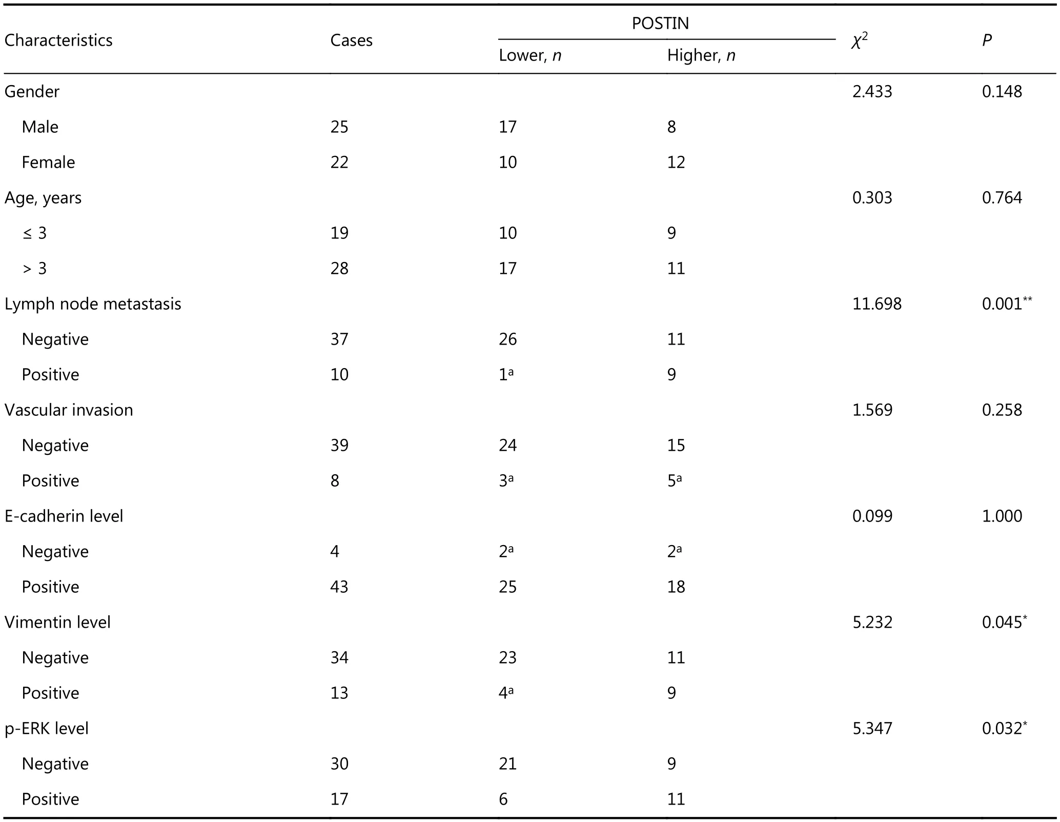
Table 1 The relationship between POSTN expression and clinical pathological characteristics in 47 HB patients
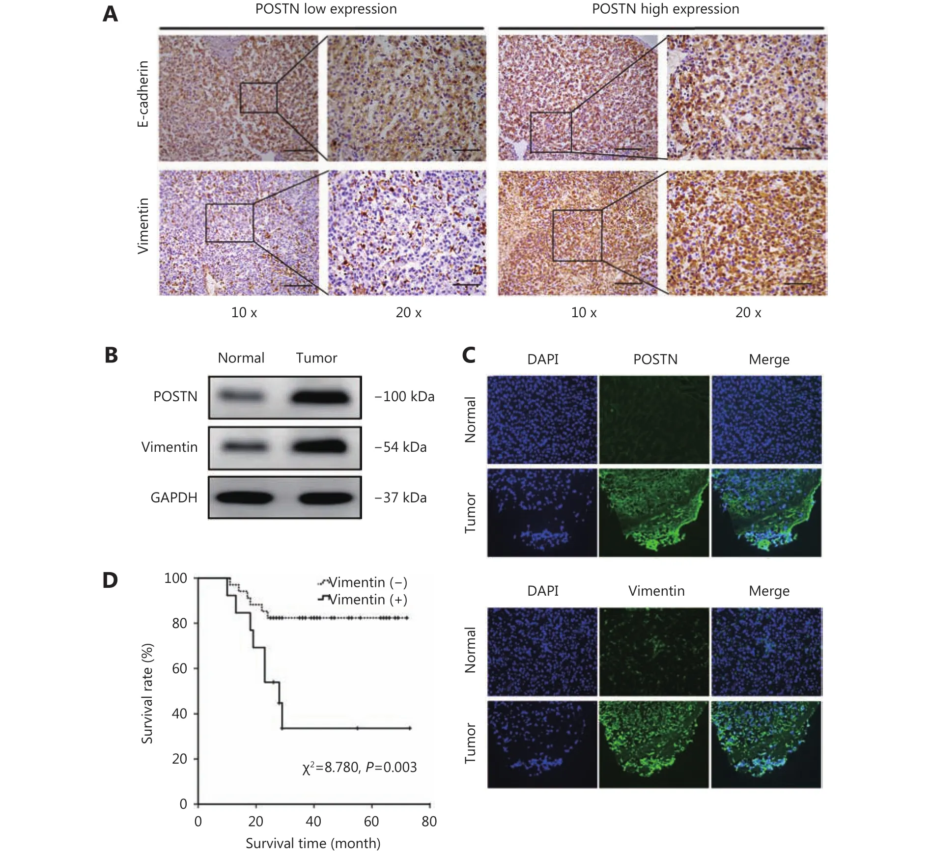
Figure 2 The expression of POSTN is positively correlated with Vimentin in HB. (A) Immunohistochemistry for E-cadherin and vimentin in HB tissues with different POSTN expression levels (scale bar, 1.0 mm). (B) Expression of POSTN and vimentin in HB and in paired normal tissue as analyzed by WB. (C) Frozen tissue immunofluorescence (IF) for the expression of POSTN and vimentin. (D) Kaplan-Meier survival curves of patients with HB stratified by vimentin expression.
The role of POSTN in HB cell migration and cell-matrix adhesion.
To investigate the role of POSTN in the biological behavior of HB cells, we stably overexpressed and reduced POSTN in the HB cell line HepG224,25. HepG2 cell line has been reported to be misidentified. It is originally thought to be a hepatocellular carcinoma cell line but shown to be from a hepatoblastoma prepared by the International Cell Line Authentication Committee (ICLAC, http://iclac.org/databases/cross-contaminations/) and the American Type Culture Collection (ATCC, https://www.atcc.org/products/all/HB-8065.aspx). The efficiencies were confirmed by WB (Figure 3A). A cell-matrix adhesion assay showed that the ability of the cells to attach to fibronectin was remarkably enhanced by the overexpression of POSTN in HepG2 cells (Figure 3B).On the contrary, the attachment rate was dramatically reduced by the downregulation of POSTN in HepG2 cells compared with the control group (Figure 3B). Furthermore,the chemotaxis assay showed that migration ability was enhanced in HepG2 cells in which POSTN was overexpressed and was inhibited in HepG2 cells in which POSTN expression was depressed compared with the control group(Figure 3C). All these results verified the positive role of POSTN in promoting the malignant potential of HB cells.
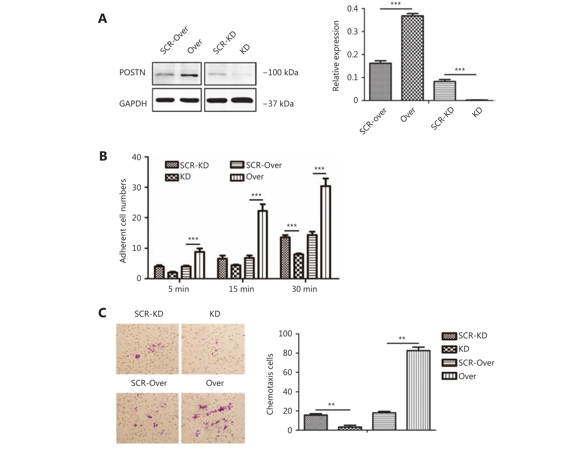
Figure 3 The role of POSTN in HB cell migration and cell-matrix adhesion. (A) Assessment of transfection efficiency of POSTN expression after retroviral infection of HepG2 cells. (B) The number of adherent cells was counted in three random fields under microscopy after 5, 15 and 30 minutes. (C) The chemotactic potential of HepG2 cells with different POSTN expression levels (Giemsa stainning, 20 ×). **P < 0.01 and ***P < 0.001. Data are expressed as the mean ± SD. All experiments were repeated at least three times.
POSTN induces HB cells to undergo EMT,which is mediated by the MAPK/ERK pathway
Next, we sought to confirm that EMT was mediated by POSTN in HB cells and to further determine the underlying mechanisms involved in the EMT process. The WB results showed that overexpression of POSTN in HepG2 cells remarkably increased the expression of the mesenchymal marker vimentin, while it decreased the expression of the epithelial marker E-cadherin. However, when POSTN was downregulated in HepG2 cells, we observed the opposite result (Figure 4A).
The detection of EMT-associated transcription factors was followed. We found that the overexpression of POSTN in HepG2 cells caused the overexpression of Snail, which is known to play a vital role in EMT (Figure 4B). In contrast,the expression of OVOL2, which is a known MET promoter,was downregulated by the overexpression of POSTN in HepG2 cells (Figure 4B). Similarly, the opposite result was observed when POSTN expression was depressed in HepG2 cells (Figure 4B).
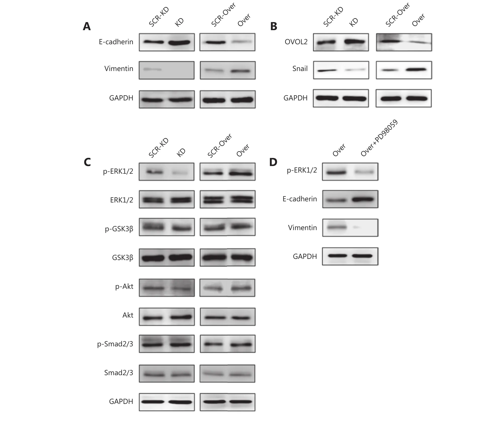
Figure 4 POSTN induces HB cells to undergo EMT, which is mediated by the MAPK/ERK pathway. (A) Expression of EMT-associated markers. (B) Expression of EMT-associated transcription factors. (C) Expression of EMT-associated signaling pathways as detected by WB in HepG2 cells in which POSTN was overexpressed or depressed. (D) WB analysis of EMT-associated markers when POSTN was overexpressed in HepG2 cells cultured with the ERK specific inhibitor PD98059.
To identify the signaling pathway involved in the POSTN-mediated EMT process, we examined the expression of components of the PI3K/Akt, MAPK/ERK and Smad signaling pathways, which are closely associated with the EMT process26. The results showed that phosphorylation of ERK1/2 was dramatically elevated in HepG2 cells in which POSTN was overexpressed, while phosphorylation of Akt,GSK3β and Smad2/3 remained no significant difference to that of the control group (Figure 4C). Then, a specific inhibitor of the ERK1/2 pathway, PD98059, was exogenously added to the culture medium of POSTN-overexpressing HepG2 cells. WB assays showed that the overexpressed level of vimentin in POSTN-overexpressing cells was balanced(Figure 4D).
In summary, all these results reveal that the overexpression of POSTN in HepG2 cell can mediate EMT in HB cells through the MAPK/ERK pathway. These results were consistent with those of previous studies, which reported that POSTN promoted metastasis and invasion via ERK signaling27.
The phosphorylation of MAPK/ERK pathway components is activated in HB samples with high POSTN expression
We further sought to estimate the expression level of ERK in HB tissues by IHC. A considerable difference in the p-ERK expression level was observed between HB and normal liver tissues (Figure 5A); POSTN expression was also found to significantly affect the expression level of p-ERK (χ2= 5.347,P = 0.032, Table 1). Moreover, the univariate analysis indicated that the expression level of p-ERK (χ2= 16.158,P < 0.001, Figure 5B) significantly affected OS. Therefore, we speculated that ERK signaling plays an important role in HB progression.
Discussion
Although HB is the third most common abdominal neoplasm in infants and toddlers, it remains rare in the spectrum of malignant tumors, and radical resection remains the preferred treatment5. Tumor metastasis and recurrence are considered crucial obstacles in the development of efficient remedies3. Due to the low incidence of this disease,limited data were available to analyze the relationship between HB metastasis and EMT in a relatively large series of HB.
The extracellular matrix-secreted protein POSTN was originally cloned from a mouse osteoblastic cell line28.Previous studies have identified its potential involvement in a number of physiological and pathological processes, which could facilitate cancer cell invasion, metastasis, and angiogenesis and enhance resistance to chemotherapy29,30.High POSTN expression is also detected in numerous cancers, and most published studies have demonstrated that POSTN is associated with a poor prognosis31. In our present study, we verified existing evidence stated that POSTN expression levels predict survival.
In addition to POSTN expression, lymph node metastasis,vascular invasion and the vimentin expression level were independent prognostic indicators of HB. Further analysis demonstrated that POSTN expression in tumor tissues significantly affected lymph node metastasis. Nevertheless,limited studies have explored the function of POSTN in the progression of HB in detail. EMT, which is an important process through which epithelial cells become mesenchymal cells, promotes malignancy in tumor progression20,32. Given that POSTN expression significantly influenced lymph node metastasis in HB and that emerging evidence indicates that POSTN is closely associated with oncogenesis and tumor progression, we analyzed the relationship between POSTN expression and metastatic potential in HB. We first validated the relationship between POSTN expression and EMT-related factors in HB tissue samples by IHC, WB and IF.Additionally, POSTN upregulation was positively correlated with the vimentin expression level and lymph node metastasis, which suggests that POSTN plays a crucial role in EMT in HB.
To further explore the biological effect of POSTN on HB cells, we investigated the effects of POSTN knockdown and upregulation on the adhesion and migration in HepG2 cells.The results of the in vitro experiments showed that the knockdown of POSTN inhibited HB cell adhesion and migration. However, when POSTN was overexpressed, the opposite result was observed, which was consistent with the results of a previous study12.
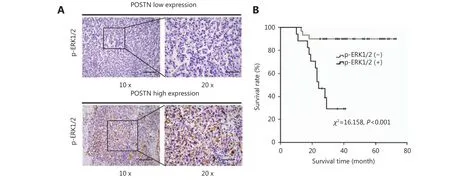
Figure 5 The phosphorylation of the MAPK/ERK pathway is activated in HB samples with high POSTN expression. (A)Immunohistochemical staining for p-ERK in HB tissues with different POSTN expression levels (scale bar, 1.0 mm). (B) Kaplan-Meier survival curves of patients with HB stratified by p-ERK expression.
Although the role of POSTN in cell biology has been sporadically reported, the mechanisms remain unclear.Emerging research has shown that POSTN promotes cancer progression via the ERK/VEGF and Akt, Wnt/β-catenin signaling pathways16,17. As has been reported, during EMT,the loss of E-cadherin and the acquisition of vimentin expression are two critical steps33. In the current study, we found that a high POSTN expression level was always accompanied by a rise in vimentin expression. We therefore further analyzed the expression level of relevant signaling genes involved in EMT, including ERK1/2, Gsk3β, Akt and Smad2/3. We found that the ERK1/2 expression level was significantly affected by POSTN expression. Therefore, our data suggest that POSTN might modulate EMT via the ERK signaling pathway, which promotes cellular adhesion and migration of HB, and that POSTN may be a novel biomarker for HB therapy.
Acknowledgements
This work was supported by grants from Key Project of Tianjin Natural Science Foundation (Grant No.18JCZDJC35200), The Science & Technology Development Fund of Tianjin Education Commission for Higher Education (Grant No. 2017KJ202).
Conflict of interest statement
No potential conflicts of interest are disclosed.
 Cancer Biology & Medicine2019年1期
- Cancer Biology & Medicine的其它文章
- Application of next-generation sequencing technology to precision medicine in cancer: joint consensus of the Tumor Biomarker Committee of the Chinese Society of Clinical Oncology
- The breakthrough in primary human hepatocytes in vitro expansion
- Circular RNAs and human glioma
- Qidong: a crucible for studies on liver cancer etiology and prevention
- The PI3K/Akt/GSK-3β/ROS/eIF2B pathway promotes breast cancer growth and metastasis via suppression of NK cell cytotoxicity and tumor cell susceptibility
- Estrogen and insulin synergistically promote endometrial cancer progression via crosstalk between their receptor signaling pathways
