Anti-cancer effects of hydro-alcoholic extract of pericarp of pistachio fruits
Hamidreza Harandi, Ahmad Majd, Soudeh Khanamani Falahati-pour, Mehdi Mahmoodi
1Department of biochemistry, North Tehran branch, Islamic Azad University, Tehran, Iran
2Department of Biology, Faculty of Science, Tehran North Branch, Islamic Azad University, Tehran, Iran
3Pistachio Safety Research Center, Rafsanjan University of Medical Sciences, Rafsanjan, Iran
4Department of Clinical Biochemistry, Afzalipoor Faculty of Medicine, Kerman University of Medical Sciences, Kerman, Iran
5Molecular Medicine Research Center, Research Institute of Basic Medical Sciences, Rafsanjan University of Medical Sciences, Rafsanjan, Iran
Keywords:Apoptosis Cytotoxicity Gene expression Pistacia vera
ABSTRACT Objective: To investigate the cytotoxicity and anti-cancer effects of hydro-alcoholic extract of pistachio pericarp on hepatocellular carcinoma cells (HepG2) and mouse fibroblast L929 cells as normal and control group cell. Methods: MTT assay was performed to investigate the cytotoxicity effects of the extract at 0-4 000 μg/mL on the cells after 24 and 48 h. The expressions of some genes involved in apoptosis including Bax, Bcl-2 and P53 were investigated by real time PCR. Results: Our results showed that after 24 and 48 hours of treatment of cells with this extract, the viability of HepG2 and L929 cells was reduced.Therefore, this extract had the cytotoxicity effect on both cells. The IC50 concentration of extract for HepG2 cells after 24 and 48 hours of treatment was 1 500 and 1 000 μg/mL and for L929 cells was 2 000 and 1 500 μg/mL, respectively. The expressions of Bax and P53 genes were up-regulated after treatment in the HepG2 and L929 cells and the expression of Bcl-2 gene was down-regulated after treatment of extract in HepG2 cell. Conclusions: According to the results of MTT assay and real time PCR, this extract can be considered as a potential candidate for use in the production of anti-cancer drugs for the treatment of patients with liver cancer in future.
1. Introduction
Medicinal herbs have been used to treat the diseases for centuries[1].Pistachio [Pistacia vera L. (P. vera)], belongs to the Anacardiaceae family, and is mainly found in Iran, Greece, Italy, Syria, Turkey,and USA[2]. Different parts of plant, including leave, resin, seed and fruit, have properties such as anti-inflammation and antimicrobial activities[3]. Pistachios fruits are widely used in the traditional medicine for treatment of kidney, liver, heart and respiratory system diseases[4]. Pistachio (P. vera L.) pericarp is part of pistachio fruit which is waste of pistachio processing and has low price. It is full of anti-oxidant compounds such as gallotannins, gallic acid, penta-O-galloyl-β-D-glucose, myricetin, quercetin and anacardic acid[5].One of the main countries in pistachio production is Iran. Therefore,using natural compounds with low cost and high anti-oxidant activity like pistachio pericarp makes a good attention to reuse[6].Management of cancer is one of the main problems in the world[7].Nowadays, due to complications of chemotherapy, medicinal plants have been considered by researchers for the treatment of cancer[8].According to IRIC (International Agency for Research on Cancer)74 600 deaths in the world are related to liver cancer that is the sixth cancer in the world and the second cause of death among cancers.This cancer involved many people from Asia and Africa[9,10].Morphologically, apoptosis process is characterized by cellular wrinkling, cell membrane damage, chromatin condensation, and fragmentation of DNA[11]. Stimulation of pro-apoptotic molecules or inhibition of anti-apoptotic factors plays an important role in apoptosis. The two main pathways of apoptosis include the external pathway or route dependent on the death receptor and the internal pathway or mitochondrial pathway[12]. Among the genes involved in apoptosis, Bcl-2, Bax and P53 genes can be mentioned. Bcl-2 proteins with the anti-apoptotic function, Bax, and P53 play an important role in apoptosis[13]. In this study, we examined the anticancer effect of hydro-alcoholic extract of pistachio (P. vera) pericarp on HepG2 cells (as a cancerous cell) and mouse fibroblast L929 (as normal cell) and its effects on the expression of some apoptotic genes including Bax, P53 and Bcl-2.
2. Materials and methods
2.1. Compounds and reagents
3-(4,5-dimethyl-2-thiazolyl)-2,5-diphenyl-2tetrazolium bromide(MTT) and dimethyl sulfoxide (DMSO) were prepared from Sigma,USA. Fetal bovine serum, trypsin, RPMI-1640, RPMI-phenol red,penicillin-streptomycin were purchased from Shellmax, China. Total RNA extraction kit and easy cDNA synthesis kit were prepared from Bioneer (South Korea) and Syber green color from Takara (RR820Q,Japan).
2.2. Sample preparation
In late summer (September 2017), pistachio (P. vera) was collected from a pistachio garden in Rafsanjan, Iran and approved by the botanist at the Vali-Asr University of Rafsanjan. Pericarps from pistachio were separated and dried at room temperature then powdered by mixer (Snijders Scientific, Holland) and stored in -20 ℃until use.
2.3. Preparation of the hydro-alcoholic extract of pistachio pericarp
To prepare the extract, 50 g of powder was mixed with 1 000 mL of 75% ethanol and incubated at room temperature for 24 h.The mixture was then incubated in dark area for 48 h and filtered by paper filter (0.2 μm). The filtered solution was freeze dried by VaCo5-D model Zirbus freeze-dryer (Germany). The obtained crystals were stored at -20 ℃ until use and dissolved in PBS buffer(pH 7.4)[14].
2.4. Cell culture
Cells were cultured in RPMI1640 medium containing penicillin antibiotics (100 U/mL) and streptomycin (100 mg/mL), 10% fetal bovine serum, and incubated at 37 ℃ in incubator with 5% CO2and 95% O2. After reaching the required number of cells (over 70%coverage), the adherent cells were trypsinized and removed from the flasks[15].
2.5. Treatment by different doses of extract
In order to determine the effect of pistachio pericarp extract on HepG2 and L929 cells, 5×10⁵cells were cultured and then treated with different concentrations (0-4 000 μg/mL) of extract for 24 h and 48 h. The concentrations of extract in gene expression and morphological analysis experiment were 1 500 μg/mL for HepG2 cells and 2 000 μg/mL for L929 cells (IC50value for these cells after 24 hours of treatment with the pistachio pericarp extract). All experiments were done in triplicate and repeated three times[16].
2.6. MTT assay
The colorimetric reaction is direct, using the color of MTT. Ten μL of MTT (5 mg/mL) were added to the well containing 100 μL of the culture medium and 5 000 cells, and the plate was placed between 2 and 4 hours in a 37 ℃ incubator. Afterwards, purple deposits could be seen by eyes in the wells, and the larger the number of live cells is in the well, more violet the deposition is.
After 4 h, 100 μL of DMSO solution was added to each well. The plate was placed at 25 ℃ for 10 min. Afterwards, optical absorption or optical density (OD) was read by the ELISA reader (Biotech ELX808, USA), based on the intensity of the Formazan blue at a wavelength of 570 nm[17]. Cell viability in this study was determined using the following formula: Cell viability = (Asample/Acontrol) × 100.
2.7. RNA extraction
All solutions, glass and plastic containers used in RNA extraction process were washed in a diethylpyrocarbonate solution and then autoclaved[18]. After treatment of HepG2 and L929 cells with hydroalcoholic extract of pistachio pericarp at IC50concentration for 24 h, the total RNA of the cells was extracted according to the RNA extraction kit (Bioneer, A101231, South Korea) instructions[18].To quantify and qualify the extracted RNA, RNA solution was determined by spectrophotometer at 260 and 280 nm. The 260/280 nm absorbance ratio was found around 2 nm and the best quality was between 1.8-2 nm (Denovix, USA). Finally, the reaction products were examined by 1.5% agarose gel electrophoresis[18].
2.8. cDNA synthesis
cDNA was synthesized according to cDNA synthesis kit (Bioneer,A101161) instructions, with a reverse transcriptase enzyme and a primer hybridized with RNA. The primer used in this test can be a specific gene or primers that are hybridized with all mRNAs(Oligo-dT oligothymidine primers). This cDNA was used to detect expressions of Bax, P53, Bcl-2 and β-actin genes. To ensure that DNA was not interrupted, all RNA samples were treated with the DNaseⅠ enzyme[19].
2.9. Primer designing
The primers used in this study were shown in Table 1. NTI vector software was used to design primers. All primers were designed with the BLAST software to ensure that primers were unique to the reference gene, β-actin gene which was used as an internal control[19].

Table 1 Primers sequences used in this study.
2.10. Real-time PCR reaction
Two major methods of real-time PCR include the use of SYBER GreenⅠ fluorescence and fluorescence probes are common. In this study, SYBER Green color (Takara, RR820Q, Japan) was performed. At first, mixed primer and mixed Syber green were synthesized according to kit instruction. Then according to protocol,mixed Syber and cDNA were added to each well. At this stage, the plate was inserted into the real-time PCR of the ABI model Biorad(United States) and the PCR reaction was performed. After the end of this step, the replication pattern for the samples was plotted,followed by subsequent analyzes. β-actin was used as the internal standard. The data were analyzed using the BIORAD CFX manager software. Finally, the reaction products were examined by 1.5%agarose gel electrophoresis[18].
2.11. Statistical analysis
Statistical analysis was performed by SPSS Version 16.0 statistic software package. One way ANOVA was used to compare differences between groups, and Dunnett test was used to compare different doses of extracts with control group. A value of P<0.05 was considered statistically significant.
3. Results
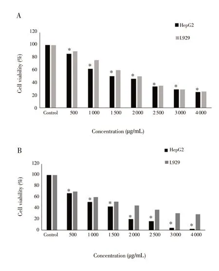
To evaluate the effect of extract on HepG2 and L929 cells at different concentrations, MTT test was used and the results were shown in Figure 1. IC50value for HepG2 and L929 cells was 1 500 and 2 000 μg/mL respectively for 24 h (Figure 1) and 1 000 and 1 500 μg/mL for 48 h, respectively (Figure 1). The morphology of HepG2 and L929 cells was observed by an inverted microscope(Hund, Germany) at IC50concentration after 24 hours of treatment with the pistachio pericarp extract (1 500 μg/mL and 2 000 μg/mL for HepG2 and L929, respectively) and cell shrinkage was found (Figure 2). In order to find out the effect of the extract on the expression of some apoptosis genes, real time PCR was performed and the mRNA expression levels of three apoptotic genes including Bax, Bcl-2 and P53 were examined. The results of the present study showed that the mRNA expression levels of P53 and Bax genes were up-regulated at IC50concentrations (1 500 μg/mL and 2 000 μg/mL for HepG2 and L929, respectively). Surprisingly, the mRNA expression level of Bax gene was down-regulated at a dose higher than IC50concentration(2 000 μg/mL and 2 500 μg/mL for HepG2 and L929, respectively).This may be the result of cell death at doses higher than IC50concentration in both HepG2 and L929 cells. However, the mRNA expression level of Bcl-2 gene was down-regulated after 24 hours of treatment with pistachio pericarp extract at IC50concentrations(1 500 μg/mL and 2 000 μg/mL for HepG2 and L929, respectively)compared to control (Figure 3). In fact, P53 promoted the expression of Bax gene. According to our results, we can say that the extract has a potential to promote intrinsic pathway of apoptosis.

Figure 1. Result of treating with different concentrations of pistachio vera pericarp extract on HepG2 and L929 after 24 (A) and 48 (B) h by MTT method.*The mean difference is significant at the 0.05 level in Dennett’s test for HepG2 and L929.

Figure 2. Morphological changes on HepG2 (A-B) and L929 cells (C-D)after exposure with pistachio pericarp extract at 0 μg/mL (untreated), and IC50 concentrations for HepG-2 (1 500 μg/mL) and L929 (2 000 μg/mL) cells,respectively that were observed with an inverted microscope under ××400 magnification.Untreated HepG2 cells (A), HepG2 cells after treatment with the extract at IC50 concentration (1 500 μg/mL) (B). Untreated L929 cells (C), L929 cells after treatment with extract at IC50 concentration (2 000 μg/mL) (D).

Figure 3. mRNA expression levels of some apoptotic genes in HepG2 and L929 cells treated with hydro-alcoholic extract of P. vera pericarp after 24 hours of treatment at IC50 (1 500 and 2 000 μg/mL for HepG2 and L929 cells,respectively) and two doses, higher and lower than IC50 concentrations by Real Time PCR method.Expression levels of P53 mRNA in HepG2 cell line (a); Expression levels of P53 mRNA in L929 cell line (b); Expression levels of Bcl2 mRNA in HepG2 cell line (c); Expression levels of Bcl2 mRNA in L929 cell line (d);Expression levels of Bax mRNA in HepG2 cell line (e); Expression levels of Bax mRNA in L929 cell line (f).
4. Discussion
Despite the report by the American Cancer Society about reduction of cancer rates, the number of deaths from liver cancer is increasing in the world. Many different anti-cancer compounds are used for cancer treatment. Some of these compounds are derived from medicinal plants[20] or microorganisms[21] and some of them are chemically synthesized such as proline-derived compounds[22],metallodrugs[23-25], indole derivatives[26,27] and etc. However,finding new compounds with anti-cancer properties has always been one of the major concerns for researchers.
Many previous studies reported the cytotoxicity effect of several medicinal plants which involved high amounts of phenolic compounds against HepG2, MCF-7, A375, and B16 cell lines[28].Also, the anti-cancer activities of pistachio (Pistacia lentiscus and Pistacia integerrima) have been reported[29]. In pistachio, the phenolic compounds are higher in hulls than in seeds.
Also, previous studies have shown anti-bacterial, anti-cancer and anti-angiogenesis[30] effects of some components of pistachio (P.vera L.) like hull, leaves, seed, essential oil and gum[31-33]. One of the route that a cancer cell became a malignant cell is evading apoptosis[34]. So designing new drugs against cancer is very important because defects in the regulation of apoptotic pathways can result in resistance to cancer chemotherapy[35]. In the present study, the hydro-alcoholic extract of P. vera pericarp was used for determination of anti-cancer and cytotoxicity effects against HepG2 and L929 cells. In our study, the IC50of the hydro-alcoholic extract for L929 cells was higher than for HepG2 cells, so we can say that the extract in dose of IC50cannot kill a lot of normal cells but kills many malignant cells. In other words, this extract selectively inhibits proliferation of HepG2 cancerous cells in a dose dependent manner. Apoptosis is occurred by the regulation of the balance between the expression of pro- and anti-apoptosis genes such as Bcl-2, P53 and Bax. P53 and Bax are pro-apoptotic genes and Bcl-2 is an anti-apoptotic gene[36]. To better understand the molecular mechanism of apoptosis, real time PCR was performed.Real-time PCR is a method based on the polymerase chain reaction and illustrates the amplification of a wanted DNA molecule. Three genes involved in apoptosis including P53, Bcl-2 and Bax were checked. The results of gene expression showed that in HepG2 and L929 cells, the expressions of P53 and Bax were up-regulated in IC50concentration but the expression of Bcl2 gene was down-regulated in this concentration for both cells. The results of the present study showed that the up-regulation of P53 can be the essential factor to promote of Bax gene expression in the malignant cells after treatment with extract. A significantly increase of P53 expression could demonstrate that P53 triggered intrinsic or mitochondrial apoptotic pathway via the up-regulation of Bax gene and downregulation of Bcl-2 gene. In HepG2 cells, the intrinsic apoptosis pathway is regulated by the balance between the expression of Bcl-2 and Bax genes. Bcl-2 protein binds to the mitochondrial outer membrane and cytochrome C channel is blocked. In contrast, Bax causes realizing of cytochrome C thus apoptosis occurs[37]. In fact,up-regulation of pro-apoptotic genes such as P53, Bax and P21 as well as the suppression of Bcl-2 gene expression as an anti-apoptotic gene can induce apoptosis in HepG-2 cells. P53 protein is a key element to promote the expression of Bax gene. Bax promotes and activates the Bax transcription directly, inducing cell cycle arrest or apoptosis. Also, Bax/Bcl-2 ratio is an important factor promoting apoptosis[38]. We also determined the expression of the genes at two doses, higher and lower than IC50concentrations (1 500 and 2 000 μg/mL for HepG2 and L929 cells, respectively). We found that in the low concentration, the expressions of P53 and Bax genes were lower than that observed at IC50. At the high concentration, the expressions of P53 and Bax genes were also reduced compared with that observed at IC50. According to the results of the present study,the pistachio pericarp extract has the potential of apoptosis induction and cytotoxic activity against HepG2 cells. Therefore, this extract can be considered as a potential candidate for use in the production of anti-cancer drugs for the treatment of patients with liver cancer in future.
Conflict of interest statement
All authors declare that they have no conflict of interest.
Acknowledgements
This article is an excerpt from Hamidreza Harandi’s PhD’s thesis in department of biochemistry, North Tehran branch, Islamic Azad University, Tehran, Iran.
 Asian Pacific Journal of Tropical Biomedicine2018年12期
- Asian Pacific Journal of Tropical Biomedicine的其它文章
- A survey of biochemical and acute phase proteins changes in sheep experimentally infected with Anaplasma ovis
- Gac fruit extracts ameliorate proliferation and modulate angiogenic markers of human retinal pigment epithelial cells under high glucose conditions
- Anti-hemolytic, antibacterial and anti-cancer activities of methanolic extracts from leaves and stems of Polygonum odoratum
- Anti-cancer and anti-inflammatory activities of aronia (Aronia melanocarpa) leaves
- Antidiabetic effects of Tetracarpidium conophorum seed on biomarkers of diabetesinduced nephropathy in rats
- Anti-insulin resistant effect of ferulic acid on high fat diet-induced obese mice
