Endophytic actinobacteria of medicinal plant Aloe vera: Isolation, antimicrobial,antioxidant, cytotoxicity assays and taxonomic study
Ahmed Nafis, Ayoub Kasrati, Asma Azmani, Yedir Ouhdouch, Lahcen Hassani
1Laboratory of Biology and Biotechnology of Microorganisms, Faculty of Sciences Semlalia, Cadi Ayyad University, PO Box 2390, Marrakech,Morocco
2Department of Biology, Faculty of Sciences Semlalia, Cadi Ayyad University, Marrakech, Morocco
Keywords:Actinobacteria Medicinal plant Aloe vera Antimicrobial activity Cytotoxicity Antioxidant assays Molecular identification
ABSTRACT Objective: To explore the new sources of novel bioactive compounds having pharmaceutical and agricultural interest and to search the endophytic actinobacteria from medicinal plants.Methods: NAF-1 an endophyte actinobacteria was isolated from leaves of medicinal plant Aloe vera collected in Marrakesh, Morocco using Bennett agar as selective medium. NAF-1 was tested for its antimicrobial activity against five pathogenic bacteria such as Staphylococcus aureus PIC 53156, Micrococcus luteus ATCC381, Bacillus subtilis ATCC 14579, Pseudomonas aeruginosa DSM 50090 and Escherichia coli ATCC 8739 and four human clinic fungi belonging to the Candida, Aspergillus and Microsporum genera. Several antioxidant activities were studied such as DPPH free radical scavenging, β-carotene and linoleic acid and reducing power assays. The total of phenol and flavonoid was also calculated. Using Artemia salina shrimp assay, the cytotoxicity of NAF-1 crude extract was determined. Results: The results revealed that the actinobacteria showed a high activity (≥20 mm) against only Gram positive bacteria but it had a moderate activity (between 13 and 15 mm) against Human clinic fungi. The isolate also exhibited a LD50 of 14.20 µg/mL in the cytotoxicity assay. The result showed that the crude extract presented an interesting free radical-scavenging activity with IC50 value of (5.58± 0.26) µg/mL and a high value of phenolic and flavonoid compounds with (15.41 ± 0.18) µg GAE/mg extract and (11.41± 0.06) µg QE/mg extract respectively. Moreover, the taxonomic position of our endophyte actinobacteria using the morphological and physiological criteria and using 16S rRNA gene sequence (polyphasic approach) showed that the NAF-1 isolate was similar to Streptomyces hydrogenans which was never described as an endophyte actinobacteria.Conclusions: This isolated strain appears promising resources of bioactive agents and can be exploited to produce therapeutic agents active against pathogenic disease.
1. Introduction
Pathogens multi-drug resistance capacity becomes more and more severe due to abusive use of antibiotics. One way to solve this problem is to look permanently for new bioactive compounds especially from natural resources[1].Endophytic actinobacteria are antagonists microorganisms that act inside the internal tissues of plants without having any negative influence on the plant host[2]. Medicinal plants constitute huge diversity of endophytic actinobacteria, which are filamentous Grampositive bacteria, with important economic value because of their ability to produce high potential bioactive substances including antimicrobial, anti-cancer and other pharmaceutical compounds[3,4].Among members of actinobacteria, Streptomyces genus remains the richest sources of valuable natural product with 7 600 (76%)bioactive metabolites[5,6].
To date, few are known about the antibiotics produced by endophytic actinobacteria. Thus, the present study is focused on isolating actinobacteria from the leave tissues of medicinal plant Aloe vera (A. vera), on evaluating the antimicrobial, antioxidant and cytotoxic activities of its secondary metabolites and on identifing it on the basis of the morphological, physiological and 16S rRNA gene analysis.
2. Materials and methods
2.1. Sample collection and isolation of endophytic actinobacteria
Strain NAF-1 was isolated from the leaves of a medicinal plant A.vera collected in Marrakech, located in the north of the foothills of the Atlas Mountains, Morocco. Firstly, the samples of leaves were washed by distillated water for 1-2 min to remove soil particles and then surface sterilized with 70% ethanol for 10 min and 1%sodium hypochlorite for 15 min. In order to reduce the opportunity of endophytic fungi emergence, the leaves were soaked 10 min in 10% NaHCO3solution to inhibit the growth of fungi. Subsequently,the plant materials were rinsed three times with sterilized distillated water[7-9] and the leaves of plant were cut into small fragments.Finally, the pieces were transferred to Petri dishes of Bennett agar medium containing 50 µg/mL of cycloheximide and 100 µg/mL of nalidixic acid, and the endophytic actinobacteria were observed after incubation at 28 ℃ for 1-4 weeks.
2.2. Screening for antimicrobial activity of endophytic actinobacteria
Strain NAF-1 was grown on Bennett agar medium during 14 days.Agar cylinders were cut and placed on Muller-Hinton medium for bacteria and Sabouraud medium for fungi (yeasts and molds),previously inoculated by the test microorganisms. Plates were kept during 4 h at 4 ℃ for a good diffusion of secondary metabolites produced, then incubated at 37 ℃ for bacteria and 30 ℃ for fungi.Antibacterial activity was evaluated in vitro using three Gram positive bacteria, Staphylococcus aureus PIC 53156, Micrococcus luteus ATCC381 and Bacillus subtilis ATCC 14579 and two Gran negative bacteria, Pseudomonas aeruginosa DSM 50090, Escherichia coli ATCC 8739. While the antifungal activity was achieved against four clinic fungi species: Candida albicans CCMM-L4,Candida tropicalis DSM11953, Aspergillus niger CCMM-M100 and Microsporum canis CCMM-M103.
2.3. Cytotoxicity assay of NAF-1 crude extract
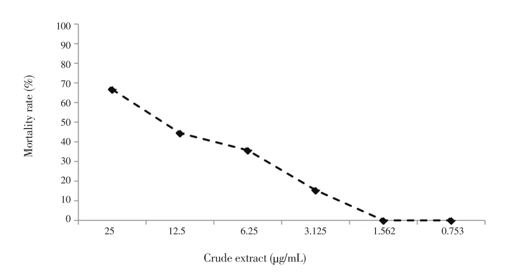
Artemia salina brine shrimp was used as a model to study cytotoxicity of crude extract of strain NAF-1. After hatching of 5 mg of brine shrimp eggs in 500 mL of sterilized seawater under constant aeration at 37 ℃ for 24 to 48 h, the active nauplii were collected and used for the test. Several concentrations were prepared by dilution from 100 µg/mL to 0.753 µg/mL of crude extract obtained by extraction from fermentation culture using ethyl acetate and concentrated under vacuum. A total of 0.5 mL of each dilution were added to 10 to 20 larvae (0.5 mL) of mature nauplii into separate well of plastic 24-well tissue culture plate. After 24 h, the LD50was calculated on the basis of the mortality rate[10].
2.4. Antioxidant activity of NAF-1 crude extract
2.4.1. Determination of 2,2-diphenyl-1-picrylhydrazyl(DPPH) radical scavenging and reducing power activities
To determine the antioxidant activity (IC50) of the crude extract obtained from the culture fermentation of strain NAF-1, we used the stable radical DPPH according to Şahin et al.[11]. Concerning the reductive potential, it was determined by the Fe3+to Fe2+transformations in the presence of the crude extract, using the method of Gülçin et al.[12]. The BHT and quercetin were used as positive control.
2.4.2. β-carotene/linoleic acid bleaching assay
The crude extract’s ability to prevent beaching of β-carotene,by its oxidation in the presence of O2molecule, was performed by Miraliakbari & Shahidi[13] method with small modifications.The absorbance values were measured at 470 nm to calculate the inhibitions percentages (I %) of the sample.
2.4.3. Determination of total phenols and flavonoids content
Total phenolic content of the crude extract of our strain which was expressed in µg of gallic acid equivalent/mg extract (µg GAE/mg extract) was determined by the Folin-Ciocalteu micro-method as described by Ali et al.[14]. On the other hand, the total flavonoid content was determined by the method of Zengin et al.[15] and expressed in µg of quercetin equivalent/mg extract (µg QE/mg extract).
2.5. Polyphasic taxonomy of endophytic actinobacteria
2.5.1. Morphological and physiological characterization
The cultural features of the strains NAF-1 were characterized following the directions given by the International Streptomyces Project (ISP) media[16,17] and the Bergey’s Manual of Systematic
Bacteriology[18]. Our promising isolate was tested for their ability to grow at pH 5 to 9 and at a temperature range from 27 ℃ to 37 ℃[19].The production of melanoide pigments, considered a useful criterion for taxonomic studies, was carried out on ISP6 and ISP7 agar[20].
The study of the physiological criteria such us using different carbon and nitrogen sources has been carried out on the ISP9 base medium to which carbon and nitrogen sources were added to a final concentration of 1%[21]. The plates were kept at 28 ℃ and the growth was evaluated visually.
2.5.2. DNA extraction, polymerase chain reaction (PCR)amplification and sequencing of 16S rRNA
Strain NAF-1 was grown under orbital shaking during 4 days at 28 ℃ in Erlenmeyer flasks containing 50 mL of Bennett medium liquid. Biomass was separated using centrifugation at 8 000 rpm for 10 min and washed several times with sterile distillated water. The genomic DNA of the pellet was extracted using Bacterial DNA kit according to the manufacturer instructions.
A PCR using primers 27 Fbac (5’AGAGTTTGATCMTGGCTCAG3’)and 1492 Runi (5’ TACGGYTACCTTGTTACGACTT 3’) was used to amplify the 16S rDNA gene. The conditions used for thermal cycling were as follows: denaturation at 95 ℃ for 5 min, followed by 30 cycles of amplification at 95 ℃ for 1 min, annealing at 54-55 ℃ for 1 min,extension at 72 ℃ for 2 min and extra extension at 72 ℃ for 10 min and then cooling to 4 ℃[22].
The purified 16S rRNA was sequenced directly using the same primers and was compared for similarity with those contained in genomic database banks, using NCBI BLAST. The phylogenetic tree was constructed using the neighbor-joining method with the Mega 6 software[23].
3. Results
3.1. Antimicrobial activity of the isolated NAF-1 strain
In this study, the antimicrobial activity was screened on agar medium and the results revealed that the maximum zone of inhibition was recorded against Staphylococcus aureus (25 mm) and Micrococcus luteus (25 mm) followed by Bacillus subtilis (20 mm) but no activity against the Gram negative bacteria. Furthermore, our actinobacteria was active against all yeasts and filamentous fungi used in our test.The maximum of inhibition was observed against Aspergillus niger(15 mm) followed by Candida tropicalis (14 mm), Candida albicans(13 mm) and Microsporum canis (13 mm).
3.2. Cytotoxicity assay
The crude extract of strain NAF-1 was subjected to Brine shrimp lethality assay to find the cytotoxicity expressed in LD50. The extract studied showed significant lethality against mature nauplii with 14.20 µg/mL as LD50(Figure 1).

Figure 1. Effect of various concentrations of crude extract (strain NAF-1)against Artemia salina shrimp larvae.
3.3. Antioxidant activity
To evaluate the antioxidant activity of NAF-1 crude extract, several tests were performed, and the IC50of crude extract and those of BHT and quercetin were given in Table 1. The result showed that our crude extract exhibited an interesting free radical-scavenging activity with IC50value of (5.58 ± 0.26) µg/mL better than those in BHT and quercetin. Concerning β-carotene acid bleaching test, IC50value of the crude extract was less effective than BHT and more effective than quercetin. Furthermore, the reducing power IC50values were significantly lower than those obtained for synthetic antioxidant agents. Our crude extract showed a high value of phenolic and flavonoid compounds with (15.41 ± 0.18) µg GAE/mg extract and(11.41 ± 0.06) µg QE/mg extract respectively.

Table 1IC50 values of crude extract, BHT and quercetin.
3.4. Taxonomic study of Endophytic actinobacteria NAF-1
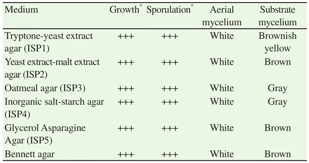
The promising strain NAF-1 was the subject of a taxonomic study based on the determination of its morphological, biochemical and molecular criteria. The phenotypic appearance and cultural characteristics were determined on ISP culture media. The importance of growth, the development of aerial mycelium on each medium, the color of the aerial and substrate mycelium as well as the presence of diffusible or non-diffusible pigments in the agar were observed and the all results were noted in the Table 2.
Strain NAF-1 showed a good growth and sporulation and abundant mycelium in all media used. The color of the aerial mycelium was white but the coloration of substrate mycelium was variable, it was brownish yellow in ISP1, brown in ISP2, ISP5 and Bennett and gray in ISP3 and ISP4 (Table 2). The diffusible pigments and melanoids were produced on ISP6 and ISP7 respectively.
In the case of the use of carbon sources, the NAF-1 isolate was capable to degrade the glucose, fructose, arabinose, galactose,rhamnose and mannitol but concerning the use of amino acids, the NAF-1 strain was able to use asparagine and arginine. Regarding the pH, we found that the strain NAF-1 was able to develop at different pH values (5, 6, 7, 8 and 9) so it tolerated neutral, slightly acidic and basic pH. In addition, the results showed that our strain grew at temperatures ranging from 27 ℃ to 37 ℃ with an optimum at 30 ℃so it preferred mesophilic growth temperatures.
According to these morphological, physiological and biochemical studies, it is impossible to determine exactly the species to which our isolate belongs, so it is necessary to make recourse to the molecular identification of 16S RNA.
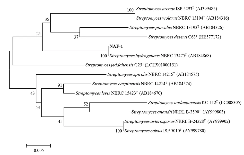
Indeed, the sequencing of the NAF-1 16S RNA yielded a nucleotide sequence of 1 395 bp having 100% of similarity with Streptomyces hydrogenans (S. hydrogenans) NBRC 13475T(Figure 2). In addition,the comparison of the profiles using some carbon sources and nitrogen substrates showed that NAF-1 had a great resemblance to S. hydrogenans except that this latter is unable to use fructose and mannitol.

Table 2Phenotypic characteristics of NAF-1 in ISP media after 2 weeks of incubation at 28 ℃.

Figure 2. Phylogenic tree based on 16S rRNA gene sequence showing the relations between NAF-1 strain and type species of the genus Streptomyces.
4. Discussion
Actinobacteria from medicinal plants are the new source of several bioactive substances with high therapeutic potential[3], and their molecules produced may be associated with the properties of the host medicinal plant[24]. We investigate the application of endophytic actinobacteria, especially from A. vera, in order to evaluate the species richness and biological properties to decrease the problem of antibiotic resistance emergence.
According to the results of the antimicrobial activity, we noticed that our NAF-1 strain showed a high activity against the Gram positive bacteria and was not active against the negative ones.This different sensitivity could be attributed to the high level of lipopolysaccharides that contain the Gram positive bacteria membrane, which could make the cell wall impermeable to bioactive compounds[25].
On the other hand, the isolated actinobacteria showed a very interesting activity against the clinic fungi. These data suggest that NAF-1 strain produces metabolites with a broad spectrum activity which are required to treat multidrug resistant pathogens, such as MRSA, Pseudomonas aeruginosa, and Acinetobacter baumannii that are considered one of the most urgent issues in modern healthcare[26-28].This result also confirms that the endophytic actinobacteria are often associated with a high antimicrobial properties[29,30].
The brine shrimp lethality assay was considered one of the most useful tests used to study the toxicity of bioactive substances[31]. In our bioassay, the cytotoxicity was similar to those obtained from actinobacteria by many research works using Artemia salina as model[32,33]. Several studies reported that, if the toxicity using brine shrimp larvae displayed LD50lower than 1 000 µg/mL of natural extract, it implies that it contains physiologically active principles[34].That idea was confirmed by the highest antioxidant activity and the values of total phenolic and flavonoid calculated. Those activities can be explained by the presence of some class of compounds,for example the fatty acids. Moreover, the benzyl alcohol and esters were previously reported to have an interesting antioxidant activity[35].
To confirm the reliability of the preliminary taxonomic characterization, the bioactive endophytic actinobacteria NAF-1 was subjected to 16S rRNA gene sequence analysis. The sequences BLAST suggest that our isolate is S. hydrogenans with 100% of similarity. This result demonstrates that the most abundant isolates of the endophytic community belong to the genus Streptomyces which was consistent with various reports on other plant hosts[7,36,37]. We reported for the first time the isolation of strain closely related to the species S. hydrogenans as endophyte. S. hydrogenans was originally isolated from soil in several works[29,38,39].
Several strains of endophytic actinobacteria show a different range of bioactivities which can be exploited therapeutically. For example,43.4% of endophytic actinobacteria recovered from leaves and roots of maize showed an antibacterial and antifungal activity against at least one of bacteria and yeast tests[40]. Similarly, another study revealed that 55% of endophytic actinobacteria isolated from leaves of Paeonia lactiflora and Trifalium repens inhibited the growth of Rhizoctonia solani a significant pathogen of plants[41]. Streptomyces longisporoflavus an endophytic actinobacteria was isolated from Lysimachia ciliate in India which has anti-diabetic activity[42].
The study conducted by Zhang et al.[43], allows to obtain 12 actinobacteria stains which are able to inhibit the growth of penicillin resistant Staphylococcus aureus, the majority of them were belonging to the genus Streptomyces and they isolated from Artemisia argyi, Radix platycodi, Achyranthes bidentata and Paeonia lactiflora.Searching for novel bioactive substances with interesting biological activity in a diverse environment has attracted more attention in recent decade due to the increased incidence of multiple resistances to the used drugs[44].
A. vera provides a new source for potential endophytic actinobacteria with promising and new biologically secondary metabolites. The study shows that the isolation from a unique niche (medicinal plants)to explore endophytic actinobacteria will facilitate the discovery of new antimicrobial agents with significant biological activity.
Conflict of interest statement
The authors declared no conflict of interest.
Acknowledgements
The corresponding author thanks a lot all the co-authors for help.
 Asian Pacific Journal of Tropical Biomedicine2018年10期
- Asian Pacific Journal of Tropical Biomedicine的其它文章
- Beneficial effects of anthocyanins against diabetes mellitus associated consequences-A mini review
- Does prospective permutation scan statistics work well with cutaneous leishmaniais as a high-frequency or malaria as a low-frequency infection in Fars province, Iran?
- Effect of extracts from Stachys sieboldii Miq. on cellular reactive oxygen species and glutathione production and genomic DNA oxidation
- Eicosane, pentadecane and palmitic acid: The effects in in vitro wound healing studies
- Phytochemical composition of Caesalpinia crista extract as potential source for inhibiting cholinesterase and β-amyloid aggregation: Significance to Alzheimer's disease
