Effect of extracts from Stachys sieboldii Miq. on cellular reactive oxygen species and glutathione production and genomic DNA oxidation
JW Lee, W Wu, SY Lim✉
1Division of Marine Bioscience, Korea Maritime and Ocean University, Busan, Korea
2Department of Marine Bio-Pharmacology, Shanghai Ocean University, Shanghai, China
Keywords:Stachys sieboldii Miq.Reactive oxygen species Glutathione DNA oxidation Antioxidant
ABSTRACT Objective: To evaluate the antioxidant activity of extracts and fractions from Stachys sieboldii Miq., and to examine its effect on the cellular reactive oxygen species (ROS) and glutathione(GSH) production and genomic DNA oxidation in HT-1080 cells. Methods: The ROS generation induced by H2O2 was measured by the dichlorofluorescein-diacetate assay. GSH levels were measured using a fluorescent method with mBBr. Genomic DNA oxidative damage was measured with levels of oxidative DNA induced by the reaction of ferritin with H2O2. Results: The n-hexane, 85% aqueous methanol and n-butanol fractions (0.05 mg/mL concentrations) inhibited H2O2-induced ROS generation by 63%, 35% and 45%, respectively.GSH levels were significantly increased in both acetone+methylene chloride and methanol extracts (P<0.05). Supplementation of cells with n-hexane significantly increased GSH levels at concentrations of 0.05 mg/mL (P<0.05). Both the acetone+methylene chloride and methanol extracts, as well as all fractions significantly inhibited oxidative DNA damage (P<0.05).Conclusions: These results indicate that cellular oxidation was inhibited by the n-hexane fraction and this fraction may contain valuable active compounds.
1. Introduction
Stachys sieboldii (S. sieboldii) Miq. is a herbaceous plant in the family Labiatae and is widely used as a vegetable in China, Japan,and Korea. Additionally, S. sieboldii Miq. has also been used as a Chinese folk medicine to treat ischemic brain injury, dementia, and various gastrointestinal related diseases[1]. S. sieboldii Miq. contains various oligosaccharides, which could stimulate the growth of beneficial microorganisms in the human intestine and is considered as a wellness-promoting food in Asian countries[2]. S. sieboldii Miq.has been reported to contain several active compounds including terpenes[1], flavonoids[3], and phenolic compounds[4], which are highly associated with its antimicrobial[5], antioxidant[3-7], and antitumor properties[8].
Free radicals are oxygen-containing molecules with one or more unpaired electrons, making them highly reactive towards other molecules. In most macromolecules, oxidative stress damage is induced by free radicals[9]. Therefore, oxidative stress is considered as a critical pathophysiological mechanism in different courses and processes of diseases, including cardiovascular diseases, cancer,diabetes, rheumatoid arthritis, and various neurological disorders[10].Antioxidants are used for reducing agents, and attenuating oxidative damage to critical biological structures by protecting them from free radicals. Antioxidants are found in many kinds of food, and several studies have recommended that continuous intake of vegetables and fruits in one’ diet leads to a positive regulation against oxidative stress induced damage in the body[11]. Our previous studies presented that acetone+methylene chloride extract from S. sieboldii Miq. had higher levels of flavonoids than the methanol extract[12], which was associated with a higher free radical scavenging activity. In this present study, we investigated the effect of extracts and fractions from S. sieboldii Miq. on cellular reactive oxygen species and glutathione production and genomic DNA oxidation to determine its potential use as a natural antioxidant supplement.
2. Materials and methods
2.1. Materials and cell line culture
Dulbeco’s modified Eagle’s medium, fetal bovine serum, phosphate buffered saline (PBS), dimethylsulfoxide, penicilline-strectomycin,and 2’-7’ dichlorofluorescein-diacetate (DCFH-DA) were purchased from Sigma-Aldrich (St. Louis, MO). A human fibroblast cell line HT-1080 was acquired from the Korea Cell Line Bank. The cells were maintained at 37 ℃ in 5% CO2in Dulbeco’s modified Eagle’s medium with 10% fetal bovine serum and 100 units/mL penicillinstreptomycin.
2.2. Sample extracts and fractions
S. Sieboldii Miq. was purchased from the Misan herb farm (Daegu,Korea). Dried S. Sieboldii Miq. (3 kg) was extracted with acetone/methylene chloride (A+M, 0.6 g) and methanol (MeOH, 12.9 g) to obtain the maximum amount of extracts. Then the combined crude extracts (6.8 g) were fractioned with n-hexane (0.82 g) and 85%aqueous MeOH (1.82 g), and the aqueous layer was also further fractioned with n-butanol (n-BuOH, 1.38 g) and water (2.0 g), resulting in the n-hexane, 85% aqueous MeOH, n-BuOH and water fractions[13].All extracts and fractions were vacuum dried at 40 ℃ using rotary vacuum evaporator (N-100, EYELA, Japan) and the residue was kept at 4 ℃ until further analysis. For the test samples all extracts and fractions were dissolved in dimethylsulfoxide at various concentrations.
2.3. Intracellular reactive oxygen species (ROS) measurement
Cellular oxidative stress due to ROS generation from H2O2was measured using the DCFH-DA method[14]. HT-1080 cells were planted in 96-well plates (5 × 10⁵/well) for 24 h. After rinsing with PBS, 20 µM DCFH-DA was treated and pre-incubated for 20 min.Then the test samples were added and set for 1 h. After the DCFHDA was removed and rinsed with PBS, 500 µM H2O2were added and set for 120 min. The control was treated with H2O2and distilled water and blank was treated with distilled water without H2O2.Dichlorofluorescein fluorescence was assessed at 485 nm and at 535 nm by a fluorometric plate reader (VICTRO3, Perkin Elmer,Wellesley, MA).
2.4. Determination of intracellular glutathione (GSH)concentration
GSH levels were measured using a fluorescent method with mBBr[15]. HT-1080 cells were planted in 96-well plates (5 × 10⁵/well)for 24 h. The cells were treated with samples at various concentrations and incubated for 30 min. After rinsing with PBS, 40 µM mBBr was added and incubated for 30 min. The fluorescence was assessed at 360 nm and at 465 nm by the fluorometric plate reader (VICTRO3,Perkin Elmer, Wellesley, MA).
2.5. Genomic DNA isolation
High molecular weight genomic DNA was separated from the HT-1080 cells through standard phenol/proteinase K procedure[16].Concisely, cultured cells were rinsed with PBS and aliquoted into 1 mL of PBS with 10 mM EDTA. After centrifugation the cells were mixed in RNase (0.03 mg/mL), NaOAc (0.175 M), proteinase K (0.25 mg/mL) and sodium dodecyl sulfate (0.6%). The mixture was set for 30 min at 37 ℃ and 1 h at 55 ℃. Then, phenol:chloroform:isoamyla lcohol (25:24:1) was diluted at 1:1 ratio and mixture was centrifuged at 6 000 rpm for 5 min at 4 ℃. The upper was blended with 100%ice cold ethanol at a 1:1.5 ratio and set for 15 min at -20 ℃. After centrifugation at 16 000 rpm for 5 min at 4 ℃, the lower pellet was stirred in tris-ethylenediaminetetraacetic acid buffer and the purity of DNA was measured at 260/280 nm.
2.6. Determination of radical mediated DNA oxidation
H2O2induced DNA oxidation was assessed according to Kang’s method[17]. Concisely, 100 µL of DNA reaction mixture was prepared by adding a pre-determined concentrations of the sample,a 200 µM final concentration of FeSO4, a 2 mM final concentration of H2O2and a 50 µg/mL of genomic DNA. Then, the mixture was set at room temperature for 30 min and the reaction was withdrawn by adding 10 mM EDTA. Aliquots (20 µL) of the reaction mixture containing about 1 µg of DNA were electrophoresed on a 1% agarose gel for 30 min at 100 V. After staining with 1 mg/mL ethidium bromide, the gels were imaged through UV light by AlphaEase®gel image analysis software (Alpha Innotech, CA, USA). The control was treated with H2O2and distilled water and blank was treated with distilled water without H2O2.
2.7. Statistical analysis
Data were presented as a mean ± standard deviation (SD). The significance of differences observed between the control and experiment groups used the standard Student’s t test at P<0.05.Analyses were conducted using the STATISTICA package (TopCo,Palo Alto, CA, USA).
3. Results
3.1. Effect of fractions of S. sieboldii Miq. on production of cellular ROS
Figure 1 shows the inhibitory effects of the solvent fractions(n-hexane, 85% aqueous MeOH, n-BuOH and water) of S. sieboldii Miq. on ROS production induced by H2O2in HT-1080 cells. All tested fractions decreased H2O2induced ROS production in a concentration-dependent manner. Adding with n-hexane, 85%aqueous MeOH and n-BuOH fractions (0.05 mg/mL concentration)resulted in 63%, 35%, and 45% ROS inhibition, respectively. These results suggested that the n-hexane fraction had the strongest effect on reducing H2O2-induced ROS production.
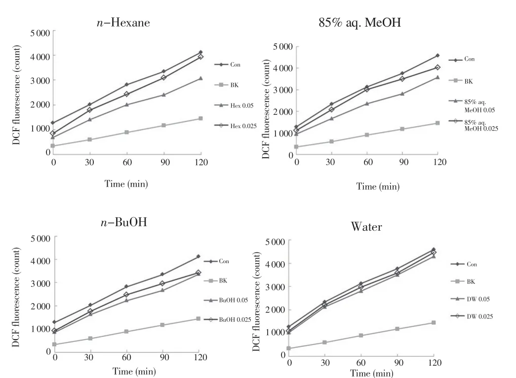
Figure 1. Effect of fractions from S. sieboldii Miq. on the amounts of ROS in HT-1080 cells.
3.2. Effect of extracts and fractions of S. sieboldii Miq. on GSH production
Total GSH levels were determined on the lysates of HT-1080 cells treated with A+M and MeOH extracts from S. sieboldii Miq.at various concentrations (Figure 2). Supplementation of cells with A+M and MeOH extracts significantly increased total GSH levels relative to that in the control experiment (P<0.05). Figure 3 presents the effects of S. sieboldii Miq. fractioned in various solvents on GSH levels. Four fractions significantly increased GSH levels at a low concentration (0.025 mg/mL) (P<0.05). Among these fractions, the n-hexane fraction showed greater levels of GSH.
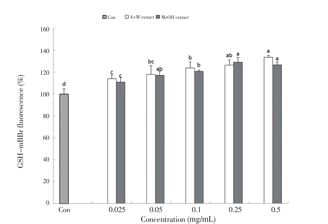
Figure 2. Effect of acetone+methylene chloride (A+M) and methanol (MeOH)extracts from S. sieboldii Miq. on the contents of GSH in HT-1080 cells.
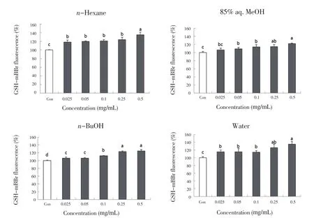
Figure 3. Effect of fractions from S. sieboldii Miq. on the contents of GSH in HT-1080 cells.
3.3. Effect of extracts and fractions of S. sieboldii Miq. on genomic DNA oxidation
The ability of extracts and fractions from S. sieboldii Miq. to inhibit DNA oxidative damage was assessed using genomic DNA isolated from HT-1080 cells. Figure 4 and 5 present the inhibition rate (%)of DNA oxidation related to the blank without Fe(Ⅱ)-H2O2. Both the A+M and MeOH extracts significantly inhibited Fe(Ⅱ)-H2O2induced oxidative stress DNA damage (P<0.05). All fractions including the n-hexane, 85% aqueous MeOH, n-BuOH and water fractions significantly inhibited oxidative DNA damage by > 90%(P<0.05). These results suggest that S. sieboldii Miq. extracts and fractions provide an inhibitory effect on radical-mediated DNA damage.
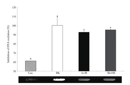
Figure 4. DNA oxidative inhibition by acetone+methylene chloride (A+M)and methanol (MeOH) extracts (0.05 mg/mL concentration) from S. sieboldii Miq. in HT-1080 cells.
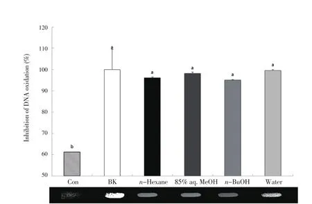
Figure 5. DNA oxidative inhibition by fractions (0.05 mg/mL concentration)from S. sieboldii Miq. in HT-1080 cells.
4. Discussion
ROS have been related to a higher risk of chronic and degenerative diseases including cardiovascular and neurodegenerative diseases,and cancer[10]. As the activity of ROS is likely inhibited by antioxidants, the damage caused by ROS attenuates presumably the risk of chronic diseases with antioxidant supplementations.Thus, consumption of vegetables and herbal plants, which are rich sources of antioxidants, could regulate biochemical pathways and prevent those chronic diseases[19]. Antioxidants attenuate ROS stress-induced damages due to their ability to donate electrons that neutralize the radicals without forming another[20]. In our previous study, extracts from S. sieboldii Miq contained flavonoids and showed a radical scavenging ability in DPPH and 2.2’-azino-bis(3-ethylbenzothiazoline-6-sulfonic acid) diammonium salt (ABTS)assays[12]. The n-hexane fraction from S. sieboldii Miq was more effective for attenuating cellular ROS production among the fractions presented in this study. According to Baek et al.[6], the ethyl acetate fraction from S. sieboldii Miq., which had higher levels of phenolic compounds, showed the strongest antioxidant radical scavenging activity among other fractions in 1,1-diphenyl-2-picryhydrazyl assay, ferric thiocyanate method and nitrite scavenging ability test.Na et al.[4] found that S. sieboldii Miq. contained more phenolic compounds than Korean ginseng, and that it had a higher radical scavenging capacity based on DPPH, ferric reducing antioxidant potential and Trolox equivalent antioxidant ability. In addition, Feng et al.[21] observed a high scavenging capacity towards superoxide anion, hydroxyl, and ABTS radicals but low scavenging activity towards DPPH radicals. Another study of S. sieboldii Miq. indicated that S. sieboldii Miq. powder adding white bread had the significantly scavenging effect of DPPH and ABTS radicals with increased levels of added S. sieboldii Miq. powder[7]. Khanavi et al.[22] compared the antioxidant activity and total phenolic contents in several Stachys species, suggesting that Stachys persica and Stachys fruticulosa had the highest phenol contents, which are associated with a higher antioxidant activity. Taken together, these results demonstrated that plants in the genus Stachys could be used to prevent numerous diseases related to oxidative stress.
GSH is an important antioxidant and very likely considered a detoxification of various electrophilic compounds and peroxides catalyzed by glutathione S-transferases and glutathione peroxidases[23].Abdel-Sattar et al.[24] observed that the methanolic extract from Stachys schimperi Vatke fairly protected against doxorubicin-induced damages by assessing the production of GSH and malondialdehyde.Based on our data, the n-hexane fraction increased GSH levels.These results are in the same degree with the inhibitory effects of S. sieboldii Miq. on H2O2-induced ROS production. Thus this plant species may contain certain active antioxidant compounds, such as polyunsaturated fatty acids and polyphenols. Tepe et al.[25] compared the effect of water extracts from Teucrium polium and Stachys iberica on antioxidant, potential protection against oxidative DHA damage and antiamoebic activities. They found that the water extract from Teucrium polium had a stronger biological activity than that of Stachys iberica in all test systems. This present paper is the first study to examine the DNA damage protective ability of S. sieboldii Miq.Both A+M and MeOH extracts, as well as the four fractions from S. sieboldii Miq. did show protective effects against oxidative DNA damage induced by Fe(Ⅱ). The findings of this study suggested that the n-hexane fraction from S. sieboldii Miq. decreased H2O2-induced ROS and oxidative stress-induced DNA damage and increased GSH production. Based on these results on the potential antioxidant activity and content of phenolics and flavonoids in S. sieboldii, this plant species may be rather useful as an alternative source instead of the synthetic antioxidants to delay lipid peroxidation in living organisms. However, additional studies are needed to determine the structures of the most active compounds of the plant.
Conflict of interest statement
The authors declare that there are no conflicts of interest.
Funding
Basic Science Research Program of the National Research Foundation of Korea (NRF) in the Ministry of Science, ICT and Future Planning (NRF-2017R1A2B4005915) supported this research.
 Asian Pacific Journal of Tropical Biomedicine2018年10期
- Asian Pacific Journal of Tropical Biomedicine的其它文章
- Beneficial effects of anthocyanins against diabetes mellitus associated consequences-A mini review
- Does prospective permutation scan statistics work well with cutaneous leishmaniais as a high-frequency or malaria as a low-frequency infection in Fars province, Iran?
- Eicosane, pentadecane and palmitic acid: The effects in in vitro wound healing studies
- Phytochemical composition of Caesalpinia crista extract as potential source for inhibiting cholinesterase and β-amyloid aggregation: Significance to Alzheimer's disease
- Endophytic actinobacteria of medicinal plant Aloe vera: Isolation, antimicrobial,antioxidant, cytotoxicity assays and taxonomic study
