
Persisting Angina after Successful Surgical Removal of a Large Coronary Artery Aneurysm Attached to the Proximal Portion of the Left Circumflex Artery: Role of Coronary Artery Spasm
2018-09-17 08:46:40BaharSayinAnastasiosAthanasiadisAdrianUrsulescuUdoSechtemandPeterOng
Bahar Sayin, Anastasios Athanasiadis, Adrian Ursulescu, Udo Sechtem and Peter Ong
1Department of Cardiology, Robert-Bosch-Krankenhaus, Auerbachstr. 110, 70376 Stuttgart, Germany
2Department of Cardiovascular Surgery, Robert-Bosch-Krankenhaus, Auerbachstr. 110, 70376 Stuttgart, Germany
lntroduction and Patient Description
Assessment of patients with angina pectoris is a challenge for the clinical cardiologist. Myocardial ischemia and angina pectoris can be caused by various mechanisms, such as coronary atherosclerosis, vasospasm, or coronary microvascular dysfunction [1]. Moreover, these mechanisms may overlap in a given patient, making it difficult to determine the cause of angina. We report here the case of a 57-year-old female patient with a history of angina pectoris that started 3 months previously.Her symptoms occurred predominantly at rest but also with effort. The patient was an active smoker who smoked about 15 cigarettes per day (~20 packyears). Moreover, she had hypertension treated with enalapril. Her LDL level was 75 mg/dl without any cholesterol-lowering therapy. She was sent for diagnostic coronary angiography for suspected stenosing coronary artery disease.
lnvasive Coronary Assessments
The patient had undergone diagnostic coronary angiography at another hospital in 2015, and the examination had revealed no epicardial stenoses. However,a large coronary artery aneurysm (8 mm in diameter)originating from the proximal portion of the left circumflex artery was found incidentally (Figure 1A and B). The latter was also imaged by CT coronary angiography, with no calcifications seen (Figure 1C and D, white arrow). The first admission to our hospital was in December 2015 when the case was discussed with the members of our Heart-Team. Because of the proximal location, lack of calcification, and potential risk of rupture, it was decided to surgically remove the aneurysm. The procedure was successfully performed in December 2015. The aneurysm was resected, and a venous patch was used to cover the distal portion of the left main coronary artery and proximal portion of the left circumflex artery. The patient was discharged under treatment with aspirin,clopidogrel, and enalapril. In the following months she completely recovered from the operation and started working as a nurse again. However, she continued to report recurrent resting angina similar to her symptoms before the operation.

Figure 1 (A and B) Large coronary artery aneurysm (8 mm in diameter), originating from the proximal left circumflex artery on diagnostic coronary angiography. (C and D) CT-coronary angiography revealed no calcifications (white arrows). (E and F)Coronary angiography after removal of the aneurysm showed an excellent result.
At the next admission we suspected that her symptoms might be due to coronary spasm. Thus we decided coronary angiography and intracoronary acetylcholine provocation testing should be performed[2]. Coronary angiography showed an excellent result in the area of the previous aneurysm (Figure 1E and F). The acetylcholine provocation test revealed diffuse spasm of the left anterior descending coronary artery with ischemic ECG shifts (Figure 2A and B),and the patient’s usual symptoms were fully reproduced during the test. After intracoronary nitroglycerin injection, the abnormal findings disappeared(Figure 2C and D). Thus a diagnosis of vasospastic angina due to epicardial coronary spasm was made.The patient was discharged under treatment with highdose amlodipine and nitroglycerin spray as needed.After 2 months her symptoms had markedly abated.
Discussion and Conclusion
In this case a large coronary artery aneurysm was found incidentally during diagnostic coronary angiography in a woman with resting angina.
Coronary artery aneurysms are localized dilatations of the coronary artery greater than 1.5 times the normal vessel diameter. They are rare, with a reported incidence of 0.3–5.3% [3]. Etiologically,they can be categorized into three groups: atherosclerotic, inflammatory, and noninflammatory. The latter group comprises coronary artery aneurysms due to congenital, inherited, and connective tissue disorders [4]. Assessment of our patient revealed no evidence of any rheumatoid,connective tissue, or chronic inflammatory condition. Potential complications in conjunction with coronary artery aneurysms include thrombosis,occlusion, fistula formation, rupture, myocardial infarction, and cardiac tamponade. The diagnostic techniques to visualize coronary artery aneurysms include invasive diagnostic coronary angiography (with or without intravascular ultrasonography). However, non invasive techniques such as echocardiography or CT coronary angiography have also been used to characterize coronary artery aneurysms [5]. All diagnostic modalities should aim at a thorough characterization of the aneurysm, including assessment of size, calcification, potential fistulas, and compression of other organs. On the basis of these findings, an individualized treatment decision should be made as there is currently no standardized treatment recommendation for coronary artery aneurysms available. Depending on the individual situation,the management options include surveillance,anticoagulant therapy, percutaneous stent or coil placement [6, 7], surgical resection, and coronary artery bypass grafting. Recent data have shown that surgical treatment can have good short-term and long-term results [8].
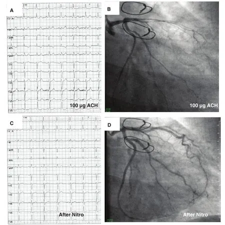
Figure 2 The ACH-test revealed diffuse spasm of the left anterior descending coronary artery with ischemic ECG shifts(A, B). The patient had reproduction of her symptoms during the test. The abnormal findings disappeared after intracoronary nitroglycerin injection (C, D).
The decision for the operation in our patient was justified given the proximal location without any calcification in a young patient. However, the aneurysm was unlikely to be responsible for the patient’s symptoms. In patients with angina pectoris and unobstructed coronary arteries it is important to look for other mechanisms of myocardial ischemia, such as vasospasm or microvascular dysfunction [9]. However, this is not yet part of standard workup in most centers, especially not in Europe or the United States. Yet, as shown in this case, coronary artery spasm is an important cause of resting angina. The confirmation of coronary spasm allows targeted pharmacological treatment with calcium channel blockers and nitrates, which often relieves symptoms. The demonstration of a coronary cause of the symptoms should also lead to meticulous treatment of risk factors, including lowering of LDL level (although the patient’s LDL level was already relatively low), which will ultimately improve prognosis.
Conflict of lnterest
The authors declare that they have no conflicts of interest.
 Cardiovascular Innovations and Applications2018年3期
- Cardiovascular Innovations and Applications的其它文章
- Cardiovascular Innovations and Applications
- The Contemporary Role of Femoral Artery Access
- Speckle Tracking Echocardiography ldentifies lmpaired Longitudinal Strain as a Common Deficit in Various Cardiac Diseases
- Bioresorbable Vascular Scaffold in the Midportion of the Left Anterior Descending Artery for Cardiac Allograft Vasculopathy in a Cardiac Transplant Patient
- The Use of Direct Oral Anticoagulants for Prevention of Stroke and Systemic Embolic Events in East Asian Patients with Nonvalvular Atrial Fibrillation
- Current Status of Coronary Atherectomy